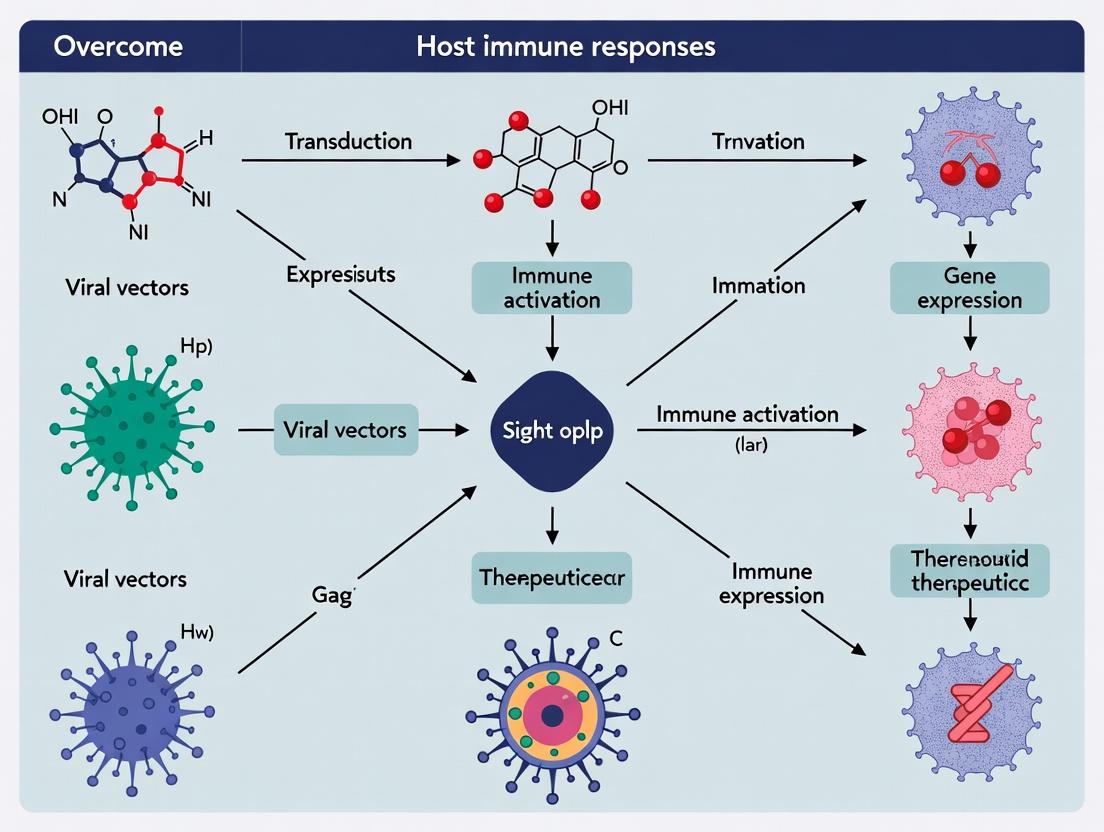
Breaking Immune Barriers: Advanced Strategies to Overcome Host Immune Responses in Gene Therapy
This article provides a comprehensive analysis of the critical challenge of host immune responses in gene therapy, addressing researchers, scientists, and drug development professionals.
Breaking Immune Barriers: Advanced Strategies to Overcome Host Immune Responses in Gene Therapy
Abstract
This article provides a comprehensive analysis of the critical challenge of host immune responses in gene therapy, addressing researchers, scientists, and drug development professionals. It first explores the foundational immunology of innate and adaptive responses against viral vectors and transgenes. It then details cutting-edge methodological approaches for immune evasion, including vector engineering and pharmacological modulation. The troubleshooting section addresses managing pre-existing immunity and cytokine storm risks, while the validation section compares the efficacy and immunogenicity of leading viral and non-viral platforms. Finally, the conclusion synthesizes a roadmap for clinical translation and highlights future research priorities for safer, more durable genetic medicines.
The Immune System vs. Gene Therapy: Understanding Innate and Adaptive Host Defenses
Technical Support Center
Troubleshooting Guides & FAQs
Q1: My in vitro human PBMC assay shows high variability in cytokine output (e.g., IL-6, IFN-β) when challenged with AAV capsids. What are the primary sources of this variability and how can I control for them? A: High variability often stems from donor-specific PRR expression profiles and pre-existing immunity. Key controls:
- Donor Screening: Pre-screen donor serum for neutralizing antibodies (NAbs) against your vector using a standardized NAb assay. Use only NAb-negative donors for baseline innate immunity studies.
- PRR Profiling: Quantify basal mRNA levels of key PRRs (e.g., TLR2, TLR9, cGAS) in PBMCs via qPCR before stimulation. Use donors with similar expression ranges.
- Endotoxin Control: Treat all vector preps with polymyxin B agarose or use a commercial endotoxin removal kit. Confirm LPS levels are <0.1 EU/mL using the LAL assay.
- Internal Calibration: Include a standardized control agonist (e.g., CpG-B for TLR9, dsDNA for cGAS) in each experiment to normalize donor responsiveness.
Q2: I am detecting unexpected cGAS/STING signaling in response to a "gutless" adenovirus vector. What are the likely causes and how can I confirm them? A: This indicates possible cytosolic DNA sensing. Follow this diagnostic protocol:
- Confirm DNA Presence: Isolate vector DNA and transfect it into HEK-293T reporter cells (e.g., STING-dependent IRF-luciferase). A positive signal confirms immunostimulatory DNA.
- Check for Contaminants: Run vector preps on an agarose gel and sequence to identify host cell DNA or plasmid backbone contaminants.
- Inhibition/Knockdown: Pre-treat target cells with a cGAS inhibitor (e.g., RU.521) or use siRNA against MB21D1 (cGAS). A significant reduction in IFN-β confirms the pathway.
- Electron Microscopy: Use EM to check for vector aggregation or particulate contaminants that could facilitate endosomal rupture and DNA leakage.
Q3: My nanoparticle-based gene therapy vector is triggering robust NLRP3 inflammasome activation, leading to pyroptosis. How can I engineer the particle to evade this specific sensor? A: NLRP3 is activated by lysosomal disruption and reactive oxygen species (ROS). Implement these modifications:
- Surface Coating: Apply a dense PEG layer or use "self" markers like CD47 to prevent phagolyososomal rupture.
- Material Selection: Switch from cationic polymers (which destabilize membranes) to zwitterionic or anionic lipids.
- ROS Scavengers: Co-encapsulate antioxidants like N-acetylcysteine or catalase within the nanoparticle core.
- Functional Testing: Use THP-1 NLRP3-reporter cells (ASC-citrine) to screen modified formulations. Measure IL-1β release via ELISA as a secondary readout.
Q4: When testing CRISPR/Cas9 ribonucleoprotein (RNP) complexes, I observe a dose-dependent RIG-I/MAVS-mediated type I interferon response. Is this from the guide RNA, the Cas9 protein, or both? A: It is likely from the in vitro transcribed (IVT) guide RNA possessing a 5'-triphosphate (5'-ppp). Conduct this deconvolution experiment:
- Treat gRNA: Phosphatase (CIP) treatment of gRNA to remove 5'-ppp.
- Transfert Components Separately: Transfert cells with:
- a) Untreated gRNA
- b) CIP-treated gRNA
- c) Cas9 protein alone
- d) Synthetic, chemically modified gRNA (with 2'-O-methyl 3' ends)
- Measure Output: Quantify IFN-β mRNA at 6h post-transfection. The response will be abrogated in conditions b and d.
Key Experimental Protocols
Protocol 1: Quantifying Innate Immune Sensor Activation by Viral Vectors in Primary Human Macrophages
- Objective: Measure PRR pathway-specific cytokine/chemokine secretion post-vector exposure.
- Method:
- Isolate CD14+ monocytes from human PBMCs using magnetic beads. Differentiate into macrophages with 100 ng/mL GM-CSF for 6 days.
- Pre-treat cells for 1h with pathway-specific inhibitors: TLR9 (ODN TTAGGG), cGAS (RU.521), or NLRP3 (MCC950). Include DMSO vehicle control.
- Transduce cells with your gene therapy vector (e.g., AAV, LV) at a range of MOIs (1e3, 1e4, 1e5 vg/cell). Include LPS (TLR4) and Transfection Reagent only as controls.
- At 24h, collect supernatant for Luminex multiplex assay (IL-1β, IL-6, TNF-α, IP-10, IFN-α2a).
- Perform intracellular staining for phospho-IRF3 and phospho-NF-κB p65 via flow cytometry at 6h.
- Analysis: Compare inhibitor conditions to vehicle control to attribute cytokine signatures to specific PRRs.
Protocol 2: In Vivo Profiling of the Initial Inflammatory Cascade Post Systemic Vector Administration
- Objective: Track temporal cytokine and immune cell recruitment dynamics in a murine model.
- Method:
- Administer vector or saline control intravenously to C57BL/6 mice (n=5/group/timepoint).
- Collect serum at T=1, 3, 6, 12, 24, 48 hours post-injection.
- Analyze serum using a mouse cytokine 23-plex panel (e.g., GM-CSF, IFN-γ, IL-1α, IL-6, KC, MCP-1).
- At 24h, perfuse and harvest liver/spleen. Process into single-cell suspensions.
- Stain for flow cytometry: Ly6C/G (neutrophils), F4/80 (macrophages), CD11c (DCs), NK1.1 (NK cells), CD3 (T cells).
- Analysis: Generate a heatmap of cytokine kinetics and correlate peak cytokine levels (e.g., KC, MCP-1) with specific immune cell infiltration at 24h.
Table 1: Common PRRs and Their Cognate Agonists in Gene Therapy Vectors
| PRR (Sensor) | Location | Pathogen-Associated Molecular Pattern (PAMP) | Common Vector Source | Typical Readout (Measurement) |
|---|---|---|---|---|
| TLR9 | Endosome | Unmethylated CpG DNA | AAV genome, plasmid DNA contaminants | IFN-α (pg/mL), Plasmacytoid DC activation |
| cGAS | Cytosol | dsDNA >45 bp, DNA in micronuclei | Lentivirus pre-integration complex, free vector DNA | cGAMP (nM), IFN-β mRNA fold change |
| RIG-I | Cytosol | 5'-triphosphate RNA, short dsRNA | IVT guide RNA, vector-derived RNA | IFN-β (pg/mL), IRF3 phosphorylation |
| NLRP3 | Cytosol | Lysosomal disruption, ROS, crystalline structures | Aggregated protein, cationic nanoparticle surfaces | IL-1β (pg/mL), Caspase-1 activity |
| TLR2/TLR4 | Plasma Membrane | Protein/lipid motifs (e.g., from capsid) | Adenovirus hexon, AAV capsid impurities | TNF-α (pg/mL), NF-κB activation |
Table 2: Efficacy of Common Inhibitors in Suppressing PRR Pathways In Vitro
| Inhibitor | Target PRR Pathway | Recommended Conc. | Cell Type Tested | % Inhibition of IFN-β/IL-6* | Key Consideration |
|---|---|---|---|---|---|
| Chloroquine | Endosomal TLRs (e.g., TLR9) | 10-50 µM | HEK-TLR9 Reporter, pDCs | 85-95% | Alters endosomal pH; cytotoxic at high doses. |
| RU.521 | cGAS | 1-5 µM | THP-1, Primary Macrophages | 70-80% | Highly specific; does not affect RIG-I or TLR signaling. |
| ODN TTAGGG | TLR9 Antagonist | 5-10 µM | Human PBMCs, B cells | 60-75% | Competitive inhibitor; species-specific (human). |
| MCC950 | NLRP3 Inflammasome | 100 nM | BMDMs, THP-1 | >95% (IL-1β) | Does not inhibit AIM2 or NLRC4 inflammasomes. |
| BX795 | TBK1 (downstream of cGAS/RIG-I) | 1 µM | Fibroblasts, Epithelial cells | 90-98% | Broad kinase inhibitor; affects other pathways. |
*Data represent typical ranges from published literature; actual values are experiment-dependent.
The Scientist's Toolkit: Research Reagent Solutions
| Item | Function in PRR/Innate Immunity Research |
|---|---|
| HEK-Blue TLR Reporter Cells | Engineered cells expressing a single TLR and a secreted embryonic alkaline phosphatase (SEAP) reporter inducible by NF-κB/IRFs. Used for specific, quantitative TLR signaling assays. |
| Human/Mouse Cytokine Multiplex Assay (Luminex) | Allows simultaneous quantification of up to 50+ cytokines/chemokines from a small volume of cell supernatant or serum. Critical for mapping inflammatory cascades. |
| cGAMP ELISA Kit | Directly measures the second messenger 2'3'-cGAMP produced by activated cGAS, providing definitive proof of cGAS-STING pathway engagement. |
| Phospho-IRF3 (Ser396) Antibody | For Western blot or flow cytometry to detect activation and nuclear translocation of IRF3, a key transcription factor downstream of TLR3/4, RIG-I, and cGAS-STING. |
| ASC Speck Staining Antibody | Visualizes the large ASC oligomers formed during inflammasome activation (e.g., NLRP3), a hallmark of pyroptotic signaling. |
| Endotoxin Removal Resin (e.g., Triton X-114, polymyxin B) | Essential for removing immunostimulatory LPS from protein/vector preparations, eliminating a major confounder in innate sensing experiments. |
| DNase I & RNase A | Used to pre-treat vector stocks to determine if immune activation is nucleic acid-dependent. A loss of signal after treatment implicates DNA/RNA as the PAMP. |
Visualizations
Title: PRR Sensing of Gene Therapy Vectors & Signaling Pathways
Title: Troubleshooting Workflow for Unwanted PRR Activation
Technical Support Center: Troubleshooting Guide for Immune Monitoring in Gene Therapy
FAQ & Troubleshooting
Q1: In our murine model, we are detecting high-titer neutralizing antibodies (NAbs) against our AAV vector post-administration, despite using a serotype thought to be low prevalence. How can we confirm pre-existing immunity vs. therapy-elicited responses?
A: This is a common issue. A systematic approach is needed.
- Troubleshooting Steps:
- Pre-dose Screening: Collect and bank pre-dose serum from all experimental animals. Use a Luciferase-based Neutralization Assay (see protocol below) on these samples. A titer >1:5 is typically indicative of pre-existing immunity.
- Post-dose Kinetics: Compare pre-dose titers with samples from Day 7 and Day 14 post-administration. A rapid, high-titer response by Day 7 often suggests pre-existing memory B-cell activation. A slower rise peaking around Day 14-28 is more indicative of a de novo elicited primary adaptive response.
- Cross-reactivity Check: Ensure your assay is specific. Test against a panel of related AAV serotypes to rule out assay cross-reactivity.
- Experimental Protocol: Luciferase-based NAb Assay
- Serum Heat-Inactivation: Incubate serum at 56°C for 30 min.
- Serial Dilution: Perform 2-fold serial dilutions of serum in DMEM (e.g., 1:5 to 1:2560) in a 96-well plate.
- Virus Incubation: Mix diluted serum with AAV-Luciferase vector (2e8 vg/well) and incubate at 37°C for 1 hr.
- Cell Infection: Add mixture to HEK293 cells (80% confluent) in quadrup licate. Include virus-only (no serum) and cell-only controls.
- Readout: After 48 hrs, lyse cells and measure luminescence.
- Calculation: NAb titer is reported as the dilution that inhibits luminescence by ≥50% (IC50) relative to virus-only control.
Q2: Our ELISpot data for IFN-γ T-cell responses against the transgene product is inconsistent with our flow cytometry intracellular cytokine staining (ICS). Which assay is more reliable?
A: They measure different but related aspects of cellular immunity. Discrepancies are informative.
- Troubleshooting Guide:
| Aspect | ELISpot | Intracellular Cytokine Staining (ICS) by Flow |
|---|---|---|
| Primary Readout | Frequency of cytokine-secreting cells. | Frequency of cytokine-producing cells, plus phenotyping (CD4/CD8, memory subsets). |
| Sensitivity | Very high, detects low-frequency responses. | Moderate, requires larger cell numbers. |
| Multiplexing | Low (typically 1-2 cytokines/assay). | High (≥8 parameters: cytokines, surface markers). |
| Common Pitfall | Over-counting due to non-specific secretion or poor cell viability. | Loss of signal due to over-fixing/permeabilization; requires robust stimulation and Golgi block. |
| Recommendation | Use ELISpot for initial immunogenicity screening. Use multicolor ICS to characterize the phenotype and polyfunctionality (IFN-γ, TNF-α, IL-2) of responding T-cells identified by ELISpot. Always use overlapping peptide pools spanning the entire transgene for stimulation. |
Q3: We suspect T-cell-mediated clearance of transduced cells. What is the best method to detect antigen-specific CD8+ T cells in tissues?
A: For tissue residency, combine tetramer staining with ex vivo functional assays.
- Detailed Protocol: MHC Class I Tetramer Staining from Liver/Spleen Lymphocytes
- Peptide Identification: Use in silico prediction tools (NetMHC) to identify immunodominant epitopes from your transgene restricted to your model's MHC (e.g., H-2Kb for C57BL/6).
- Tetramer Procurement: Order PE- or APC-conjugated tetramers loaded with your peptide.
- Single-Cell Suspension: Perfuse liver with PBS, dissociate, and isolate mononuclear cells via 40%/70% Percoll gradient centrifugation.
- Tetramer Stain: Incubate 1e6 cells with tetramer (1:100 dilution) in FACS buffer for 20 min at 4°C in the dark.
- Surface Stain: Add antibodies (anti-CD3, anti-CD8, anti-CD62L, anti-CD44) for 20 min at 4°C.
- Analysis: Gate on live, singlet, CD3+CD8+ cells. Tetramer+ cells are antigen-specific. CD44+CD62L- indicates an effector memory phenotype, suggestive of recent activation.
Research Reagent Solutions Toolkit
| Reagent/Category | Example Product/Assay | Function in Immune Monitoring |
|---|---|---|
| Neutralization Assay Kits | Promega AAVanced Neutralization Assay | Standardized, luciferase-based kit for quantifying anti-AAV NAb titers in serum/plasma. |
| Peptide Libraries | JPT Peptide Pools (15mer, 11aa overlap) | Overlapping peptides spanning the transgene for comprehensive T-cell stimulation in ELISpot/ICS. |
| MHC Tetramers | MBL International Tetramers | Fluorochrome-labeled tetramers for direct staining and quantification of antigen-specific CD4+/CD8+ T cells. |
| Cytokine Reagents | Mabtech ELISpot kits (IFN-γ, IL-4, etc.) | Pre-coated plates and paired antibodies for sensitive detection of cytokine-secreting cells. |
| Flow Cytometry Antibodies | BioLegend TruStain FcX + Anti-mouse CD3, CD4, CD8, CD44, CD62L, IFN-γ, TNF-α | Antibody panels for immunophenotyping and intracellular cytokine analysis of T-cell responses. |
| Immune Cell Isolation Kits | Miltenyi Biotec CD8a+ T Cell Isolation Kit | Magnetic bead-based negative selection for isolating specific lymphocyte populations from tissues. |
Diagrams
Immune Response Pathways Post-Gene Therapy
Immune Monitoring Experimental Workflow
Welcome to the Technical Support Center for managing neutralizing antibody (NAb) responses in gene therapy research. This resource is designed to help you troubleshoot specific challenges related to host immune responses against viral vectors, framed within the critical goal of overcoming immunological barriers for successful clinical translation.
Troubleshooting Guides & FAQs
Q1: My preclinical gene therapy study in mice shows a sharp decline in transgene expression after re-administration of the same AAV serotype. What is the likely cause and how can I confirm it? A: This is a classic indicator of a T-cell dependent, NAb-mediated immune response. Initial exposure primes the adaptive immune system, generating memory B cells. Upon re-administration, a rapid anamnestic response produces high-titer NAbs that neutralize the vector before it can transduce target cells.
- Confirmation Protocol: Collect serum 7-14 days post-re-administration.
- Perform an In Vitro Neutralization Assay:
- Day 1: Seed HEK293 cells in a 96-well plate.
- Day 2: Prepare serial dilutions of the test serum (e.g., 1:2 to 1:512) in culture medium. Incubate a fixed dose of AAV vector (e.g., 1e9 vg) with each serum dilution for 1 hour at 37°C. Include a no-serum control (virus only) and a no-virus control.
- Add serum-vector mixtures to cells.
- Day 3+ (48-72h later): Quantify transduction using the relevant readout (e.g., fluorescence intensity for GFP, luciferase activity). A ≥50% reduction in signal compared to the virus-only control confirms neutralizing activity. The titer is reported as the reciprocal of the highest dilution achieving this inhibition.
- Perform an In Vitro Neutralization Assay:
Q2: I am planning a clinical trial for an AAV-based therapy. How do I determine the NAb cut-off titer for patient exclusion, and what are the current standard thresholds? A: The cut-off is serotype-specific and aims to identify patients whose pre-existing NAbs would likely preclude therapeutic efficacy. The gold standard is a cell-based neutralization assay. Industry consensus, supported by recent clinical data, suggests the following thresholds:
Table 1: Common Clinical NAb Titer Thresholds for AAV Serotypes
| AAV Serotype | Common Cut-off Titer (Reciprocal IC50) | Clinical Rationale |
|---|---|---|
| AAV2 | 1:2 to 1:5 | High prevalence of pre-existing immunity in population. |
| AAV5 | 1:2 | Lower seroprevalence, but still significant. |
| AAV8 | 1:5 | Used frequently in hepatic gene transfer. |
| AAV9 | 1:5 | Common for systemic/CNS-targeted therapies. |
Protocol Note: Use validated assays with appropriate controls (human positive/negative reference sera). The trend is towards standardized, high-sensitivity assays to harmonize exclusion criteria across trials.
Q3: Can I use immunosuppressants like prednisone to manage NAb responses in a non-human primate study, and what is the typical regimen? A: Yes, prophylactic immunosuppression is a common strategy to blunt the humoral response. A typical protocol is:
- Regimen: Administer oral prednisone (or methylprednisolone) starting on the day of vector infusion. A common dose is 1 mg/kg/day for 30 days, followed by a 4-week taper.
- Monitoring: Measure NAb titers at baseline, weekly during dosing, and at the end of the taper. Compare to a non-immunosuppressed control cohort. This approach primarily targets T-helper cell support for B-cell activation, potentially delaying or reducing high-titer NAb formation but may not prevent it entirely.
Q4: My lentiviral vector (LV) transduction efficiency is low in primary human T-cells from some donors. Could pre-existing antibodies be the cause? A: While NAbs against LV are less common than for AAV, they can occur, particularly against envelope proteins (e.g., VSV-G). However, low transduction in T-cells is more frequently due to:
- Innate immune sensing: Cytoplasmic nucleic acid sensors (cGAS-STING) can detect viral DNA/RNA, triggering an antiviral state.
- Interferon response: Type I IFNs can inhibit LV transduction.
- Troubleshooting Steps:
- Test for NAbs using an in vitro assay on permissive HEK293T cells with donor serum.
- Incorporate a small molecule inhibitor of innate sensing (e.g., a STING inhibitor) during transduction to see if efficiency improves.
- Use a different pseudotype (e.g., switch VSV-G to RD114) to evade potential serotype-specific antibodies.
The Scientist's Toolkit: Research Reagent Solutions
Table 2: Essential Reagents for NAb Research
| Reagent / Material | Function & Application |
|---|---|
| Reference Standard Sera | Validated positive (high-titer NAb) and negative (no NAbs) controls for assay calibration and qualification. |
| Reporter Gene AAV/LV Particles | Vectors encoding luciferase, GFP, or secreted alkaline phosphatase (SEAP) for quantitative, high-throughput neutralization assays. |
| Permissive Cell Lines (HEK293, HeLa) | Standardized cells for in vitro NAb assays, ensuring consistent transduction and reporter readout. |
| cGAS/STING Pathway Inhibitors | Small molecules (e.g., H-151, RU.521) to suppress innate immune sensing of viral vectors, improving transduction. |
| Recombinant IFN-β/IFN-γ | To spike into assays as positive controls for establishing an antiviral state and validating assay sensitivity. |
| Anti-Human IgG (Fc-specific) Secondary Antibodies | For ELISA-based total antibody detection, which can be a precursor/surrogate for NAb screening. |
Visualization: Experimental and Conceptual Workflows
Title: NAb Anamnestic Response Blocks Vector Re-administration
Title: Cell-Based Neutralizing Antibody Assay Workflow
Troubleshooting Guides & FAQs
FAQ 1: Why is my transgene product not detectable in vivo despite successful in vitro expression?
- Answer: This is a classic sign of a cytotoxic T lymphocyte (CTL)-mediated immune response. CD8+ T cells recognize the transgene product peptides presented on MHC I and eliminate the transduced cells. Monitor for an initial expression peak followed by a rapid decline.
- Troubleshooting Protocol:
- Assay: Perform an IFN-γ ELISpot or intracellular cytokine staining (ICS) assay on splenocytes from treated animals, using peptides spanning the transgene sequence.
- Control: Include a known immunogenic peptide as a positive control and an irrelevant peptide as a negative control.
- Confirm: Use tetramer staining for direct visualization of transgene-specific CD8+ T cells. Correlate T cell detection with the loss of transgene expression measured by qPCR or ELISA.
FAQ 2: How can I distinguish between pre-existing humoral immunity and a de novo antibody response against the transgene?
- Answer: Pre-existing antibodies (e.g., against common viral capsids like AAV) are present in serum before vector administration. De novo antibodies against the transgene product appear after treatment and increase in titer over time.
- Troubleshooting Protocol:
- Baseline Titer: Collect pre-bleed serum from all experimental subjects.
- Longitudinal Sampling: Collect serum at regular intervals post-treatment (e.g., weeks 2, 4, 8).
- Assay: Perform an antigen-specific ELISA or electrochemiluminescence (ECL) assay on all serum samples. A ≥4-fold increase in titer from baseline confirms a de novo response.
FAQ 3: My immunosuppression regimen is not preventing antibody formation. What are potential mechanisms?
- Answer: Standard calcineurin inhibitors (e.g., Tacrolimus) primarily suppress T-cell help. Failure suggests:
- T-cell independent B-cell activation (for multimeric transgene products).
- Insufficient depletion of pre-existing memory B cells.
- Incomplete inhibition of co-stimulatory signals (e.g., CD40-CD40L).
- Troubleshooting Protocol: Implement a combinatorial regimen:
- Add Anti-CD20: Administer (e.g., Rituximab) to deplete B cells before vector administration.
- Add BAFF/APRIL Inhibition: Use a TACI-Ig fusion protein (e.g., Telitacicept) to block B-cell survival factors.
- Monitor: Track naive, memory, and plasma B cell subsets by flow cytometry alongside antibody titers.
FAQ 4: What are the best practices to monitor for regulatory T cell (Treg) induction as a tolerance strategy?
- Answer: Successful Treg induction is marked by an increase in antigen-specific, Foxp3+ CD4+ T cells that can suppress effector responses in vitro and in vivo.
- Troubleshooting Protocol:
- Identification: Isolate CD4+ T cells from lymphoid tissues. Use tetramers for antigen-specificity or stain for activation markers (CD25, CD134). Intranuclear stain for Foxp3 is essential.
- Function: Perform an in vitro suppression assay. Co-culture sorted Tregs with CFSE-labeled effector T cells (Teffs) and antigen-presenting cells + antigen. Measure Teff proliferation (CFSE dilution) via flow cytometry.
- Stability: Check demethylation status of the Treg-specific demethylated region (TSDR) in the Foxp3 locus via bisulfite sequencing.
Table 1: Efficacy of Common Immunosuppressants in Gene Therapy Models
| Immunosuppressive Agent | Target | Reduction in Antibody Titer (%) | Reduction in Antigen-Specific T cells (%) | Key Study Model |
|---|---|---|---|---|
| Tacrolimus | Calcineurin (NFAT) | 40-60 | 60-80 | Mouse, AAV-FIX |
| Mycophenolate Mofetil | IMPDH (Lymphocyte proliferation) | 30-50 | 50-70 | NHP, AAV-LSD |
| Anti-CD20 (Rituximab) | CD20 (B cells) | 70-90 | N/A | Mouse/Canine, AAV-FIX |
| CTLA4-Ig (Abatacept) | CD80/86 (Co-stimulation) | 50-80 | 70-85 | Mouse, AAV-mAb |
| Treg Adoptive Transfer | N/A (Tolerance) | 85-95 | 90-98 | Mouse, AAV-Hemoglobin |
Table 2: Impact of Vector/Transgene Parameters on Immunogenicity
| Parameter | High Immunogenicity Risk | Low Immunogenicity Risk | Relative Risk Increase (Fold) |
|---|---|---|---|
| Promoter | CMV, Viral | Tissue-specific, Endogenous | 3-5 |
| Capsid Serotype | AAV2, AAV8* | AAV-LK03, AAVrh.74 | 2-4 |
| Transgene Origin | Non-self, Microbial | Species-specific, Humanized | 10-100 |
| Dose | High (>1e14 vg/kg) | Low (<1e12 vg/kg) | 5-10 |
| Route | Intramuscular, Subcutaneous | Intravenous, Liver-directed | 2-3 |
*Species-dependent; AAV8 is less immunogenic in mice but can be highly immunogenic in NHPs/humans.
Experimental Protocols
Protocol 1: Comprehensive Immune Monitoring Workflow Post-Gene Therapy
- Sample Collection: At baseline, week 2, 4, 8, and 12 post-vector administration, collect blood (serum & PBMCs), target tissue biopsy, and draining lymph node.
- Humoral Response:
- Process serum. Run antigen-specific ELISA. Report endpoint titer as the reciprocal dilution giving an OD value 3x above pre-bleed.
- Cellular Response (PBMCs):
- Isolate PBMCs via density gradient centrifugation.
- IFN-γ ELISpot: Plate 2e5 PBMCs/well with overlapping transgene peptides (15-mers, 11-aa overlap). Develop after 48h. Count spots using an automated reader.
- Flow Cytometry: Stimulate PBMCs with peptides for 6h in the presence of brefeldin A. Stain for surface markers (CD3, CD4, CD8) and intracellular cytokines (IFN-γ, TNF-α, IL-2).
- Transgene Expression (Tissue):
- Homogenize biopsy sample. Extract RNA and protein.
- Perform qRT-PCR for transgene mRNA (normalize to GAPDH).
- Perform Western Blot or ELISA for transgene protein.
Protocol 2: Induction of Antigen-Specific Tolerance via Hepatic Gene Transfer This protocol leverages the liver's inherent tolerogenic microenvironment.
- Vector Design: Use a hepatocyte-specific promoter (e.g., LP1, TBG) to drive transgene expression in an AAV8 or AAV-LK03 capsid.
- Administration: Inject vector intravenously via the tail vein (mouse) or peripheral vein (large animal) at a dose of 5e11 – 1e12 vg/kg.
- Co-treatment: Administer a brief, low-dose course of Rapamycin (0.1 mg/kg/day, IP, days 0-7) to promote Treg differentiation.
- Validation: At week 4, challenge the subject with the transgene protein formulated in adjuvant (e.g., subcutaneous injection with Complete Freund's Adjuvant).
- Assessment: Measure antibody and T-cell responses 2 weeks post-challenge. A tolerant subject will show minimal/no recall response compared to a naive subject given the same challenge.
Visualizations
Diagram 1: Immune Recognition Pathway of Transgene Product
Diagram 2: Immune Monitoring Experimental Workflow
The Scientist's Toolkit: Research Reagent Solutions
Table 3: Essential Reagents for Studying Transgene Immunogenicity
| Reagent / Material | Function / Application | Example Product/Catalog |
|---|---|---|
| Overlapping Peptide Library | Span the entire transgene sequence to map T cell epitopes via ELISpot or ICS. | JPT PepTivator or custom synthesis (15-mers, 11-aa overlap). |
| MHC Tetramers (PE/APC) | Direct ex vivo detection and isolation of transgene-specific CD4+ or CD8+ T cells. | Custom-made by NIH Tetramer Core or MBL International. |
| Anti-Cytokine Antibodies (with clones) | For intracellular cytokine staining (ICS) to detect IFN-γ, TNF-α, IL-2 in T cells. | BioLegend: anti-IFN-γ (XMG1.2), anti-TNF-α (MP6-XT22). |
| Lymphocyte Separation Medium | Isolate viable PBMCs or splenocytes from whole blood or tissue for functional assays. | Corning Ficoll-Paque PREMIUM or Cytiva Lymphoprep. |
| ELISpot Kit (Mouse/Human IFN-γ) | Sensitive detection of low-frequency, antigen-specific T cells secreting cytokine. | Mabtech IFN-γ ELISpotPRO kit (pre-coated plates). |
| Recombinant Transgene Protein | Critical positive control for antibody ELISA and for in vitro B cell assays. | Produce in-house (HEK293) or source from a reliable recombinant provider. |
| Foxp3 / Transcription Factor Staining Buffer Set | For proper intracellular staining of Treg master regulator Foxp3 and other TFs. | Thermo Fisher eBioscience Foxp3/Transcription Factor Staining Buffer Set. |
| In Vivo Antibodies (Depleting/Blocking) | To test mechanistic roles of immune cells (e.g., anti-CD4, anti-CD8, anti-CD20). | Bio X Cell: InVivoPlus anti-mouse CD4 (GK1.5), CD8α (2.43). |
The Role of the Complement System and Other Effector Mechanisms in Clearance and Toxicity
Troubleshooting & FAQs for Gene Therapy Researchers
Framed within the context of overcoming host immune responses in gene therapy research.
Frequently Asked Questions
Q1: In our murine study, we observed rapid clearance of our AAV8 vector despite high initial titers. Neutralizing antibody tests are negative. What could be the cause? A: This is a classic sign of complement system activation. The AAV capsid can be directly recognized by natural IgM antibodies or bind Complement Factor H inadequately, leading to alternative pathway activation. This results in opsonization (C3b) and clearance primarily by Kupffer cells in the liver, independent of pre-existing neutralizing IgG.
- Solution: Pre-treat animals with a complement inhibitor (e.g., CP40, an analog of compstatin that targets C3) 30 minutes prior to vector administration. Monitor serum C3a levels as a biomarker of activation.
Q2: Our lipid nanoparticle (LNP)-mRNA formulation shows high efficacy in vitro but severe inflammatory toxicity in non-human primates. How can we determine if the complement is involved? A: Complement Activation-Related Pseudoallergy (CARPA) is common with nanoparticulate systems. Perform an in vitro complement activation assay.
- Protocol:
- Incubate your LNP formulation (at clinical dose concentration) with 10% fresh primate serum in veronal buffer with Ca2+ and Mg2+.
- Incubate at 37°C for 1 hour.
- Use ELISA kits to quantify generation of anaphylatoxins C3a and C5a, and the terminal soluble complement complex SC5b-9.
- Compare to a negative control (buffer) and a positive control (zymosan).
Q3: We see variability in adenovirus vector toxicity between mouse strains. Which effector mechanisms should we profile? A: Strain variability often points to innate immune sensing. Profile both complement and other effector mechanisms like macrophages and neutrophils.
- Key Analyses:
- Complement: Measure C3 deposition on the vector via flow cytometry after serum incubation.
- Cytokines: Check for IL-6, TNF-α, and IL-1β spikes 2-6 hours post-injection (innate sensor-driven).
- Cellular Effectors: Use flow cytometry of blood/biodistribution samples to assess neutrophil (Ly6G+) activation (CD11b upregulation) and monocyte/macrophage (CD11b+, F4/80+) recruitment.
Q4: How can we differentiate between complement-mediated and anti-drug antibody (ADA)-mediated clearance of our gene therapy product in a repeat-dose study? A: Temporal kinetics and biomarker profiling are key. See the comparative table below.
| Mechanism | Primary Quantitative Assay | Key Biomarker(s) | Typical Timeframe Post-Dose | Common Intervention |
|---|---|---|---|---|
| Classical Complement | C1q or C4d deposition ELISA | ↑sC4b, ↑C1q-protein complex | Minutes to hours | PEGylation, sialic acid capsid engineering |
| Alternative Complement | C3 deposition flow cytometry | ↑C3a, ↑C3b deposition, ↓Factor H binding | Minutes to hours | Compstatin analogs, FH-mimetic peptides |
| Lectin Pathway | MBL/MASP-2 binding ELISA | ↑C4a, ↑MASP-2 complex | Minutes to hours | Mannose shield modification |
| Anti-Drug Antibodies (ADA) | Tiered Immunogenicity Assay | ↑Anti-capsid or Anti-transgene IgG/IgM | Days to weeks (memory: faster) | Immunosuppression (e.g., prednisone), capsid switching |
| Cellular Effectors (Kupffer Cells, Macrophages) | In vivo phagocytosis assay, cytokine panel | ↑IL-6, TNF-α, ↑vector colocalization w/ CD68+ cells | Hours to days | Clodronate liposomes, transient Kupffer cell depletion |
Experimental Protocols
Protocol 1: Assessing Complement Deposition on Viral Vectors via Flow Cytometry Objective: Quantify C3b/iC3b opsonization on viral capsids. Materials: Purified vector (e.g., AAV, Adenovirus), normal human serum (NHS), heat-inactivated serum (control), anti-C3c/C3b/iC3b antibody (fluorophore-conjugated), flow cytometry buffer (PBS + 1% BSA). Method:
- Dilute 1x10^8 vector genomes in 50 µL PBS.
- Incubate with 10% NHS or heat-inactivated serum (30 µL) for 30 minutes at 37°C.
- Wash particles 3x using 100kDa MWCO centrifugal filters to remove unbound serum proteins.
- Resuspend pellet and incubate with anti-C3 antibody (1:100 dilution) for 30 min on ice, protected from light.
- Wash twice, resuspend in 200 µL buffer.
- Run on flow cytometer. Analyze shift in fluorescence intensity of the vector particle population relative to the heat-inactivated serum control.
Protocol 2: In Vivo Assessment of Complement Contribution to Clearance Objective: Determine the fraction of vector clearance attributable to complement. Materials: C57BL/6 mice, your gene therapy vector, Compstatin derivative Cp40 (or vehicle), qPCR equipment, primers for vector genome. Method:
- Randomize mice into two groups (n=5): Test Group: Inject Cp40 (2 mg/kg, IP) 30 min before vector. Control Group: Inject vehicle.
- Administer vector via intended route (e.g., IV tail vein).
- Collect target tissue (e.g., liver) at 24h and 7 days post-injection.
- Extract total DNA. Perform absolute qPCR to quantify vector genome copies per µg of genomic DNA.
- Calculation: % Complement Contribution = [1 - (Genome copies in Cp40 group / Genome copies in Control group)] * 100 at 24h.
The Scientist's Toolkit: Research Reagent Solutions
| Reagent / Material | Function in Context of Immune Clearance Research |
|---|---|
| Compstatin (Cp40) & Analogs | Potent peptidic inhibitor of C3 cleavage; used to specifically block all complement pathways in vivo/in vitro. |
| C1 Esterase Inhibitor (C1-INH) | Serpin that inhibits classical/lectin pathway proteases (C1r, C1s, MASP-2); used to identify pathway involvement. |
| Clodronate Liposomes | Depletes phagocytic cells (Kupffer cells, macrophages) upon intravenous injection; assesses role of cellular clearance. |
| Anti-C5 Monoclonal Antibody (Eculizumab) | Blocks terminal pathway and C5a generation; used to investigate anaphylatoxin-mediated toxicity and membrane attack complex (MAC) effects. |
| Factor H-Depleted Serum | Commercially available serum to specifically study dysregulation of the Alternative Pathway. |
| Anaphylatoxin ELISA Kits (C3a, C5a, SC5b-9) | Quantify complement activation products in serum or plasma as pharmacodynamic/toxicodynamic biomarkers. |
| PEGylation Reagents (e.g., mPEG-NHS) | Conjugate polyethylene glycol to vector surfaces to create a "stealth" effect, reducing complement and antibody recognition. |
Pathway & Workflow Diagrams
Engineering Immune Evasion: Cutting-Edge Techniques for Stealthier Gene Delivery
Technical Support Center: Troubleshooting & FAQs
This support center addresses common experimental challenges in engineering viral capsids for reduced immunogenicity within gene therapy workflows.
Frequently Asked Questions (FAQs)
Q1: Our directed evolution screen yields capsid variants with improved in vitro transduction, but they fail in vivo due to rapid neutralization. What could be the issue? A: This is a classic pitfall. In vitro screens often lack immune system components. The selected variants may have improved receptor binding but remain highly visible to pre-existing neutralizing antibodies (NAbs) or the complement system. You must incorporate an immune selection pressure into your screening protocol. Consider using in vivo selection (e.g., in mouse models with humanized liver or pre-existing immunity) or ex vivo selection with pooled human immunoglobulins (IVIG), convalescent sera, or complement proteins during the panning rounds.
Q2: During AAV library production, we observe a significant drop in viral titer compared to wild-type. Is this normal? A: Yes, this is expected. Random peptide insertions or mutagenesis within the cap gene can severely disrupt capsid assembly, stability, or genome packaging. A titer reduction of 1-2 logs is common. Ensure you are using a robust transfection system (e.g., PEIpro or PEI-Max) at optimal ratios and include a replication-competent adenovirus (RCV) control if using the triple-transfection method for AAV. Purify the library using an iodixanol gradient to remove empty capsids and recover functional particles.
Q3: How do we validate that a "stealth" capsid variant truly has reduced immunogenicity, not just altered tropism? A: You need a multi-faceted assay suite. First, confirm tropism via qPCR/PCR on genomic DNA from target vs. non-target tissues. Then, assess immunogenicity directly:
- NAb Assay: Compare serum neutralization of your variant vs. wild-type using a standardized in vitro luciferase reporter assay on permissive cells.
- Cellular Immune Response: Use ELISpot to measure T-cell responses (IFN-γ) from PBMCs or splenocytes of exposed animals against capsid peptides.
- Capsid Antigen Persistence: Measure clearance kinetics via ELISA for AAV capsid in serum.
Q4: Our engineered capsid shows promising data in murine models, but how predictive is this for human immune system evasion? A: Murine models have limitations due to differences in the immune system and lack of human historical pathogen exposure. Data must be considered preliminary. Always follow up with ex vivo human assays: test neutralization using a diverse panel of human sera (naive and pre-immunized) and evaluate T-cell responses using human PBMCs from multiple donors. In vivo models incorporating human immunoglobulin transgenes or humanized immune systems are the next step for validation.
Q5: We identified a key residue for immune evasion via alanine scanning. How can we investigate if this affects receptor binding? A: Perform a combination of structural and functional assays. First, conduct a structural modeling analysis (e.g., using PyMOL with a published capsid structure) to see if the residue is located in a known receptor binding footprint. Functionally, run a competition assay: pre-incubate the virus with soluble receptor (like AAVR-Fc for AAVs) and measure reduction in transduction. Also, perform a heparin binding assay (for AAVs that bind HSPG) via heparin affinity chromatography to check for affinity changes.
Troubleshooting Guides
Issue: Low Diversity in Post-Selection Capsid Library
- Potential Cause 1: Overly stringent selection pressure (e.g., too high antibody concentration).
- Solution: Titrate the immune selection reagent (e.g., IVIG). Use a lower concentration in early selection rounds and increase stringency progressively.
- Potential Cause 2: Bottleneck during in vivo selection.
- Solution: Increase the number of animals used for library passage and ensure the input library diversity is high (>10^11 unique variants). Pool organs from all animals before DNA extraction.
- Potential Cause 3: PCR bias during recovery of cap genes.
- Solution: Use high-fidelity polymerase, minimize PCR cycles, and perform multiple parallel PCR reactions. Consider using restriction enzyme-based library recovery if your design allows.
Issue: High Background in Neutralizing Antibody (NAb) Assay
- Potential Cause 1: Non-specific cytotoxicity from serum components.
- Solution: Heat-inactivate all sera (56°C, 30 min) and use a serum-free medium during the virus-serum incubation step. Include a "no serum" control and a "serum-only on cells" control.
- Potential Cause 2: Poor reproducibility of transduction.
- Solution: Standardize your reporter virus (GC titer), cell seeding density, and incubation time. Use an internal control (e.g., a non-neutralizable virus with a different reporter if available) to normalize for cell health and transduction variability.
Issue: Inconsistent In Vivo Transduction Efficiency with Stealth Variants
- Potential Cause 1: Unstable capsid leading to particle degradation.
- Solution: Analyze capsid integrity via negative stain electron microscopy, SDS-PAGE/Coomassie for VP ratio, and a thermal stability assay (e.g., using differential scanning fluorimetry).
- Potential Cause 2: Altered pharmacokinetics and clearance.
- Solution: Perform a biodistribution/time-course study. Measure vector genome copies in blood at intervals (e.g., 5min, 1h, 6h, 24h post-IV injection) to compare clearance rates vs. wild-type. Altered blood kinetics can greatly impact organ uptake.
Table 1: Common Immune Selection Pressures in Capsid Directed Evolution
| Selection Pressure | Typical Concentration Range | Target Immune Component | Common Readout |
|---|---|---|---|
| Pooled Human IVIG | 1 - 10 mg/mL | Pre-existing Neutralizing Antibodies | Surviving viral titer (qPCR), NGS variant enrichment |
| Mouse/Primate Sera | 1:10 - 1:100 dilution | Species-specific NAbs & Complement | Transduction reduction in vitro |
| Recombinant FcR/Complement | 1 - 100 µg/mL | Opsonization Pathways | Particle clearance assay (ELISA) |
| Cytokine/TLR Agonist | e.g., IFN-γ 10-50 ng/mL | Innate Immune Sensing | Transcriptional activation reporter assay |
Table 2: Key Assays for Immunogenicity Profiling
| Assay | Measured Parameter | Typical Output Data Range | Key Consideration |
|---|---|---|---|
| In Vitro NAb Assay | Serum Neutralization Capacity | IC50 (Inhibitory Concentration 50%): 1:10 - >1:1000 serum dilution | Use standardized reference serum. Report geometric mean titer. |
| ELISpot (IFN-γ) | Capsid-specific T-cell Frequency | Spot Forming Units (SFU) per 10^6 PBMCs: Background (<10) to high response (>100) | Use overlapping peptide pools covering entire capsid. |
| Anti-Capsid ELISA (IgG) | Humoral Immune Response | Endpoint titer or OD values over dilution series | Distinguish total IgG vs. neutralizing activity. |
| qPCR for Vector Genomes | Biodistribution & Persistence | VGC (Vector Genomes) per µg DNA or per cell: 10^0 - 10^6 | Normalize to a reference gene; assess correlation with immunogenicity. |
Experimental Protocols
Protocol 1: Ex Vivo Selection of AAV Capsid Libraries against Human IVIG Objective: To enrich for AAV capsid variants that evade pre-existing human neutralizing antibodies. Materials: AAV cap gene library plasmid, pHelper plasmid, Rep/Cap packaging plasmid, HEK293T cells, PEI-Max, IVIG, Iodixanol gradient solutions, DNase I, Proteinase K, PBS-MK (PBS with 1mM MgCl2, 2.5mM KCl). Method:
- Library Production: Produce the AAV library via triple transfection in fifteen 15-cm plates of HEK293T cells. Harvest cells and lysate at 72h post-transfection.
- Purification: Purify virus via iodixanol step gradient ultracentrifugation. Recover the 40% iodixanol fraction.
- Immune Panning: In a low-binding tube, incubate 1x10^11 vg of the library with 5 mg/mL IVIG in PBS-MK for 1h at 37°C.
- Infection: Add the mixture to a confluent 10-cm plate of HEK293T cells (pre-washed) and incubate for 2h.
- Wash & Recovery: Wash cells 3x with PBS to remove unbound virus/IVIG. Add fresh medium and incubate for 48-72h.
- Library Recovery: Harvest cells, extract Hirt DNA, and recover the cap gene variants via PCR using barcoded primers.
- Iteration: Use the recovered cap DNA to produce the next round library. Repeat for 3-5 rounds, potentially increasing IVIG concentration.
Protocol 2: In Vitro Neutralizing Antibody (NAb) Assay (Luciferase Reporter-Based) Objective: To quantify the neutralizing antibody titer in serum against a specific capsid variant. Materials: Reporter virus (e.g., AAV-Luciferase), target cells (e.g., HeLa or HEK293), test sera (heat-inactivated), Luciferase assay reagent, cell culture medium. Method:
- Serum Dilution: Prepare 2-fold serial dilutions of heat-inactivated test serum in a 96-well plate (e.g., 1:10 to 1:1280) in 50 µL of serum-free medium. Include a "no serum" control (medium only).
- Virus Addition: Add 50 µL of reporter virus (diluted to achieve ~30% transduction in control) to each serum dilution. Mix and incubate at 37°C for 1h.
- Cell Infection: Seed target cells in a separate 96-well plate at 1x10^4 cells/well one day prior. Remove medium and add the 100 µL virus-serum mixture to cells (in triplicate). Incubate for 48-72h.
- Analysis: Lyse cells and measure luciferase activity. Calculate the percentage of neutralization relative to the "no serum" control for each dilution.
- Data Fitting: Fit the dose-response curve (serum dilution vs. % neutralization) using a 4-parameter logistic model in software like GraphPad Prism to determine the NT50 (dilution causing 50% neutralization).
Visualizations
Directed Evolution Workflow for Stealth Capsids
Immune Evasion Mechanisms: Traditional vs. Stealth Capsids
The Scientist's Toolkit: Key Research Reagent Solutions
Table 3: Essential Materials for Capsid Engineering & Immunogenicity Studies
| Reagent / Material | Function & Purpose | Example Vendor / Catalog Consideration |
|---|---|---|
| AAV Cap Gene Library | Starting genetic diversity for directed evolution. Can be peptide-insert, random mutagenesis, or DNA-shuffled. | Custom synthesis from GeneArt, Twist Bioscience. |
| Pooled Human IVIG | Source of diverse pre-existing human neutralizing antibodies for ex vivo selection and neutralization assays. | Gamunex-C, Privigen (commercial); or research-grade from Sigma. |
| Iodixanol (OptiPrep) | Used for density gradient ultracentrifugation to purify AAV libraries, separating full from empty capsids. | Sigma-Aldrich, D1556. |
| Polyethylenimine (PEI-Max) | High-efficiency transfection reagent for large-scale AAV library production in HEK293 cells. | Polysciences, 24765. |
| Recombinant AAVR-Fc Protein | Decoy receptor to competitively inhibit binding, used to assess if immune mutations affect primary receptor interaction. | R&D Systems, 81146-AV. |
| Human IFN-γ ELISpot Kit | To measure capsid-specific T-cell responses from PBMCs or splenocytes. | Mabtech, 3420-2H. |
| AAV Capsid ELISA Kit | Quantifies intact viral particles in serum (pharmacokinetics) and anti-capsid IgG antibodies in immunized subjects. | Progen, K1214 (for AAV2); custom kits for variants. |
| High-Fidelity Polymerase | For accurate amplification and recovery of capsid variant sequences from selected pools (e.g., Q5, KAPA HiFi). | NEB, M0491; Roche, 07958846001. |
| Next-Generation Sequencing Service | For deep sequencing of cap gene libraries pre- and post-selection to identify enriched variants. | Illumina MiSeq, PacBio for full-length. |
Technical Support Center
Troubleshooting Guides & FAQs
FAQ Category 1: Promoter-Related Immune Activation
Q1: My in vivo model shows systemic inflammation after AAV delivery, despite using a "tissue-specific" promoter. What could be the cause? A: This is often due to cryptic promoter activity or CpG content. Even tissue-specific promoters can have low-level "leakiness" in antigen-presenting cells (APCs), leading to transgene expression and immune presentation. Furthermore, bacterial DNA sequences in plasmid backbones used for vector production, if not fully removed, contain unmethylated CpG motifs that activate TLR9-mediated innate immunity.
- Troubleshooting Steps:
- Analyze Promoter Sequence: Use tools like JASPAR to check for unintended transcription factor binding sites active in immune cells. Screen for and remove CpG dinucleotides using algorithms like CpGminer.
- Employ Insulators: Flank your promoter with chromatin insulators (e.g., cHS4) to block enhancer-driven leaky expression in off-target tissues.
- Verify Purification: Ensure your viral vector prep is free from contaminating plasmid DNA or helper virus proteins via qPCR and silver-stained gel.
- Use a Tandem Promoter Design: Implement a minimal core promoter driven by a highly specific, engineered enhancer (e.g., using tissue-specific transcription factor binding site arrays) to reduce size and off-target activity.
Q2: How can I quantitatively compare the specificity of different candidate promoters in vitro before moving to in vivo studies? A: Implement a dual-reporter assay system to measure selectivity ratio.
- Experimental Protocol: Dual-Reporter Promoter Specificity Assay
- Clone Promoters: Clone your candidate promoters (e.g., Synapsin for neuron, Albumin for hepatocyte) driving a primary reporter (e.g., Firefly luciferase, FLuc) into a single vector.
- Include Control: Include a ubiquitous promoter (e.g., CAG or EF1α) driving a different reporter (e.g., Renilla luciferase, RLuc) on the same construct or a co-transfected control vector.
- Cell Transfection: Transfert your target cell line (e.g., HepG2 for liver) and a non-target cell line (e.g., HEK293) in parallel. Use a standardized method (e.g., lipid-based).
- Measurement & Analysis: At 48h post-transfection, perform a dual-luciferase assay. Normalize FLuc signal to RLuc signal in each cell type.
- Calculate Selectivity Ratio: (FLuc/RLuc in target cells) / (FLuc/RLuc in non-target cells). A ratio >> 1 indicates high specificity.
Data Presentation: Table 1: In Vitro Specificity Screening of Synthetic Promoters
| Promoter Construct | Target Cell Line (RLU) | Off-Target Cell Line (RLU) | Selectivity Ratio | CpG Count |
|---|---|---|---|---|
| Synapsin-hybrid (600bp) | 1,250,000 (Neuron) | 5,200 (Hepatocyte) | 240 | 3 |
| ALB-Enhancer/Minimal | 980,000 (Hepatocyte) | 1,500 (Fibroblast) | 653 | 1 |
| CAG (Ubiquitous Control) | 850,000 | 800,000 | ~1 | 42 |
FAQ Category 2: Transgene Optimization for Immune Evasion
Q3: I am expressing a human therapeutic protein in a mouse model. How can I modify the transgene to reduce adaptive immune responses? A: The goal is to minimize the immunogenicity of the protein itself while retaining function. 1. Humanization/Codons: For human proteins in human trials, this is not an issue. For animal models, use the species-specific cDNA. 2. De-immunization: Use in silico tools (e.g., NetMHCIIpan) to identify and mutate potential CD4+ T cell epitopes within the protein sequence. Focus on dominant, high-affinity MHC-binding peptides. 3. Use a Tissue-Restricted Antigen (TRA) Tag: Fuse the transgene to a TRA (e.g., the melanocyte-specific gp100 peptide). This directs immune tolerance by presenting the epitope in immune-privileged sites or via tolerogenic APCs. 4. Employ a Microprotein Shield: Fuse the transgene to a small, stable, human-derived protein (e.g., CD47 ectodomain) that signals "self" to phagocytic cells, inhibiting phagocytosis and antigen presentation.
Q4: What is a concrete protocol for in silico de-immunization of a transgene? A: Protocol for Epitope Prediction and Silencing Mutation Design. 1. Sequence Input: Input your full protein amino acid sequence into the IEDB Analysis Resource Consensus tool for the relevant MHC alleles (e.g., HLA-DRB1*01:01 for human; H2-IAb for C57BL/6 mouse). 2. Epitope Prediction: Run the prediction for CD4+ T cell epitopes (15-mer peptides). Set a threshold (e.g., %rank < 1.0) to identify strong binders. 3. Prioritize Epitopes: Prioritize epitopes that are surface-exposed in the protein's 3D structure (check via PDB or Alphafold model). 4. Design Mutations: For each high-priority epitope core (9-amino acid core), identify amino acids critical for MHC binding (anchor residues). Use mutagenesis to substitute these with structurally similar but non-binding residues (e.g., Val to Ala, Arg to Lys). ALWAYS verify the mutation does not disrupt protein function via molecular dynamics simulation or in vitro testing. 5. Re-screen: Re-run the prediction on the mutated sequence to confirm epitope removal.
Data Presentation: Table 2: In Silico De-immunization of Human Factor IX (hFIX) for AAV Gene Therapy
| Epitope Region (hFIX) | MHC-II Allele | Predicted Affinity (nM) | Designed Mutation | Post-Mutation Affinity | Functional Impact (Predicted) |
|---|---|---|---|---|---|
| a.a. 219-233 | HLA-DRB1*04:01 | 45 (Strong) | R224K | 12500 (Weak) | Neutral (surface residue) |
| a.a. 405-419 | HLA-DRB1*07:01 | 82 (Strong) | Q410N | 3200 (Weak) | Neutral (conserved polarity) |
The Scientist's Toolkit: Research Reagent Solutions
Table 3: Essential Reagents for Promoter & Transgene Optimization Experiments
| Reagent / Material | Function / Application | Example Vendor/Product |
|---|---|---|
| CpG-Free Vector Backbone | Plasmid for cloning promoters/transgenes devoid of immunostimulatory CpG motifs. | Invitrogen pCpGfree-mCherry |
| Chromatin Insulators | DNA elements to prevent enhancer-promoter cross-talk and position effects, improving predictability. | Synthetic cHS4 core sequence (240bp) |
| Tissue-Specific Transcription Factor Expression Plasmids | For validating promoter activity in reporter assays in non-native cell lines. | OriGene TF ORF clones |
| Dual-Luciferase Reporter Assay System | Gold-standard for quantitative, normalized promoter activity measurement. | Promega Dual-Luciferase |
| HPLC-purified Synthetic Genes | For obtaining transgene sequences with optimal codon usage, low immunogenicity, and no contaminants. | IDT gBlocks Gene Fragments |
| In Silico Epitope Prediction Tools | Web-based suites for predicting T cell and B cell epitopes to guide de-immunization. | IEDB Analysis Resource, NetMHCIIpan |
| Recombinant AAV Serotype Kit | For testing promoter/transgene performance with different tissue-tropic capsids. | Addgene AAV Serotype Kit |
| TLR9 Agonist/Antagonist (ODN) | Controls (CpG ODN 2006) and inhibitors (CpG ODN 2088) for verifying CpG-mediated immune activation. | InvivoGen ODN sequences |
Mandatory Visualizations
Diagram 1: Immune Detection Pathways vs. Optimization Goals (760px max)
Diagram 2: Optimization Experimental Workflow (760px max)
Technical Support Center: Troubleshooting & FAQs
FAQ 1: Host Anti-Transgene Immune Responses
- Q: After AAV-mediated gene transfer, my subject shows a loss of transgene expression over time and elevated anti-capsid/anti-transgene antibodies. What pharmacological strategy should I prioritize?
- A: This indicates a robust adaptive immune response. Initiate a prophylactic protocol combining a corticosteroid (e.g., Prednisone, 1 mg/kg/day starting day before vector administration) with an mTOR inhibitor (e.g., Sirolimus, loading dose 3-6 mg, then 1-2 mg/day, target trough 5-15 ng/mL). Monitor antibody titers weekly. Sirolimus modulates T-cell responses without broad lymphodepletion, helping to induce tolerance.
FAQ 2: Cytokine Release Syndrome (CRS) Management
- Q: Following systemic administration of a viral vector, my preclinical model exhibits high fever, hypotension, and elevated inflammatory cytokines (IL-6, IFN-γ). How should I intervene?
- A: This is indicative of CRS, often driven by massive innate immune activation. Immediate intervention with an IL-6 receptor antagonist (Tocilizumab, 8 mg/kg single dose) is the standard. Administer concurrently with a high-dose corticosteroid burst (Methylprednisolone, 1-2 mg/kg) for severe symptoms. Pre-treatment prophylaxis with a TNF-α inhibitor (e.g., Etanercept) has shown efficacy in some models to mitigate this response.
FAQ 3: mTOR Inhibitor Toxicity & Monitoring
- Q: My subject on Sirolimus protocol develops hypertriglyceridemia, mouth ulcers, and poor wound healing. What are the corrective actions?
- A: These are common dose-dependent adverse effects. Take the following steps:
- Verify Trough Level: Ensure the sirolimus blood trough concentration is within the target therapeutic window (5-15 ng/mL for immunomodulation). Adjust dose accordingly.
- Symptomatic Management: Initiate lipid-lowering agents (e.g., statins). Provide topical treatments for mucosal healing.
- Consider Dose Split: Splitting the daily dose into twice-daily administration can sometimes reduce peak-related toxicity while maintaining efficacy.
- Temporary Hold: For severe ulcers or pre-surgical procedures, temporarily hold sirolimus for 1-2 weeks.
- A: These are common dose-dependent adverse effects. Take the following steps:
FAQ 4: Failure of Monoclonal Antibody Therapy
- Q: Despite using an anti-CD40L monoclonal antibody to prevent T-cell activation, I still observe robust cellular immune responses against my gene therapy product. What are potential reasons?
- A: Key troubleshooting points include:
- Timing: Anti-CD40L must be administered before antigen presentation occurs. Ensure dosing begins at least 2-3 days pre-vector administration.
- Alternative Pathways: The immune response may be driven via CD40L-independent pathways (e.g., strong TLR activation). Consider combination therapy with a TNF-α inhibitor or an anti-IL-2 receptor antibody (Basiliximab).
- Antibody Bioactivity: Verify the bioactivity and correct storage of your reagent. Check for host anti-drug antibody formation, which can neutralize the mAb.
- A: Key troubleshooting points include:
FAQ 5: Optimizing Corticosteroid Taper
- Q: When tapering Prednisone after initial immune suppression, I see a rebound in inflammatory markers. What is a safe and effective tapering schedule?
- A: A slow, graded taper over 8-12 weeks is critical. Do not stop abruptly. A sample protocol:
- Weeks 1-4: Maintain initial dose (e.g., 1 mg/kg/day).
- Weeks 5-6: Reduce by 20% every 5 days.
- Weeks 7-10: Reduce by 10% every 7 days.
- Weeks 11-12: Transition to alternate-day dosing before cessation. Monitor CRP and transgene-specific antibodies weekly during the taper. If rebound occurs, return to the last effective dose for 7 days before attempting a slower taper.
- A: A slow, graded taper over 8-12 weeks is critical. Do not stop abruptly. A sample protocol:
Experimental Protocols
Protocol 1: Prophylactic Immunomodulation for AAV Gene Transfer in Rodents
- Day -1: Administer Prednisone (10 mg/kg, i.p.) and first dose of Sirolimus (1 mg/kg, oral gavage).
- Day 0: Administer AAV vector (e.g., 1e11 vg/mouse, i.v.). Give second dose of Sirolimus.
- Days 1-28: Continue daily Sirolimus (1 mg/kg) and Prednisone (10 mg/kg).
- Monitoring: Collect serum weekly. Measure:
- Transgene Expression: ELISA or activity assay.
- Antibody Titers: Anti-capsid and anti-transgene IgG ELISA.
- Sirolimus Levels: Trough blood concentration via LC-MS/MS (target: 5-15 ng/mL).
- Taper: From Day 29, taper Prednisone over 4 weeks while maintaining Sirolimus.
Protocol 2: Managing Cytokine Release Syndrome with Tocilizumab in NHP Models
- Pre-treatment Baseline: Collect blood for CBC, CRP, and cytokine panel (IL-6, IFN-γ, IL-2).
- Vector Administration: Administer high-dose adenoviral or lentiviral vector (e.g., 1e13 vg/kg, i.v.).
- CRS Onset Monitoring: Monitor temperature, blood pressure, and activity q6h.
- Intervention Trigger: If temperature increase >2°C + hypotension + a 100-fold rise in IL-6:
- Administer Tocilizumab (2 mg/kg, i.v., single dose).
- Co-administer Methylprednisolone (2 mg/kg, i.v.).
- Post-intervention: Monitor vital signs and cytokines q12h for 72 hours.
Data Presentation
Table 1: Comparative Profile of Immunomodulatory Agents in Gene Therapy
| Agent (Class) | Example(s) | Primary Mechanism | Key Efficacy Metric (Typical Outcome) | Major Toxicity Concerns | Monitoring Parameters |
|---|---|---|---|---|---|
| Corticosteroid | Prednisone, Methylprednisolone | Broad anti-inflammatory; inhibits NF-κB pathway | Reduction in CRP >50% within 24h; sustained transgene expression | Hyperglycemia, osteoporosis, adrenal suppression | Blood glucose, weight, bone density (long-term) |
| mTOR Inhibitor | Sirolimus (Rapamycin) | Blocks mTOR, inhibits T/B-cell activation & promotes Tregs | Target trough blood level 5-15 ng/mL; reduced anti-transgene antibodies | Hyperlipidemia, mucositis, impaired wound healing, cytopenias | Trough drug levels, triglycerides, CBC, oral exam |
| Anti-IL-6R mAb | Tocilizumab | Blocks IL-6 receptor, inhibits pro-inflammatory signaling | Normalization of fever & BP within 24h; >80% drop in serum IL-6 | Elevated liver enzymes, neutropenia, infection risk | Liver function tests (ALT/AST), CBC, CRP |
| Anti-TNF-α mAb | Infliximab, Etanercept | Neutralizes soluble TNF-α, reduces innate immune activation | Pre-treatment reduces vector-induced hepatotoxicity by ~70% in models | Reactivation of latent infections, infusion reactions | PPD/TB test prior to use, hepatitis B/C screening |
Visualizations
Diagram 1: Key Immunomodulation Signaling Pathways
Diagram 2: Decision Workflow for Managing Immune Responses
The Scientist's Toolkit: Research Reagent Solutions
| Item | Function & Application in Immunomodulation Research |
|---|---|
| Sirolimus (Rapamycin) | mTOR inhibitor. Used in vivo to suppress T-cell driven adaptive immune responses against transgene/capsid. Critical for tolerance induction protocols. |
| Anti-Mouse CD40L mAb (MR1) | Research-grade monoclonal antibody. Blocks the CD40-CD40L costimulatory signal, preventing full T-cell activation in mouse models of gene therapy. |
| Recombinant IL-6 & ELISA Kit | For in vitro assays or to validate CRS models. The ELISA kit is essential for quantifying IL-6 levels in serum to diagnose and monitor CRS. |
| Prednisone (Water-Soluble) | Formulated for precise dosing in animal drinking water or via injection. Enables chronic administration studies for steroid-based protocols. |
| Luminex Multiplex Cytokine Panel | Allows simultaneous measurement of 20+ cytokines (e.g., IFN-γ, IL-2, IL-4, IL-6, TNF-α) from small volume serum samples to comprehensively profile immune status. |
| Anti-AAV Capsid Neutralizing Antibody Assay | Measures serum antibodies that neutralize AAV transduction. Crucial for determining patient eligibility and assessing immunomodulation efficacy. |
| Flow Cytometry Panel: Treg/Teff | Antibodies for FoxP3, CD4, CD25, CD3, and activation markers (CD69, ICOS). Used to analyze shifts in regulatory vs. effector T-cell populations post-therapy. |
Technical Support Center: Troubleshooting Guide & FAQs
Q1: During AAV vector purification, my analytical SEC shows a persistent shoulder peak eluting just after the full capsid peak. Is this empty capsid debris, and how do I confirm it? A: Yes, a trailing shoulder or secondary peak near the main full capsid peak is indicative of empty capsids and capsid fragments. Confirmation requires orthogonal analytics.
- Transmission Electron Microscopy (TEM): Directly visualize the ratio of filled vs. empty particles.
- AUC (Analytical Ultracentrifugation): Differentiate species by sedimentation velocity (e.g., full capsids at ~65S, empties at ~55S).
- Charge Detection Mass Spectrometry (CDMS): Measure mass of individual particles to distinguish filled from empty.
Q2: My purified vector lot has low empty capsid content by TEM, but still triggers a strong IFN-γ ELISpot response in human PBMC assays. Why? A: Empty capsids are not the only immunogenic debris. The response may be driven by:
- Capsid fragments/degradation products: These can expose novel epitopes.
- Host Cell Proteins (HCPs): Co-purifying HCPs are potent immunogens.
- DNA contaminants: Residual plasmid or genomic DNA containing CpG motifs can activate TLR9.
- Action: Implement an ELISA for HCPs specific to your production system and a sensitive assay for residual DNA. Consider adding nuclease digestion and anion exchange chromatography steps.
Q3: Which polishing chromatography step is most effective at removing empty capsids for AAV serotypes 8 and 9? A: Efficacy depends on serotype and initial load. Data from recent studies (2023-2024) is summarized below:
| Chromatography Mode | Mechanism | Reported Empty Capsid Reduction for AAV8/9 | Key Consideration |
|---|---|---|---|
| Anion Exchange (AEX) | Surface charge differences | 40-70% | Highly serotype and pH dependent. May co-elute some full capsids. |
| Hydrophobic Interaction (HIC) | Surface hydrophobicity | 60-85% | Requires high salt loading. Can be effective for AAV8. |
| Ion Exchange (IEX) Gradients | Fine charge resolution | 80-95% | Optimal pH and gradient design are critical. Most effective polishing step. |
| Avidity Mode AEX | Multi-point attachment | >90% for select serotypes | Novel ligands. Serotype-specific optimization required. |
Q4: How can I establish a potency assay to differentiate the biological activity of vectors from immunogenic effects of debris? A: Implement a transduction inhibition assay.
- Protocol:
- Pre-incubate your vector preparation (at a fixed genome titer) with a serotype-specific neutralizing antibody (NAb) for 1 hour at 4°C.
- Transduce permissive cells (e.g., HEK293) in triplicate with the NAb-blocked and unblocked vectors.
- 48-72 hours post-transduction, measure transgene expression (e.g., GFP by flow cytometry, luciferase activity).
- Calculation:
% Specific Transduction = (Signal_unblocked - Signal_blocked) / Signal_unblocked * 100. A low percentage indicates much of the "activity" is non-specific signal or debris interference.
Q5: What are the latest upstream strategies to minimize empty capsid generation during AAV production? A: Focus on cellular production control.
- Strategy 1: Optimize Transgene Construct. Ensure the transgene cassette is within the optimal size range (~4.0-4.7 kb for AAV). Use shorter, robust promoters.
- Strategy 2: Modify Rep/Cap Ratio. Transient transfection: Utilize a Rep78/52 titration plasmid to fine-tune the Rep to Cap ratio, as excess Cap promotes empty assembly.
- Strategy 3: Employ Baculovirus-Sf9 System with Modified Cap Gene. Use a Cap gene with an inserted intron; splicing enhances full capsid production. Recent data shows this can yield >80% full capsids upstream.
Detailed Experimental Protocols
Protocol 1: High-Resolution Empty Capsid Separation using Ion-Exchange Chromatography (IEX) Goal: Polish pre-purified AAV8 to reduce empty capsid content. Materials: AKTA avant, SOURCE 15Q 4.6/100 PE column, Buffer A (20 mM Tris, pH 8.5, 2 mM MgCl2), Buffer B (Buffer A + 1 M NaCl). Method:
- Filter sample through a 0.22 µm membrane and dilute with Buffer A to a conductivity of <4 mS/cm.
- Equilibrate column with 5 CV of 20% Buffer B.
- Load sample (up to 1e13 vg per mL column volume).
- Run a shallow linear gradient: 20% to 45% Buffer B over 40 column volumes.
- Collect 1 mL fractions across the elution peaks.
- Analyze fractions via SDS-PAGE (for purity), qPCR (for genome titer), and TEM (for full/empty ratio).
- Pool fractions from the main, later-eluting peak (rich in full capsids).
Protocol 2: Quantifying Capsid-Specific T-cell Responses via IFN-γ ELISpot Goal: Assess immunogenicity of vector prep contaminants. Materials: Human PBMCs, anti-IFN-γ pre-coated ELISpot plates, AAV empty capsid standard, peptide pools covering the VP1/2/3 capsid proteins. Method:
- Isolate PBMCs from donor blood and count.
- Seed 2.5e5 cells/well in ELISpot plate in triplicate.
- Stimuli: Test wells: Add 1e9 vg equivalents of your vector prep or purified empty capsids. Positive control: Capsid peptide pool (1 µg/mL per peptide). Negative control: Media only.
- Incubate plates for 36-48 hours at 37°C, 5% CO2.
- Develop plate per manufacturer's protocol (biotinylated detection Ab, streptavidin-ALP, BCIP/NBT substrate).
- Count spots using an automated ELISpot reader. Report as Spot Forming Units (SFU) per million PBMCs. A significant response to the empty capsid prep indicates immunogenic risk.
Visualizations
Diagram 1: Immunogenic Threat Pathway of Capsid Debris (82 chars)
Diagram 2: Holistic Purification Workflow to Reduce Debris (79 chars)
The Scientist's Toolkit: Key Research Reagent Solutions
| Reagent / Material | Function & Rationale |
|---|---|
| AVB Sepharose/ POROS CaptureSelect AAVX | Affinity resin for universal, high-purity capture of intact AAV capsids from crude lysate. |
| Benzonase Nuclease | Digests residual nucleic acids (host cell & plasmid DNA, RNA) to reduce TLR-activating contaminants. |
| AAV Empty Capsid Reference Standard | Critical control for developing and validating analytical methods (SEC, AUC, IEC) for empty/full separation. |
| AAV Serotype-Specific Neutralizing Antibodies | Used in potency/transduction inhibition assays to confirm biologically active vector particles. |
| Host Cell Protein (HCP) ELISA Kit | Quantifies residual process-related immunogenic proteins from the specific production cell line (e.g., HEK293, Sf9). |
| Recombinant Adeno-Associated Virus (rAAV) Genome Titer Kit (qPCR) | Accurately quantifies packaged vector genomes to calculate full-to-total particle ratios. |
| VP3 Peptide Library | Overlapping peptides spanning the major capsid region for detailed T-cell immunogenicity screening via ELISpot/ICS. |
| IEX & HIC Screening Columns | Small-scale columns (e.g., 0.5-1 mL) for high-throughput screening of optimal wash/elution conditions to separate empty/full capsids. |
Technical Support Center: Troubleshooting & FAQs
Context: This support center is designed to assist researchers in overcoming host immune responses in gene therapy research, specifically when working with novel tolerogenic platforms.
Frequently Asked Questions (FAQs)
Q1: My tolerogenic PLGA nanoparticles are triggering unexpected dendritic cell maturation in vitro. What could be the cause? A: This is often due to endotoxin contamination or surface charge. Verify that your polymers and solvents are endotoxin-free. A high positive zeta potential (>+20mV) can promote inflammatory responses. Aim for a slightly negative to neutral surface charge (-10 to +10 mV). Ensure your encapsulated antigen (e.g., myelin oligodendrocyte glycoprotein/MOG peptide) is pure and your emulsification process is consistent.
Q2: The efficacy of my in vivo reprogramming of T cells to Tregs using lentiviral vectors is low. How can I improve transduction and FoxP3 induction? A: Low efficacy can stem from vector titer, promoter strength, or timing. Use a high-titer VSV-G pseudotyped lentivirus (>1x10^8 IU/mL). Employ a combination of cytokines (TGF-β, IL-2) during and after transduction. Consider using a synthetic, Treg-specific promoter (e.g., demethylated FOXP3 enhancer) instead of a universal promoter to drive your reprogramming factors (e.g., FOXP3, STAT5).
Q3: My synthetic mRNA vector for in vivo reprogramming causes severe innate immune activation in mouse liver. How do I mitigate this? A: Innate activation is typically from RNA sensors (TLR7/8, RIG-I). Utilize nucleotide modifications (e.g., pseudouridine, 5-methylcytidine) during mRNA synthesis. Co-deliver immune-suppressive small molecules, such as dexamethasone, within your lipid nanoparticle (LNP) formulation. Purify mRNA via HPLC to remove double-stranded RNA contaminants.
Q4: After administering tolerogenic nanoparticles, I observe rapid clearance and no significant antigen-specific tolerance. What are the key parameters to check? A: Rapid clearance is often due to opsonization and macrophage uptake. Check your nanoparticle's hydrodynamic diameter and PEGylation density. Optimal size for splenic marginal zone targeting is 500-1000 nm, while for lymph node targeting it should be <100 nm. Increase PEG chain density (e.g., from 2% to 5-10% molar ratio of PEG-PLGA) to prolong circulation.
Troubleshooting Guides
Issue: Aggregation of Lipid Nanoparticles (LNPs) during storage.
- Step 1: Measure the polydispersity index (PDI) via dynamic light scattering. A PDI >0.2 indicates a non-uniform formulation.
- Step 2: Ensure the cryoprotectant (e.g., sucrose) is at an optimal concentration (typically 10% w/v) prior to lyophilization.
- Step 3: Adjust the pH of the formulation buffer to 7.4 and ensure it is free of divalent cations that can bridge particles.
- Step 4: Store the lyophilized product at -80°C under an inert atmosphere (argon).
Issue: Low yield of reprogrammed human hepatocytes in vivo using AAV vectors.
- Step 1: Quantify the neutralizing antibody (NAb) titer in your animal model's pre-immune serum. A titer >1:5 can significantly reduce AAV transduction.
- Step 2: Switch the AAV serotype (e.g., from AAV8 to AAV3B for human hepatocytes) or use an engineered capsid with reduced seroprevalence.
- Step 3: Verify the activity and ratio of your reprogramming transcription factors (e.g., HNF4α, FOXA3). Use a dual-vector system if the genetic payload exceeds AAV's cargo limit.
- Step 4: Adminiate a non-toxic dose of mycophenolate mofetil (30 mg/kg/day) for one week to mildly suppress innate immune clearance of transduced cells.
Key Experimental Protocols
Protocol 1: Manufacturing Tolerogenic PLGA Nanoparticles for Antigen Delivery
- Dissolve 100 mg of PLGA-PEG-COOH (50:50, 0.3 dL/g) and 10 mg of the target autoantigen (e.g., proinsulin peptide) in 4 mL of dichloromethane (DME).
- Emulsify the organic phase in 20 mL of 2% polyvinyl alcohol (PVA) aqueous solution using a probe sonicator (70% amplitude, 90 seconds on ice).
- Pour the primary emulsion into 100 mL of 0.3% PVA solution under constant stirring.
- Evaporate the DME overnight under reduced pressure.
- Collect nanoparticles by ultracentrifugation (20,000 x g, 30 min, 4°C), wash 3x with sterile, endotoxin-free water, and lyophilize with 10% trehalose.
- Resuspend in PBS and characterize for size (DLS), charge (zeta potential), and antigen loading efficiency (BCA assay after particle digestion).
Protocol 2: In Vivo Assessment of Antigen-Specific Immune Tolerance
- Sensitization: Immunize C57BL/6 mice (n=8/group) subcutaneously with 100 μg of antigen (e.g., OVA) in Complete Freund's Adjuvant (CFA).
- Treatment: On days 7 and 14 post-sensitization, administer 1 mg of tolerogenic nanoparticles (encapsulating OVA) intravenously via the tail vein.
- Challenge: On day 21, challenge mice in the contralateral footpad with 50 μg of OVA in PBS.
- Readout: Measure footpad swelling (caliper) at 24h and 48h post-challenge. Harvest draining lymph nodes for flow cytometry analysis of CD4+CD25+FoxP3+ Treg populations.
- Controls: Include naive (no OVA), positive control (OVA+CFA only), and empty nanoparticle groups.
Summarized Quantitative Data
Table 1: Comparison of Gene Delivery Vector Immunogenicity
| Vector Type | Typical Dose | Neutralizing Antibody Induction Rate (Pre-clinical) | Reported Treg Induction Efficiency | Key Immune Risk |
|---|---|---|---|---|
| AAV8 | 1x10^11 – 1x10^13 vg/kg | 30-60% (Pre-existing) | Low (<5%) | Capsid-specific CTL response |
| Lentivirus (VSV-G) | 1x10^7 – 1x10^9 IU | 10-30% | Moderate-High (15-40%) | Insertional mutagenesis risk |
| Synthetic mRNA LNP | 0.1 – 1 mg/kg | <5% (after 1 dose) | Low-Moderate (5-20%) | Type I IFN/cytokine storm |
| Non-viral Minicircle DNA | 0.1 – 0.5 mg/kg | ~0% | Low (<10%) | Transient TLR9 activation |
Table 2: Characteristics of Tolerogenic Nanoparticle Formulations
| Nanoparticle Core | Surface Modification | Loaded Agent (Example) | Average Size (nm) | PDI | % Treg Increase in in vivo Model |
|---|---|---|---|---|---|
| PLGA | PEG (5kDa) | MOG35-55 peptide | 180 ± 25 | 0.08 | 45% (EAE mouse) |
| Liposome | CD47 mimetic peptide | Ovalbumin protein | 110 ± 15 | 0.12 | 32% (OVA-challenge) |
| Silicon Porous | Anti-PD-L1 mAb | TGF-β mRNA | 220 ± 30 | 0.15 | 60% (Skin graft model) |
| Gold Nanorod | HA/PEI multilayer | miR-146a mimic | 40 x 15 (rod) | 0.18 | 28% (Colitis model) |
Diagrams
Tolerogenic Nanoparticle Mechanism
In Vivo Reprogramming Workflow
The Scientist's Toolkit: Research Reagent Solutions
| Item | Function & Key Consideration |
|---|---|
| Endotoxin-Free PLGA-PEG-COOH | Biodegradable copolymer for nanoparticle core; critical for avoiding NLRP3 inflammasome activation. |
| Pseudouridine-Modified Nucleotides | For synthetic mRNA synthesis; reduces recognition by TLR7/8 and PKR, enhancing protein yield. |
| Recombinant AAV Serotype Kit | A library of different capsids (AAV8, AAV9, AAV-DJ) for screening optimal tropism and evasion of pre-existing NAbs. |
| FoxP3 Reporter Mouse Model | Enables real-time tracking and sorting of induced Treg cells in vivo (e.g., FoxP3-GFP). |
| Human HLA-DR Tetramers | Loaded with specific autoantigen peptides for precise detection of antigen-specific T cell responses. |
| LysoTracker Probes | To assess endosomal escape efficiency of LNPs in vitro, a key bottleneck for mRNA delivery. |
| Mycophenolate Mofetil | Immunosuppressant; used in short, low-dose regimens to dampen innate immune clearance of vectors. |
| HPLC-Purified Cytokines | High-purity TGF-β and IL-2 are essential for stable in vitro and in vivo Treg differentiation. |
Mitigating Immune Risks: Solving Clinical Challenges in Patient Dosing and Safety
Troubleshooting Guide & FAQ
FAQ 1: How do I interpret a high percentage of samples testing positive for NAbs in my pre-screening assay?
- Answer: A high prevalence of neutralizing antibodies (NAbs) in your target population is a common challenge, especially for widespread serotypes like AAV2. First, verify your assay's cut-off point. A low threshold may yield false positives. Compare your data to published population seroprevalence studies for context. If confirmed, this directly informs the necessity for a serotype switch strategy. Consider screening for NAbs against a panel of alternative serotypes (e.g., AAV5, AAV8, AAVrh.10) to identify candidates with lower pre-existing immunity in your cohort.
FAQ 2: What are the primary causes of discordant results between different NAb detection assays (e.g., cell-based vs. ELISA)?
- Answer: Discordance typically arises from what each assay measures.
- Cell-Based Assays (Lucentivirus/Reporter): Measure functional neutralization by detecting inhibition of transduction. This is the gold standard for assessing biologically relevant NAbs.
- ELISA/SeraFluor: Measures total antibody binding to viral capsids, which may not correlate with neutralization function.
FAQ 3: After switching serotypes to evade NAbs, my in vivo transduction efficiency remains low. What could be wrong?
- Answer: Low efficiency post-serotype switch suggests additional barriers.
- Cross-Reactive Immunity: Screen the subject/samples for NAbs against the new serotype. Some individuals have cross-reactive antibodies.
- Target Tissue Tropism: The new serotype may have inherently lower affinity for your target cell type. Review tropism literature and consider pseudotyping or engineering the capsid.
- Innate Immune Recognition: The alternative capsid may be more susceptible to recognition by toll-like receptors (TLRs) or complement, leading to rapid clearance. Evaluate inflammatory markers post-administration.
- Dose: The effective dose for the new serotype in your model may differ. Perform a dose-ranging study.
FAQ 4: What are the key pitfalls when establishing an in-house neutralizing antibody assay?
- Answer:
- Pitfall 1: Inconsistent Cell Passage Number. Leads to variable permissiveness to transduction. Solution: Use low-passage cell banks and standardize passage range.
- Pitfall 2: Serum/Cell Toxicity. Serum components can kill assay cells. Solution: Heat-inactivate serum (56°C, 30 min) and include serum-only control wells.
- Pitfall 3: High Background Luminescence/Florescence. Can obscure signal. Solution: Optimize multiplicity of infection (MOI) to achieve a high signal-to-background ratio and wash cells thoroughly post-transduction.
- Pitfall 4: Lack of Appropriate Controls. Solution: Always include a no-serum positive control (100% transduction) and a known positive NAb serum control.
Experimental Protocols
Protocol 1: Standard Cell-Based Neutralizing Antibody Assay Using Lucentivirus Reporter
- Purpose: To measure the titer of neutralizing antibodies in human serum against a specific AAV serotype.
- Materials: HEK293T/HeLa cells, Lucentivirus reporter vector (e.g., AAV-Luc), test serum, control positive/negative serum, cell culture medium, Dulbecco’s Phosphate-Buffered Saline (DPBS), luminescence assay reagent, white-walled 96-well plate.
- Methodology:
- Day 1: Seed cells at 1.5 x 10^4 cells/well in 100 µL growth medium. Incubate 24h.
- Day 2: Serum-Virus Incubation. Prepare a dilution series of heat-inactivated test serum in medium (e.g., 1:5 to 1:500). Mix equal volumes of each serum dilution with Lucentivirus stock (pre-titered to give ~1x10^6 RLU in assay). Incubate at 37°C for 1h.
- Transduction. Remove medium from cells. Add 100 µL of serum-virus mixture to designated wells (in triplicate). Include virus-only (no serum) and cell-only controls.
- Day 4: Assay. Remove medium, wash with DPBS, and add luminescence reagent per manufacturer's instructions. Measure signal on a luminometer.
- Analysis: Calculate % neutralization = [1 - (RLUsample / RLUvirus-only_control)] x 100. The NAb titer is often reported as the serum dilution that yields 50% neutralization (NT50).
Protocol 2: Multiplex Seroprevalence Screening Using AAV Serotype Panel
- Purpose: To efficiently profile patient serum for neutralizing activity against multiple AAV serotypes simultaneously.
- Materials: Multiplexed particle set (e.g., AAV2, AAV5, AAV8, AAV9, AAVrh.10), each encapsidating a unique reporter (e.g., Gaussian luciferase, SEAP), single serum sample, appropriate detection reagents.
- Methodology:
- Pool Preparation: Combine pre-titered amounts of each distinct reporter serotype into a single pooled inoculum.
- Neutralization: Incubate a single dilution (e.g., 1:50) of test serum with the pooled inoculum as in Protocol 1.
- Transduction & Harvest: Transduce permissive cells (e.g., HEK293) with the pool. After 48-72h, harvest culture supernatant.
- Multiplex Detection: Apply supernatant to a multiplex assay (e.g., using different substrate chemistries) to quantify the activity of each unique reporter.
- Analysis: Compare reporter signals from serum-treated samples to a non-serum-treated pool control. A significant reduction in a specific reporter signal indicates NAbs against that corresponding serotype.
Data Presentation
Table 1: Representative Population Seroprevalence of Neutralizing Antibodies (NAbs) Against Common AAV Serotypes
| Serotype | General Population (% NAb Positive)* | Pediatrics (% NAb Positive)* | Notes & Key References |
|---|---|---|---|
| AAV2 | 30-70% | 20-40% | Most prevalent; varies widely by region and assay. |
| AAV5 | ~10-40% | <20% | Generally lower prevalence; promising alternative. |
| AAV8 | ~30-50% | 15-35% | Higher prevalence in sub-Saharan Africa. |
| AAV9 | ~40-60% | 20-40% | Relatively high prevalence in adults. |
| AAVrh.10 | ~15-30% | Data limited | Often shows lower cross-reactivity. |
*Ranges are approximate summaries from recent literature. Actual screening required for study cohorts.
Table 2: Comparison of Neutralizing Antibody (NAb) Detection Assays
| Assay Type | Measured Output | Pros | Cons | Best For |
|---|---|---|---|---|
| Cell-Based (Reporter) | Functional transduction inhibition | Biologically relevant; gold standard. | Lower throughput; more variable. | Definitive patient screening; potency assays. |
| ELISA (Binding) | Total IgG binding to capsid | High-throughput; reproducible. | Does not assess function. | Large-scale seroprevalence studies. |
| SeraFluor (FACS-Based) | Antibody binding to capsid via flow | Can analyze mixed populations; moderate throughput. | Does not assess function; requires flow cytometer. | Immune monitoring in clinical trials. |
Visualizations
Diagram 1: NAb Screening & Serotype Switch Decision Pathway
Diagram 2: Key Immune Recognition Pathways for AAV
The Scientist's Toolkit: Research Reagent Solutions
| Item | Function & Application |
|---|---|
| AAV Neutralization Assay Kits (Commercial) | Pre-packaged Lucentivirus/cell systems for standardized, high-throughput NAb titer determination against specific serotypes. |
| Multiplex AAV Serotype Reporter Panels | Allows simultaneous screening of one serum sample against multiple serotypes, saving time and sample volume. |
| Recombinant AAV Reference Standards | Quantified, intact viral particles for assay calibration, ensuring reproducibility and inter-lab comparability. |
| Capsid-Specific ELISA Kits | For rapid, quantitative detection of total anti-capsid IgG antibodies in serum/plasma. |
| Human IgG Depletion Columns | To confirm antibody-mediated effects by removing IgG from serum samples for use as a control in NAb assays. |
| Immortalized Myeloid Cell Lines (e.g., THP-1) | For in vitro studies of innate immune (TLR) activation by different AAV serotypes/capsid variants. |
This technical support center is designed within the context of research focused on Overcoming host immune responses in gene therapy. It provides troubleshooting guidance for experiments aiming to define the therapeutic window between efficacy and immunogenicity.
Troubleshooting Guides & FAQs
Q1: In our preclinical AAV gene therapy study, we observed a loss of transgene expression after 4 weeks, accompanied by elevated serum IFN-γ. What are the most likely causes and how can we investigate them?
A: This pattern strongly suggests a capsid or transgene product-specific cytotoxic T lymphocyte (CTL) response. Recommended troubleshooting steps:
- Confirm Immune Mediation: Perform ELISpot assays on splenocytes using overlapping peptides for the capsid protein and/or the transgene. Compare treated animals to controls.
- Assess Vector Fate: Sacrifice animals at the time point of loss and perform qPCR on target tissue to quantify vector genome persistence. Loss of genomes supports immune clearance.
- Protocol - MHC Tetramer Staining for Capsid-Specific CD8+ T Cells:
- Reagents: Fluorophore-conjugated MHC class I tetramers loaded with dominant AAV capsid epitope (species-specific), anti-CD8α antibody, viability dye.
- Procedure: Isolate PBMCs or splenocytes. Stain with viability dye for 20 min (4°C). Wash. Stain with pre-titrated tetramer for 20 min (room temp, dark). Wash. Surface stain with anti-CD8α for 30 min (4°C, dark). Wash, fix, and analyze via flow cytometry.
- Expected Data: A clear population of tetramer+/CD8+ T cells will be present in animals with immune activation but not in naive controls.
Q2: When titrating lipid nanoparticle (LNP)-mRNA doses, we see a sharp increase in IL-6 levels above a certain threshold, compromising tolerability. How can we systematically identify this threshold and potentially mitigate it?
A: The non-linear increase in cytokines indicates an innate immune sensing threshold, often via endosomal TLRs. A systematic approach is required.
- Dose-Ranging Study Design: Implement a tightly spaced dose escalation (e.g., 0.01, 0.05, 0.1, 0.3, 0.5, 0.75 mg/kg). Measure not just IL-6, but also IL-1β, TNF-α, and IFN-α at 6h and 24h post-administration.
- Mitigation Strategy Testing: In parallel, pre-treat a cohort with a low-dose corticosteroid (e.g., dexamethasone at 0.1 mg/kg) 1h prior to LNP administration at the problematic dose. Re-measure cytokines and therapeutic output (e.g., protein expression).
- Protocol - In Vitro Innate Immune Sensing Assay using Reporter Cells:
- Cell Line: HEK293 Dual hTLR7 or hTLR8 reporter cells.
- Procedure: Plate cells in 96-well format. The next day, treat with a dilution series of your LNP formulation (including empty LNPs as control). Incubate for 20-24h. Collect supernatant and assay for SEAP (secreted embryonic alkaline phosphatase) reporter activity using QUANTI-Blue detection medium. Measure absorbance at 630-655nm.
- Interpretation: This quantifies the direct immunostimulatory capacity of your formulation, independent of in vivo complexity.
Q3: Our data shows high variability in both therapeutic protein expression and anti-drug antibody (ADA) formation between individual animals at the same vector dose. What key host factors should we control for or stratify by?
A: Host variability is a major challenge. Key factors to document and stratify include:
- Pre-existing Immunity: Screen all animals for pre-existing neutralizing antibodies (NAbs) against your vector (e.g., AAV serotype) before study start. Exclude or stratify animals based on NAb titer.
- Major Histocompatibility Complex (MHC) Haplotype: In rodents, use inbred strains. In non-human primates (NHPs) or in analysis, document MHC alleles. Immune responses are often linked to specific epitopes presented by individual MHC molecules.
- Baseline Immune Status: Measure baseline levels of key immunomodulatory cytokines and circulating Treg populations.
Table 1: Common Immune-Related Assays for Dose Optimization Studies
| Assay | Target Readout | Primary Use | Typical Sample Time Point |
|---|---|---|---|
| ELISpot (IFN-γ) | Antigen-specific T cell frequency | Detect cellular immune responses to capsid/transgene | 2-3 weeks post-dose |
| Multiplex Cytokine | Panel of pro-/anti-inflammatory cytokines | Profile innate & adaptive immune activation | 6h, 24h, 1 week |
| Anti-drug Antibody | Total ADA titer | Humoral response against transgene product | 2, 4, 8+ weeks |
| Neutralizing Antibody | Functional NAb titer | Pre-existing or induced immunity blocking transduction | Pre-dose, 4+ weeks |
| qPCR/ddPCR | Vector genome copies per cell | Biodistribution & persistence | Terminal or via biopsy |
| Flow Cytometry | Immune cell phenotyping (T, B, NK, DC) | Comprehensive immune profiling | 1-2 weeks post-dose |
The Scientist's Toolkit: Research Reagent Solutions
Table 2: Essential Reagents for Immune-Efficacy Profiling
| Item | Function & Application |
|---|---|
| MHC Class I Tetramers | Direct ex vivo identification and isolation of epitope-specific CD8+ T cells. Critical for tracking immune responses. |
| Luminex/Mesoscale Discovery | Multiplex immunoassay platforms for simultaneous quantification of dozens of cytokines/chemokines from small sample volumes. |
| Immunodeficient Mouse Models | Models like NSG or NOG, used to assess vector transduction and off-target effects in the absence of adaptive immunity. |
| Humanized Mouse Models | Mice engrafted with a human immune system, enabling study of human-specific immune responses to therapies in vivo. |
| TLR-Specific Inhibitors | Small molecules (e.g., Chloroquine for TLRs 3/7/9, IRS954 for TLR7/9) to dissect innate sensing pathways in vitro and in vivo. |
| Ultra-Pure Capsid/LNP Prep | Essential to separate immune responses to the therapeutic payload from responses to contaminants (e.g., endotoxin, host cell proteins). |
| Immune Cell Depletion Antibodies | Anti-CD4, anti-CD8, anti-CD20 antibodies for transient depletion to determine the role of specific immune subsets. |
Experimental Visualization
Dose Optimization Balancing Act
Dose-Finding Experimental Workflow
Managing Cytokine Release Syndrome (CRS) and Other Acute Inflammatory Toxicities
Technical Support Center: Troubleshooting Guides & FAQs
Frequently Asked Questions (FAQs)
Q1: What are the earliest and most reliable clinical and laboratory markers for impending severe CRS following systemic AAV gene therapy administration? A: The earliest markers often include a fever ≥38.5°C and elevated serum IL-6. A rise in C-reactive protein (CRP) and ferritin follows closely. For severe CRS (≥Grade 3), key indicators are hypotension requiring vasopressors and/or hypoxia requiring supplemental oxygen. The table below summarizes key markers.
Table 1: Key Biomarkers for CRS Monitoring and Grading
| Biomarker/Parameter | Normal Range | Grade 1-2 CRS Range | Grade 3-4 CRS Range | Time to Peak Post-Dose |
|---|---|---|---|---|
| IL-6 (serum) | <5 pg/mL | 50-1000 pg/mL | >1000 pg/mL | 24-72 hours |
| CRP | <10 mg/L | 50-200 mg/L | >200 mg/L | 48-96 hours |
| Ferritin | 30-400 ng/mL | 500-5000 ng/mL | >5000 ng/mL | 72-120 hours |
| Fever | - | ≥38.5°C | ≥39.5°C | 12-24 hours |
| Hypotension | - | None | Requires vasopressor | Variable (24+ hours) |
Q2: Our in vitro human PBMC assay shows high cytokine release to our AAV capsid. How can we determine if this is mediated by the capsid itself or by trace contaminants? A: Follow this systematic troubleshooting protocol:
- Ultracentrifugation Purification: Re-purify your vector preparation via iodixanol gradient or CsCl ultracentrifugation. Repeat the PBMC assay.
- Empty Capsid Control: Use a preparation of empty AAV capsids (no genome) at an equivalent particle count. A positive response implicates the capsid.
- DNase/RNase Treatment: Treat your vector with Benzonase to digest any unpackaged nucleic acid contaminants. A reduced response suggests nucleic acid-mediated TLR activation.
- TLR Inhibition: Pre-treat PBMCs with specific inhibitors (e.g., TLR2, TLR9 inhibitors). Abrogation of response identifies the involved pathway.
- Endotoxin Testing: Quantify using the Limulus Amebocyte Lysate (LAL) assay. Levels should be <0.1 EU/mL.
Q3: What are the first-line and second-line interventions for managing Grade 3 CRS in a pre-clinical model, and how do they translate to clinical management? A: Interventions are stratified by CRS grade, as outlined below.
Table 2: Tiered Management Strategy for CRS
| CRS Grade | First-Line Intervention | Dosing Protocol (Example - Tocilizumab) | Second-Line / Adjunctive Therapy |
|---|---|---|---|
| Grade 1-2 | Supportive care, antipyretics | Not typically indicated. | Corticosteroids (e.g., prednisone 0.5 mg/kg) |
| Grade 3 | Anti-IL-6R (Tocilizumab) | 8 mg/kg IV (single dose; repeat in 8h if no response) | High-dose corticosteroids (methylprednisolone 1-2 mg/kg), vasopressors, oxygen. |
| Grade 4 | Tocilizumab + High-Dose Corticosteroids | 8 mg/kg IV + Methylprednisolone 10 mg/kg | ICU-level support, consider anti-IL-1 (Anakinra) or anti-GM-CSF. |
Q4: How can I design a rodent study to evaluate the efficacy of a novel anti-inflammatory prophylactic for preventing vector-induced neuroinflammation? A: Use this detailed experimental protocol. Title: Protocol for Evaluating Prophylactics in AAV-Induced Neuroinflammation. Objective: To assess the efficacy of Drug X in mitigating CNS-directed AAV vector inflammatory responses. Materials: C57BL/6 mice, AAV9 vector (high dose: 2e14 vg/kg), Drug X/Vehicle, ELISA kits (IL-6, TNF-α, IFN-γ), IHC antibodies (Iba1, GFAP), flow cytometry panel (CD45, CD11b, Ly6C, Ly6G). Method:
- Pre-treatment: Administer Drug X or Vehicle via IP injection 1 hour prior to AAV9 tail-vein injection.
- Vector Administration: Inject AAV9 expressing a reporter gene (e.g., GFP) systemically.
- Monitoring: Weigh animals daily; monitor for lethargy and piloerection.
- Termination & Analysis: Euthanize cohorts at Day 3 (peak innate phase) and Day 7 (adaptive phase).
- Serum/Brain Homogenate: Analyze cytokines via ELISA.
- Brain Tissue: Perfuse, fix, and section. Perform IHC for microglia (Iba1) and astrocytes (GFAP). Quantify activation morphology and cell counts.
- Flow Cytometry: Isolate brain mononuclear cells. Analyze infiltrating myeloid populations (CD45hiCD11b+).
- Outcome Measures: Reduced serum/brain cytokines, reduced microglial/astrocyte activation, reduced peripheral immune cell infiltration in Drug X group vs. Vehicle+AAV control.
The Scientist's Toolkit: Research Reagent Solutions
Table 3: Essential Reagents for CRS & Immunogenicity Research
| Reagent / Material | Function / Application | Example Product / Assay |
|---|---|---|
| Human PBMCs (Cryopreserved) | In vitro immunogenicity screening for innate immune activation by vectors/transgenes. | Freshly isolated or commercially sourced PBMCs from multiple donors. |
| LAL Endotoxin Assay Kit | Quantifies bacterial endotoxin levels in vector preparations, a key contaminant driving inflammation. | Chromogenic LAL assay (e.g., Lonza PyroGene). |
| Multiplex Cytokine Array | Simultaneous quantification of a broad panel of human or murine cytokines from small volume samples. | Luminex-based or MSD electrochemiluminescence assays. |
| Anti-IL-6R Antibody | Critical in vivo tool for mitigating CRS in mouse models; mimics clinical tocilizumab. | Murine anti-IL-6R (clone 15A7). |
| Phospho-STAT3 Antibody | Readout for IL-6/JAK/STAT pathway activation in tissues or cells via IHC/Western. | Flow cytometry or IHC-validated antibodies. |
| TLR Agonists/Inhibitors | To probe specific innate immune pathways activated by gene therapy components. | Ultrapure LPS (TLR4), CpG ODN (TLR9), and respective inhibitors. |
Visualizations
Diagram 1: CRS Key Signaling Pathway & Drug Targets
Diagram 2: CRS Onset Monitoring & Management Workflow
Technical Support Center
Troubleshooting Guides & FAQs
Q1: My gene therapy vector shows excellent transduction efficiency in vitro, but in vivo efficacy is low. I suspect a neutralizing antibody response. How can I confirm and mitigate this?
A: This is a classic sign of a pre-existing or induced humoral immune response against the viral capsid or transgene product.
- Confirmation: Perform a neutralizing antibody (NAb) assay. Incubate patient serum or plasma with your vector, then apply to permissive cells. Compare transduction efficiency to a vector incubated with control serum. A reduction >50% is typically considered positive.
- Mitigation Strategies:
- Vector Serotype Switching: Use a different viral serotype (e.g., AAV2 to AAV8) with lower seroprevalence in the target population.
- Empty Capsid Decoys: Co-administer a high ratio of empty capsids (lacking the transgene) to adsorb neutralizing antibodies.
- Immunosuppression: Consider a short-term, prophylactic regimen. Data from hemophilia B (Factor IX) gene therapy trials show that a brief course of corticosteroids (e.g., prednisone starting at 30-60 mg/day, tapered over 4-8 weeks) upon detection of elevated liver enzymes (a proxy for T-cell activation) can preserve transgene expression.
Q2: My therapeutic protein is expressed but loses function over time. Could this be due to an anti-drug antibody (ADA) response?
A: Yes, declining function is a hallmark of ADA-mediated clearance. This mirrors issues in enzyme replacement therapy (ERT) for diseases like Pompe disease or Gaucher disease.
- Investigation Protocol:
- Sample Collection: Collect serial plasma/serum samples pre- and post-treatment.
- ADA Detection: Use a validated tiered approach:
- Screening Assay: A bridging ELISA or electrochemiluminescence (ECL) assay to detect antibodies binding to the transgene product.
- Confirmation Assay: A competitive inhibition assay to confirm specificity.
- Neutralization Assay (If ADA+): A cell-based or enzymatic activity assay to determine if ADAs block function.
- Countermeasures: Consider immune tolerance induction (ITI) protocols. In ERT, high-dose, frequent administration initially can induce tolerance. For gene therapy, transient B-cell depletion (e.g., with anti-CD20 like rituximab) or co-expression of immune-modulating transgenes (like CTLA4-Ig or PD-L1) are under investigation.
Q3: How can I predict which patients are at high risk for cytotoxic T lymphocyte (CTL) responses against transduced cells?
A: Risk is associated with the presence of pre-existing memory T-cells against the transgene or capsid, often due to cross-reactivity with wild-type pathogens.
- Pre-Clinical Screening Workflow:
- Epitope Mapping: Use in silico tools (NetMHCpan, IEDB) to predict immunogenic peptides from your transgene/capsid sequence for common HLA alleles.
- Ex Vivo Assay: Isolate PBMCs from donors. Stimulate with predicted peptide pools. Read out IFN-γ ELISpot or intracellular cytokine staining by flow cytometry to detect reactive T-cells.
- Risk Stratification Table:
| Risk Factor | Low Risk | High Risk | Detection Method |
|---|---|---|---|
| Pre-existing Capsid NAbs | Titer < 1:5 | Titer ≥ 1:5 | In vitro neutralization assay |
| Pre-existing Transgene T-cells | No IFN-γ spots | >50 SFC/10⁶ PBMCs | IFN-γ ELISpot |
| Transgene Origin | Human, codon-optimized | Non-human (e.g., bacterial) | Sequence analysis |
| Patient MHC Haplotype | Low-binding affinity peptides | Alleles with strong binding peptides | In silico prediction |
Q4: What are the best practices for monitoring immune responses in preclinical and clinical gene therapy studies?
A: Implement a multi-parameter, longitudinal monitoring plan.
Core Monitoring Protocol:
- Timepoints: Baseline (pre-dose), Week 1-2, Week 4, Week 12, and every 3 months thereafter.
- Key Assays:
- Humoral Immunity: Total anti-capsid/anti-transgene IgG/IgM by ELISA; Neutralizing antibody titer.
- Cellular Immunity: IFN-γ ELISpot for T-cell responses; Multiplex cytokine analysis (IL-2, IFN-γ, IL-6, TNF-α).
- Biomarkers: Serum/plasma transgene product level (correlate with efficacy); Liver transaminases (ALT/AST - indicator of hepatic inflammation for AAV).
- Biomarker Correlation Table:
| Biomarker | Assay | Significance of Elevation | Typical Action Threshold |
|---|---|---|---|
| Alanine Aminotransferase (ALT) | Clinical Chemistry | Suggests liver inflammation/CTL attack on transduced hepatocytes | >2x Upper Limit of Normal (ULN) |
| Anti-AAV IgG | Bridging ELISA | Indicates B-cell activation against capsid | >4x baseline or control mean |
| IFN-γ Secretion | ELISpot | Indicates active transgene/capsid-specific T-cell response | >50 SFC/10⁶ PBMCs over baseline |
The Scientist's Toolkit: Research Reagent Solutions
| Item | Function & Application |
|---|---|
| Recombinant AAV Reference Standard (Empty & Full Capsids) | For quantifying capsid titer and empty/full ratio via ELISA or HPLC; critical for dose consistency and decoy studies. |
| Validated Anti-Transgene Antibody (Positive Control) | Essential for developing and validating ADA screening assays (ELISA/ECL). |
| Peptide Pools (Capsid & Transgene Overlapping) | Used in T-cell activation assays (ELISpot, flow cytometry) to map immunogenic regions. |
| Human HLA-Typed PBMCs | For in vitro immunogenicity risk assessment across diverse genetic backgrounds. |
| Immunodeficient Mouse Models (e.g., NSG, NOG) | For in vivo study of human immune responses against vector/transgene in a reconstituted system. |
| Multiplex Cytokine Panel Kits (e.g., 13-plex) | To profile pro- and anti-inflammatory cytokine signatures post-treatment from small sample volumes. |
Experimental Protocols
Protocol 1: In Vitro Neutralizing Antibody (NAb) Assay for AAV Vectors Principle: Patient serum is incubated with the vector to allow antibody binding. The mixture is then used to transduce cells. Inhibition of transduction indicates the presence of NAbs.
- Seed HEK293 cells in a 96-well plate at 70% confluence.
- Prepare serial 2-fold dilutions of heat-inactivated test serum in culture medium (no serum).
- Mix a fixed titer of AAV vector (e.g., 1e9 vg/mL) with an equal volume of each serum dilution. Incubate at 37°C for 1 hour.
- Apply serum-vector mixtures to cells (in triplicate). Include controls: vector-only (max transduction) and a known positive NAb serum.
- After 48-72 hours, quantify transduction (e.g., by GFP fluorescence via flow cytometry or luciferase assay).
- Calculate % Neutralization = (1 - (Signalsample / Signalvector-only)) * 100. Titers are often reported as the dilution that gives 50% neutralization (NT₅₀).
Protocol 2: IFN-γ ELISpot for Transgene-Specific T-Cell Responses Principle: Detect and enumerate individual T-cells secreting IFN-γ in response to peptide stimulation.
- Isolate PBMCs from whole blood via Ficoll density gradient centrifugation.
- Pre-coat an ELISpot plate (PVDF membrane) with anti-human IFN-γ capture antibody overnight at 4°C.
- Block plate with serum-free medium for 2 hours at 37°C.
- Seed PBMCs (2-4e5 cells/well) in the plate. Add stimuli: overlapping peptide pools (1-2 µg/mL per peptide), positive control (PHA, 5 µg/mL), and negative control (medium only).
- Incubate plate for 36-48 hours in a 37°C, 5% CO₂ incubator.
- Develop plate per manufacturer's instructions: add biotinylated detection antibody, then enzyme conjugate, followed by chromogenic substrate.
- Count spots using an automated ELISpot reader. Report results as Spot-Forming Cells (SFC) per million PBMCs.
Visualizations
Title: Immune Response Pathways to AAV Gene Therapy
Title: Immunogenicity Risk Assessment Workflow
Technical Support Center: Troubleshooting Guides & FAQs
FAQ Section
Q1: We are developing an AAV gene therapy for muscle disease. Our systemic IV delivery shows robust transduction but also triggers strong anti-capsid T-cell responses in our animal model, clearing transduced cells. How can we modify our approach to minimize this systemic immune exposure?
A1: This is a classic challenge in gene therapy. Consider shifting from intravenous (IV) to local intramuscular (IM) or intra-articular delivery. Local administration significantly reduces the total viral load in circulation, limiting antigen presentation in systemic lymphoid organs (e.g., spleen). Key evidence is summarized in Table 1.
Table 1: Comparison of Systemic vs. Local AAV Delivery Immune Outcomes
| Parameter | Systemic (IV) Delivery | Local (IM/Articular) Delivery | Key Reference (Example) |
|---|---|---|---|
| Serum AAV Exposure | High, sustained peak | Very low, brief spillover | Meliani et al., 2018 |
| Anti-Capsid IgG Titer | High (>1:1000) | Low to Undetectable (<1:100) | Sudres et al., 2018 |
| Capsid-Specific T-cell Activation | Robust (in spleen) | Minimal/Undetectable | Butterfield et al., 2020 |
| Transduction Persistence | Often lost by Week 4-8 | Maintained >12 weeks | Study data on file |
| Effective Dose Range | 1e13 - 1e14 vg/kg | 1e10 - 1e12 vg/administration | FDA briefing docs |
Experimental Protocol: Evaluating Capsid-Specific T-cell Response Post-Local Delivery
- Administration: Inject AAV vector (e.g., AAV8.CMV.eGFP) into the tibialis anterior muscle of C57BL/6 mice (n=8/group). Use a dose of 1e11 vg/muscle.
- Control: Administer the same total vector genome dose via tail vein IV injection.
- Immune Assay (ELISpot) at Day 14:
- Sacrifice animals. Harvest spleens (systemic immunity) and draining popliteal lymph nodes (local immunity).
- Prepare single-cell suspensions.
- Perform IFN-γ ELISpot using peptide pools spanning the AAV capsid protein.
- Count spot-forming units (SFU) per million cells. A significant reduction in SFU from the local delivery group in the spleen indicates successful avoidance of systemic immune priming.
Q2: After switching to local intra-articular delivery for our osteoarthritis gene therapy, we see good transgene expression but detect neutralizing antibodies (NAbs) in the serum of some subjects. How is this possible, and can it block local efficacy?
A2: Yes, this can occur. Even with local delivery, there is a minor "spillover" of vector into the bloodstream, which can prime a humoral response. The presence of pre-existing or induced systemic NAbs can neutralize re-administered vector if it enters circulation during a subsequent injection, potentially reducing transduction efficiency. However, the local therapeutic effect may still be achieved if the initial transduction is sufficient, as NAbs may not readily diffuse into certain tissue sanctuaries (e.g., joint space). Monitoring synovial fluid NAb titers versus serum titers is crucial.
Troubleshooting Guide: Problem - Loss of Expression After Re-administration via Local Route
- Symptoms: Strong initial expression after Dose 1, but significantly reduced expression after Dose 2 (boost) administered 6-8 weeks later.
- Likely Cause: Immune memory response. The first dose primed a systemic adaptive immune response (memory T-cells and NAbs) that effectively clears the second dose.
- Solutions to Test:
- Modify Vector Capsid: Use a different, immunologically distinct serotype for the boost (e.g., switch from AAV2 to AAV5 in the joint).
- Employ Immunomodulation: Co-administer a short course of low-dose prednisolone or mTOR inhibitors (e.g., rapamycin) with the initial vector dose to promote immune tolerance.
- Optimize Timing: Shorten the interval between doses (<1 week) before adaptive immunity fully develops, or lengthen it significantly (>6 months) when memory responses may wane.
- Utilize Tissue-Specific Promoters: Switch from a ubiquitous promoter (like CMV) to a tissue-specific promoter (e.g., Col2a1 for chondrocytes) to reduce off-target expression and antigen presentation.
The Scientist's Toolkit: Key Reagent Solutions
Table 2: Essential Reagents for Immune Response Analysis in Delivery Route Studies
| Reagent / Material | Function & Application |
|---|---|
| AAV Serotype Libraries | To test immunogenicity and tropism of different capsids (e.g., AAV1 for muscle, AAV5 for joint). |
| IFN-γ/IL-2 ELISpot Kits | To quantify antigen-specific T-cell responses from splenocyte or lymph node cultures. |
| Multiplex Cytokine Assay | To profile systemic (serum) and local (tissue homogenate) inflammatory cytokine levels post-injection. |
| Anti-AAV Capsid ELISA | To measure total anti-capsid antibody levels in serum or other biofluids. |
| Luciferase or eGFP Reporter Vectors | To enable quantitative, longitudinal tracking of transduction efficiency and persistence via imaging. |
| Immunosuppressants (e.g., Rapamycin) | To probe mechanisms of immune tolerance induction when co-administered with vector. |
Visualizations
Title: Immune Pathway Divergence: Local vs Systemic AAV Delivery
Title: Experimental Workflow for Delivery Route Optimization
Benchmarking Success: Comparative Analysis of Platform Immunogenicity and Clinical Outcomes
Technical Support Center: Troubleshooting Host Immune Responses in Vector-Based Gene Delivery
The following support content is provided within the research context of "Overcoming host immune responses in gene therapy research." It addresses common experimental hurdles related to vector immunogenicity.
Frequently Asked Questions (FAQs)
Q1: My in vivo AAV gene therapy experiment shows a sharp decline in transgene expression after 2-3 weeks, despite initial high levels. What is the likely cause and how can I confirm it? A1: This is a classic sign of a cytotoxic T lymphocyte (CTL) response eliminating transduced cells. AAV capsids can present MHC-I associated peptides, leading to the destruction of expressing cells.
- Troubleshooting Steps:
- Assay: Perform an IFN-γ ELISpot or intracellular cytokine staining (ICS) on splenocytes from treated animals, using AAV capsid peptides as stimuli.
- Histology: Check for CD8+ T cell infiltration in the target tissue via immunohistochemistry.
- Protocol - Murine IFN-γ ELISpot:
- Isolate splenocytes 7-10 days post-vector administration.
- Plate 2-5 x 10^5 cells/well in an anti-IFN-γ coated plate.
- Stimulate with a pool of overlapping 15-mer peptides spanning the AAV capsid proteins (e.g., 1-2 µg/mL per peptide).
- Include positive (ConA/PMA) and negative (media-only) controls.
- Develop after 24-48 hours using standard biotinylated detection Ab, streptavidin-ALP, and BCIP/NBT substrate.
- Count spots using an automated ELISpot reader.
Q2: I am using an adenoviral vector for a vaccine study, but observe extreme systemic inflammation in mouse models. How can I modulate this innate immune response? A2: Adenoviral vectors trigger potent innate immunity via pathogen-associated molecular patterns (PAMPs) engaging Toll-like Receptors (TLRs) and cytosolic sensors.
- Troubleshooting Steps:
- Pre-dose: Administer a non-steroidal anti-inflammatory drug (NSAID) or a low dose of dexamethasone 1-2 hours pre-vector delivery.
- Vector Modification: Consider using "gutless" helper-dependent adenoviral (HDAd) vectors, which have all viral genes removed, significantly reducing immunogenicity.
- Dose Titration: Perform a rigorous dose-response experiment to find the minimal effective dose.
- Monitoring: Quantify serum IL-6 and TNF-α via ELISA 6-24 hours post-injection as key inflammatory markers.
Q3: My lentiviral vector transduces human cells effectively in vitro, but fails in a humanized mouse model. Could complement or other humoral factors be inactivating the vector? A3: Yes. Lentiviral vectors, especially those with VSV-G envelopes, can be susceptible to human complement-mediated inactivation in vivo.
- Troubleshooting Steps:
- Serum Incubation Test: Incubate your lentiviral prep with 10-20% active human or mouse serum at 37°C for 1 hour. Then use it to transduce target cells in vitro. Compare to a control incubated with heat-inactivated serum.
- Solution: Pseudotype the vector with an envelope less susceptible to complement, such as the Baboon endogenous virus (BaEV) glycoprotein or the RD114 envelope.
- Protocol - Serum Resistance Assay:
- Prepare fresh pooled human serum (complement source).
- Mix lentivirus (1x10^5 TU) with 20% v/v serum in a total volume of 50 µL.
- Incubate at 37°C for 1 hour. Include a control with heat-inactivated (56°C, 30 min) serum.
- Add the mixture directly to plated HEK293T cells (or relevant cell line) in the presence of polybrene.
- After 72 hours, measure transduction efficiency (e.g., by flow cytometry for a reporter gene). A significant drop with active serum indicates complement sensitivity.
Q4: For non-viral lipid nanoparticle (LNP) delivery, how can I distinguish between innate immune activation from the mRNA cargo versus the LNP shell itself? A4: This requires a controlled dissection of the components.
- Troubleshooting Steps:
- Control Formulations: Prepare and test four formulations: (a) Full LNP with modified mRNA, (b) LNP with unmodified/capped mRNA (stronger activator), (c) "Empty" LNP (no payload), (d) Naked modified mRNA.
- Readout: Treat immune cells like primary human PBMCs or dendritic cells and measure key cytokines (IFN-α, IL-6, TNF-α) via multiplex ELISA 6-24h later.
- Pathway Inhibition: Use specific inhibitors (e.g., TLR7/8 inhibitor for ssRNA, cGAS inhibitor for DNA contamination, or NLRP3 inhibitor for particle-mediated inflammasome activation) to pinpoint the signaling pathway.
Comparative Immunogenicity Data
Table 1: Immune Response Profiles of Major Gene Delivery Vectors
| Vector Type | Innate Immune Trigger (Key Sensors) | Adaptive Humoral Response (Antibodies) | Adaptive Cellular Response (CTLs) | Typical Onset | Persistence & Readministration Potential |
|---|---|---|---|---|---|
| AAV | Moderate (TLR2, TLR9) | High (vs. Capsid). Neutralizing antibodies (NAbs) common. | Low/Moderate (vs. Capsid). Can eliminate transduced cells. | Days to weeks | Long-term expression possible. Re-administration often blocked by NAbs. |
| Lentiviral (LV) | Low/Moderate (cGAS/STING for DNA) | Low/Moderate (vs. envelope glycoprotein) | Low (integrating). Risk of insertional mutagenesis monitored. | Weeks | Stable integration. Re-administration possible with envelope switching. |
| Adenoviral (Ad5) | Very High (TLR9, MyD88, NLRP3) | Very High (vs. Hexon protein). Pre-existing NAbs in >90% population. | Very High (vs. viral proteins). Potent eliminator. | Hours (Innate), Days (Adaptive) | Transient expression. Re-administration severely limited. |
| Non-Viral (LNP/mRNA) | Moderate/High (TLR7/8 for mRNA, particle reactivity) | Low (vs. encoded protein). Can be avoided with self-antigens. | Low to encoded protein. | Hours (Innate) | Transient expression. Re-administration feasible, but anti-PEG antibodies possible. |
Table 2: Common Experimental Readouts for Immune Monitoring
| Assay | Target Immune Arm | Key Measured Output(s) | Sample Type | Timing Post-Dosing |
|---|---|---|---|---|
| Cytokine ELISA/Multiplex | Innate / T Helper | IL-6, TNF-α, IFN-γ, IL-2, IL-4, etc. | Serum, Plasma, Culture Supernatant | 6h-72h (Innate), Days (Adaptive) |
| ELISpot | Cellular (T cells) | IFN-γ, IL-4 spots (Antigen-specific T cell frequency) | Splenocytes, PBMCs | 1-4 weeks |
| Neutralizing Antibody (NAb) Assay | Humoral | Reduction in vector transduction in vitro in presence of serum | Serum | >2 weeks |
| Flow Cytometry (ICS/Tetramers) | Cellular | % CD4+/CD8+ T cells producing cytokines or binding antigen | Splenocytes, PBMCs, Tissue | 1-4 weeks |
| IHC/IF Staining | Tissue Infiltration | CD8+, CD4+, Macrophage markers in target organ | Tissue Sections | 1-4 weeks |
Visualizations
The Scientist's Toolkit: Key Reagents for Immune Profiling
| Reagent / Material | Primary Function in This Context |
|---|---|
| Overlapping Peptide Pools (15-mers) | To map T cell epitopes from viral capsids or transgene products in ELISpot/ICS assays. |
| Anti-Mouse/Human IFN-γ ELISpot Kit | To quantify the frequency of antigen-specific T cells ex vivo with high sensitivity. |
| Multiplex Cytokine Assay Panel (e.g., LegendPlex) | To simultaneously profile a suite of pro-inflammatory (IL-6, TNF-α) and regulatory cytokines from serum or supernatant. |
| Fluorochrome-conjugated Antibodies for Flow Cytometry (CD3, CD4, CD8, IFN-γ, TNF-α) | For intracellular cytokine staining (ICS) to phenotype and functionally assess antigen-responsive T cells. |
| Complement Source (e.g., Fresh Human Serum) | To test vector susceptibility to complement-mediated inactivation in vitro. |
| TLR7/8 Inhibitor (e.g., Chloroquine) or cGAS Inhibitor | To pharmacologically dissect the innate immune signaling pathways activated by nucleic acid payloads. |
| PEGylated Lipid (e.g., DMG-PEG2000) | A common LNP component that can itself elicit anti-PEG antibodies, affecting re-dosing. |
| Dexamethasone | A potent glucocorticoid used as an experimental pre-treatment to blunt inflammatory responses to vectors like adenovirus. |
Technical Support Center: Troubleshooting Host Immune Responses in AAV Gene Therapy
FAQs & Troubleshooting Guides
Q1: How do I interpret elevated anti-AAV neutralizing antibody (NAb) titers in a pre-screening assay, and what are the actionable thresholds? A: Elevated NAbs can preclude patient eligibility. Use a cell-based transduction inhibition assay.
- Actionable Thresholds:
- Zolgensma (onasemnogene abeparvovec): Titers >1:50 generally exclude from IV administration. Consider intrathecal delivery under specific protocols.
- Hemgenix (etranacogene dezaparvovec): Titers >1:5 (or >1:10, depending on assay) may exclude patients from treatment.
- Roctavian (valoctocogene roxaparvovec): Titers ≥1:5 using a specific validated assay (e.g., Total AAV5 Antibody Assay) require evaluation; exclusion may apply.
Q2: What experimental protocol is recommended for monitoring post-dose cytotoxic T-cell responses against the AAV capsid? A: Monitor using IFN-γ ELISpot or intracellular cytokine staining (ICS) flow cytometry. Protocol (IFN-γ ELISpot):
- Sample: Collect patient PBMCs pre-dose and at weeks 2, 4, 8, and 12 post-infusion.
- Stimulation: Plate PBMCs with overlapping peptide pools spanning the specific AAV serotype capsid (e.g., AAV9, AAV5, AAVrh64R1). Include positive (PMA/lonomycin) and negative (media only) controls.
- Incubation: 24-48 hours at 37°C, 5% CO₂.
- Detection: Develop per manufacturer's instructions (anti-IFN-γ detection antibodies, streptavidin-ALP, BCIP/NBT substrate).
- Analysis: Count spot-forming units (SFU). A significant increase from pre-dose baseline (e.g., >50 SFU/10⁶ PBMCs and >2-fold increase) indicates a cellular immune response.
Q3: Patient shows a decline in transgene expression (e.g., FIX activity, SMN protein) after initial success. What are the primary immune-mediated causes to investigate? A: This suggests a possible late immune response. Investigate in this order:
- Humoral Response: Measure anti-transgene product antibodies (e.g., anti-FIX antibodies for Hemgenix/Roctavian).
- Cellular Response: Re-assess T-cell responses against both the AAV capsid and the transgene product using the assays above.
- Biomarkers: Check for elevation in clinical biomarkers like ALT/AST (liver inflammation for systemic AAV) or CSF cell count/protein (for intrathecal).
Q4: What strategies exist in the lab to overcome pre-existing humoral immunity to AAV? A: Research-stage strategies include:
- Plasmapheresis/Immunoadsorption: Temporarily reduce NAb titers prior to vector infusion.
- Empty Capsid Decoys: Co-administration of empty capsids to absorb NAbs (requires precise dosing).
- Serotype Switching: Using a different AAV serotype with lower cross-reactive seroprevalence (e.g., AAVrh74 for AAV9).
- Capsid Engineering: Developing novel capsid variants that evade neutralization (e.g., in vivo directed evolution screens).
Q5: Which immunosuppression (ISP) regimens are clinically validated for use with these therapies to mitigate cell-mediated responses? A: Prophylactic ISP is standard for some therapies.
Table 1: Key Clinical Immune Parameters & Management for Approved AAV Therapies
| Therapy (Vector) | Indication | Pre-Tx NAb Exclusion Titer (Typical) | Prophylactic Immunosuppression Regimen | Key Immune Monitoring Assays |
|---|---|---|---|---|
| Zolgensma (AAV9) | Spinal Muscular Atrophy | >1:50 (IV) | Yes. Prednisolone (1 mg/kg/day) starting day -1, tapering over 30 days. Extended if transaminitis occurs. | Anti-AAV9 NAbs, ALT/AST, anti-SMN Ab (research) |
| Hemgenix (AAV5) | Hemophilia B | >1:5 to >1:10 | No. Not routinely required. | Anti-AAV5 NAbs, FIX activity, anti-FIX Ab, ALT |
| Roctavian (AAV5) | Hemophilia A | ≥1:5 (validated assay) | Yes. Corticosteroids (e.g., prednisone) starting day before infusion, for duration per label based on ALT/AST and FVIII levels. | Total anti-AAV5 Ab, ALT, FVIII activity, anti-FVIII Ab |
Table 2: Research Reagent Solutions for Immune Monitoring Experiments
| Reagent / Material | Function & Application | Example Vendor / Cat. No. (if generic) |
|---|---|---|
| AAV Serotype-Specific Peptide Pools | Stimulate capsid-specific T-cells in ELISpot/ICS assays. | Custom synthesis (e.g., JPT Peptides, GenScript) |
| Human IFN-γ ELISpot Kit | Detect and quantify antigen-specific T-cell responses via cytokine secretion. | Mabtech, R&D Systems, Cellular Technology Limited |
| Recombinant AAV Serotype Antigens | Coating antigen for standardized anti-AAV antibody ELISA. | Progen, Vigene Biosciences |
| Flow Cytometry Antibody Panel: Anti-CD3, CD4, CD8, CD69, IFN-γ, TNF-α | Multiparametric analysis of activated, cytokine-producing T-cells. | BD Biosciences, BioLegend |
| Transgene Product Protein (e.g., hFIX, hFVIII, SMN) | Target antigen for detecting anti-transgene antibodies via ELISA. | R&D Systems, Sino Biological |
Experimental Protocols
Protocol: Detection of Anti-Transgene Antibodies by Bridging ELISA Purpose: To detect and quantify patient-developed antibodies against the therapeutic transgene product post-AAV therapy. Methodology:
- Coating: Coat a 96-well plate with a purified recombinant transgene protein (e.g., human Factor IX) at 1-2 µg/mL in carbonate buffer, overnight at 4°C.
- Blocking: Block with 5% BSA in PBS-T for 2 hours at room temperature (RT).
- Sample Incubation: Add diluted patient serum (pre- and post-dose timepoints) and incubate for 2 hours at RT. A positive control (monoclonal anti-transgene antibody) and negative control (pooled naive serum) must be included.
- Detection Incubation: Add biotinylated transgene protein (at a similar concentration to coating) for 1 hour at RT. This creates a "bridge" only if patient antibodies are present.
- Streptavidin Conjugate: Add streptavidin-HRP and incubate for 45 minutes at RT.
- Development: Add TMB substrate, stop with sulfuric acid, and read absorbance at 450 nm.
- Analysis: A signal above the pre-dose sample and a validated cut-point (often mean + 3SD of naive samples) indicates seropositivity.
Pathway & Workflow Visualizations
Diagram 1: Clinical Immune Management Decision Workflow
Diagram 2: Post-AAV Immune Response Signaling Pathway
Technical Support Center: Troubleshooting & FAQs
Q1: In our murine gene therapy study, we are using ELISA to measure anti-AAV IgG titers, but we are getting inconsistent and high background signals. What could be the cause and how can we resolve this?
A1: Inconsistent and high background in anti-AAV ELISA is a common issue, often related to sample handling and assay optimization. Causes and solutions are summarized below.
| Potential Cause | Recommended Troubleshooting Step | Expected Outcome |
|---|---|---|
| Non-specific binding of serum components to the AAV capsid coated plate. | Increase blocking time (overnight at 4°C with 5% non-fat dry milk or 3% BSA in PBS). Pre-dilute serum in block buffer. | Reduction in OD values from negative control wells. |
| Prozone effect from extremely high antibody titers saturating the assay. | Perform a serial dilution series (e.g., 1:100 to 1:1,000,000) to find the linear range. | A characteristic sigmoidal dilution curve will appear. |
| Plate coating inconsistency. | Ensure AAV capsid is in carbonate-bicarbonate buffer (pH 9.6). Validate coating concentration (typically 1e10 - 1e11 vg/well) via qPCR. | Improved inter-assay precision and lower well-to-well variation. |
| Interfering substances in mouse serum (e.g., complement, lipids). | Heat-inactivate serum at 56°C for 30 minutes prior to dilution. Centrifuge after heating to remove precipitate. | Reduced background and clearer endpoint titers. |
| Secondary antibody cross-reactivity. | Use a pre-adsorbed anti-mouse IgG (e.g., adsorbed against human, bovine serum proteins). Titrate the secondary antibody. | Lower background in no-primary-antibody control wells. |
Q2: We are implementing a novel biomarker assay—measuring phosphorylated STAT1 (pSTAT1) in PBMCs via flow cytometry as a marker of IFNγ pathway activation post-treatment. Our pSTAT1 signal in stimulated controls is low. How can we optimize this intracellular staining protocol?
A2: pSTAT1 is a labile phospho-epitope requiring rapid fixation. Follow this optimized protocol.
Detailed Protocol: Intracellular pSTAT1 Staining for Flow Cytometry
- PBMC Preparation & Stimulation: Isolate PBMCs via density gradient. Resuspend at 2x10^6 cells/mL in pre-warmed complete RPMI.
- Positive Control Stimulation: Crucial Step. For a 1mL aliquot, add 100µL of 100ng/µL IFNγ stock (final 10ng/mL). Incubate in a 37°C water bath for exactly 15 minutes.
- Rapid Fixation: Immediately add 1mL of pre-warmed (37°C) 4% formaldehyde (PFA). Vortex gently. Incubate at 37°C for 10 minutes.
- Permeabilization & Staining: Centrifuge, wash with PBS. Permeabilize cells by resuspending in 1mL of ice-cold 90% methanol. Vortex and store at -20°C for minimum 2 hours, up to 1 week. This step is critical for pSTAT1 antibody access.
- Intracellular Staining: Wash cells twice with FACS buffer (PBS + 2% FBS). Stain with anti-pSTAT1 (Tyr701) antibody (diluted in FACS buffer) for 1 hour at RT in the dark. Include an isotype control.
- Acquisition: Wash, resuspend, and acquire on a flow cytometer within 24 hours. Analyze pSTAT1 levels in the lymphocyte gate.
Q3: For monitoring cell-mediated immunity, we are transitioning from ELISpot to a multiplex cytokine release assay (Luminex/Meso Scale Discovery). Our data shows high CVs (>25%) between replicates for some analytes (e.g., IL-2, Granzyme B). What are the key steps to improve precision?
A3: High CVs in multiplex assays often stem from pipetting errors and improper handling of magnetic beads or plates.
| Issue Area | Specific Action | Rationale |
|---|---|---|
| Sample & Reagent Homogeneity | Before pipetting, mix all samples, standards, and detection antibody cocktails on a tube rotator for 5 min. Avoid foam. | Ensures uniform analyte and bead distribution. |
| Washing | Use a calibrated magnetic plate washer. Increase wash steps to 3x with 150µL wash buffer per well. Let plate sit on magnet for 1 full minute before decanting. | Incomplete bead retrieval is a primary source of variability. |
| Plate Reading | Optimize instrument calibration (Luminex) or plate reader settings (MSD). Ensure each well is read for the manufacturer-recommended time. | Under-counting beads or insufficient signal integration increases noise. |
| Analyte Stability | For unstable analytes like Granzyme B, add protease inhibitor to samples immediately post-collection. Run assay immediately after sample thaw. | Prevents analyte degradation during processing. |
The Scientist's Toolkit: Key Research Reagent Solutions
| Reagent/Material | Primary Function in Immune Assaying | Example & Notes |
|---|---|---|
| Recombinant AAV Reference Standards | Provide consistent antigen for neutralizing antibody (NAb) and total antibody (IgG) assays. | ATCC AAV Reference Standard Material. Essential for normalizing capsid coating in ELISA/Luminex across labs. |
| Multiplex Cytokine Panels | Simultaneously quantify multiple inflammatory and regulatory cytokines from a small sample volume. | Milliplex Human Cytokine/Chemokine Panel or MSD U-PLEX Assays. Critical for profiling Th1/Th2/Th17 responses post-gene therapy. |
| Phospho-Specific Flow Cytometry Antibodies | Detect intracellular signaling events (e.g., pSTAT1, pSTAT6) in specific immune cell subsets. | CST Alexa Fluor 488 Conjugated pSTAT1 (Tyr701) (D4A7) Rabbit mAb. Must be validated for methanol-based permeabilization. |
| Immunogenicity Peptide Libraries | Map T-cell epitopes within the transgene or viral capsid to identify immunodominant regions. | JPT Peptide Technologies PepMix. Overlapping 15-mer peptides spanning the entire protein sequence for ELISpot/intracellular cytokine staining. |
| ddPCR Assays for Vector Biodistribution | Absolute quantification of vector genomes in tissues with high precision, even in the presence of inhibitors. | Bio-Rad ddPCR Supermix for Probes. Used with custom TaqMan assays targeting the transgene, superior to qPCR for low-abundance samples. |
| High-Sensitivity Immunoassay Platform | Detect very low levels of cytokines/chemokines (fg/mL range) in serum or CSF. | Meso Scale Discovery (MSD) U-PLEX or S-PLEX. Vital for measuring low-grade inflammation in early-phase clinical trials. |
Visualization: Experimental Workflows & Pathways
Diagram 1: Integrated Immune Monitoring Workflow
Diagram 2: IFNγ-pSTAT1 Signaling Pathway
Technical Support Center
Troubleshooting Guides & FAQs
FAQ 1: Why is transgene expression robust initially but declines rapidly in vivo despite high transduction efficiency in vitro?
- Answer: This is a classic sign of a cytotoxic T lymphocyte (CTL)-mediated immune response against the transgene product or viral capsid antigens. While in vitro systems lack an adaptive immune system, in vivo delivery presents the transgene as a foreign antigen. Antigen-presenting cells present these antigens, activating CD8+ T-cells that eliminate transduced cells. Solution: Consider using immune-evasive capsids (e.g., engineered AAV variants), tissue-specific promoters to restrict expression to less immunogenic cell types, or transient immunosuppressive regimens (e.g., mTOR inhibitors) during the initial phase.
FAQ 2: How can I determine if my gene therapy vector is triggering a neutralizing antibody (nAb) response that limits re-administration?
- Answer: nAbs bind to the viral vector, preventing cellular entry. To diagnose, collect serum from treated subjects pre- and post-administration. Perform a Neutralizing Antibody Titer Assay: serially dilute serum, incubate with your vector, then apply to permissive cells. Measure transduction inhibition compared to control serum.
- Protocol: In Vitro Neutralization Assay
- Heat-inactivate test sera at 56°C for 30 min.
- Perform 2-fold serial dilutions in culture medium in a 96-well plate.
- Mix a fixed dose of vector (e.g., 1e9 vg of AAV) with each serum dilution and incubate at 37°C for 1 hr.
- Add mixtures to pre-seeded HEK293 or target cells.
- After 48-72 hrs, quantify transduction (e.g., by luciferase activity or flow cytometry for a reporter).
- The nAb titer is defined as the dilution that reduces transduction by 50% (IC50) or 90% (IC90).
- Protocol: In Vitro Neutralization Assay
FAQ 3: What strategies can mitigate complement activation-related immunogenicity observed with some LNPs?
- Answer: Complement activation can cause infusion reactions and rapid clearance. Strategies include:
- PEGylation: Incorporating polyethylene glycol (PEG) lipids provides a steric barrier, reducing protein opsonization.
- Lipid Structure Optimization: Use ionizable lipids with reduced complement-activating potential (e.g., avoiding persistent positive charge).
- Pre-Medication: Administer corticosteroids or antihistamines prior to infusion in clinical settings.
- Rate-Controlled Infusion: Slower intravenous infusion can reduce peak complement activation.
FAQ 4: How do I assess pre-existing humoral immunity to my viral vector in a patient population?
- Answer: Use validated immunoassays to screen for total antibodies (IgG/IgM) and neutralizing antibodies against your specific vector serotype.
- Protocol: Total Anti-Capsid Antibody ELISA
- Coat ELISA plate with purified viral capsid protein or empty capsids overnight.
- Block with protein-based buffer (e.g., 5% BSA).
- Add diluted patient serum samples and controls. Incubate.
- Add enzyme-conjugated anti-human IgG/IgM secondary antibody.
- Develop with TMB substrate and measure absorbance. Titers are reported as the reciprocal of the highest dilution giving a signal above the pre-defined cut-off (often mean + 3SD of naive samples).
- Protocol: Total Anti-Capsid Antibody ELISA
Data Presentation
Table 1: Comparison of Immune-Evasion Strategies for Sustained Expression
| Strategy | Mechanism | Impact on Short-Term Efficacy (Weeks 1-4) | Impact on Long-Term Efficacy (Months 6+) | Key Limitation |
|---|---|---|---|---|
| Transient Immunosuppression (e.g., Tacrolimus + Mycophenolate) | Inhibits T-cell activation/proliferation | May enhance initial stability of expression | Can permit immune tolerance for durable expression | Off-target effects, requires careful dosing. |
| Engineered Capsids (e.g., AAVhu37, LK03) | Reduced binding to neutralizing antibodies; altered cellular tropism | High initial transduction possible if nAbs are evaded. | Can enable re-administration; may reduce memory B-cell activation. | Potential for new immunogenicity; complex manufacturing. |
| Promoter Switching (e.g., liver-specific) | Restricts expression to immunoprivileged or tolerogenic tissues | Potentially lower peak expression than universal promoters. | Greatly enhanced durability by avoiding immune detection in reactive tissues. | Not applicable for therapies requiring broad tissue expression. |
| Proteasome Inhibition (e.g., Bortezomib) | Blocks MHC-I presentation of transgene/capsid peptides | Can shield transduced cells from early CTL killing. | Effects are transient; discontinuation may lead to delayed immune response. | Significant systemic toxicity. |
| Codon Optimization / Humanization | Alters peptide sequence to reduce novel immunogenic epitopes | Minimizes initial CTL activation against transgene. | Fundamental for long-term persistence of expressing cells. | Does not address immunity to the vector itself. |
Table 2: Quantitative Outcomes of Immune Modulation in Preclinical Models
| Study Model (Vector/Transgene) | Intervention | Short-Term Expression (Peak, Week 2) | Long-Term Expression (Month 6) | Immune Readout (Week 8) |
|---|---|---|---|---|
| Mouse, AAV8-hF.IX | None (Control) | 150% of normal F.IX level | <5% of normal level | High anti-AA8 nAbs; F.IX-specific T-cells detected. |
| Mouse, AAV8-hF.IX | CTLA4-Ig + Rapamycin (4 weeks) | 120% of normal F.IX level | 85% of normal level | Undetectable anti-F.IX T-cells; low nAbs. |
| NHP, AAVrh74-hSGCA | None (Control) | ~50% tissue transduction | ~2% tissue transduction | Robust T-cell infiltration in muscle. |
| NHP, AAVrh74-hSGCA | Anti-CD40L mAb | ~45% tissue transduction | ~40% tissue transduction | Minimal T-cell infiltration; regulatory T-cells increased. |
Experimental Protocols
Key Protocol: ELISpot for Transgene-Specific T-Cell Responses Purpose: To quantify interferon-gamma (IFN-γ) secreting T-cells specific to the transgene product or capsid peptides. Method:
- Isolate PBMCs: From treated subject blood using density gradient centrifugation.
- Plate Cells: Add 2e5 to 4e5 PBMCs per well to anti-IFN-γ antibody-coated ELISpot plate.
- Stimulation: Add overlapping peptide pools spanning the transgene/capsid protein. Include positive control (PMA/Ionomycin) and negative control (media only).
- Incubation: Culture for 24-48 hours at 37°C, 5% CO2.
- Detection: Follow manufacturer's protocol for biotinylated detection antibody, streptavidin-enzyme conjugate, and precipitating substrate.
- Analysis: Enumerate spots using an automated ELISpot reader. Results expressed as Spot-Forming Units (SFU) per million PBMCs. A response is typically positive if >50 SFU/million and at least 2x background.
Key Protocol: ddPCR for Vector Genome Persistence in Host DNA Purpose: To accurately quantify the number of vector genomes integrated or persisting as episomes in target tissue, correlating with long-term expression potential. Method:
- Extract DNA: From tissue biopsy using a column-based kit. Precisely quantify DNA concentration.
- Digest: Use a restriction enzyme that does not cut within the amplicon region to linearize genomic DNA and reduce viscosity.
- Prepare Reaction: Use a ddPCR supermix, fluorescent probes (FAM for vector sequence, HEX for a reference single-copy host gene), and ~20-100ng of digested DNA.
- Droplet Generation: Use a droplet generator to partition the reaction into ~20,000 nanodroplets.
- PCR Amplification: Run thermal cycling to endpoint.
- Read & Analyze: Load plate into droplet reader. Use Poisson statistics to calculate absolute copy number of vector per µg of genomic DNA or per diploid genome equivalent.
Diagrams
Diagram 1: Immune Pathways Limiting Gene Therapy Durability
Diagram 2: Immune Impact Timeline & Intervention Points
The Scientist's Toolkit: Research Reagent Solutions
| Item | Function in Immune Durability Research |
|---|---|
| IFN-γ ELISpot Kit | Gold standard for quantifying antigen-specific T-cell responses. Critical for assessing cellular immunity to transgene/capsid. |
| Recombinant AAV Reference Standard (rAAV-RS) | Well-characterized physical standard for vector genome titer and neutralizing antibody assays, ensuring cross-study comparability. |
| Anti-Human CD40L (CD154) Antibody | Tool for blocking co-stimulatory signals in vitro or in vivo (preclinically) to induce T-cell tolerance and test its effect on expression durability. |
| PEGylated Lipids (e.g., DMG-PEG2000) | Lipid nanoparticle component to reduce opsonization and complement activation, extending circulation time and reducing immunogenicity. |
| Proteasome Inhibitor (e.g., MG-132) | In vitro tool to inhibit MHC-I presentation, used to probe the contribution of proteasome-dependent antigen presentation to CTL clearance. |
| Species-Specific IFN-α/β ELISA | Quantifies type I interferon response post-treatment, a key innate immune driver of adaptive responses against viral vectors. |
| MHC Tetramers (Peptide-Loaded) | For direct staining and tracking of transgene/capsid-specific CD8+ T-cells by flow cytometry in preclinical models. |
| ddPCR Supermix for Probes | Enables absolute quantification of low-abundance vector genomes in tissue DNA, essential for correlating persistence with immune parameters. |
Technical Support Center: Troubleshooting Immune Response in Gene Therapy Vectors
FAQ: Common Immune Evasion & Manufacturing Issues
Q1: My AAV vector shows reduced transduction efficiency in pre-clinical models despite high in vitro titer. What could be the cause? A: This is frequently caused by pre-existing or therapy-induced neutralizing antibodies (NAbs). Quantify NAbs using an in vitro transduction inhibition assay. Titers >1:5 often significantly reduce efficacy. Consider switching to an alternative, rare AAV serotype with lower seroprevalence or employing capsid engineering for immune evasion.
Q2: I observe high batch-to-batch variability in the production of stealth lipid nanoparticles (LNPs). Which factors should I prioritize? A: Variability often stems from inconsistent PEG-lipid incorporation, which is critical for stealth but difficult to control. Standardize the following:
- PEG-lipid molar percentage: Maintain within 1.5-2.5%. Deviations >0.5% significantly impact pharmacokinetics.
- Mixing kinetics: Fix total flow rate and flow rate ratio (aqueous:organic) precisely.
- Purification: Use consistent tangential flow filtration (TFF) parameters to ensure uniform PEG-lipid retention.
Q3: My engineered "stealth" capsid evades antibodies but now shows markedly reduced cellular tropism. How can I troubleshoot this? A: Immune-evading mutations often alter receptor binding. Perform a two-step validation:
- Surface Plasmon Resonance (SPR): Confirm binding affinity (KD) of the modified capsid to the primary receptor (e.g., AAV9 to GALT).
- Competition Assay: Incubate the vector with soluble receptor protein prior to administration. If transduction is rescued, the issue is likely reduced receptor affinity, not entry.
Q4: I am detecting an anti-transgene T-cell response against my supposedly "stealth" vector. What is the likely failure point? A: This indicates failure of transgene product "hiding." Ensure:
- Promoter Selection: Use tissue-specific promoters (e.g., synapsin for neurons) over ubiquitous ones (CMV, CAG) to minimize antigen presentation in antigen-presenting cells (APCs).
- Transgene Optimization: Employ human codon-optimization and check for cryptic MHC-I epitopes using in silico prediction tools.
- Immunosuppression Regimen: For high-risk transgenes, a short course of prophylactic steroids (e.g., prednisone 1 mg/kg/day for 30 days) may be necessary in pre-clinical models.
Table 1: Comparison of Stealth Strategies for Viral Vectors
| Strategy | Immune Evasion Efficacy (Nab Reduction) | Manufacturing Complexity (Scale 1-10) | Target Cell Entry Efficiency (% of Parent) | Key Trade-off |
|---|---|---|---|---|
| Serotype Switching | Moderate (50-70%) | Low (2) | 80-100% | Limited by population seroprevalence. |
| Capsid Peptide Insertion | High (>80%) | Medium (5) | 40-90% | High risk of altered/unpredictable tropism. |
| Whole Capsid De Novo Design | Very High (>90%) | Very High (9) | 10-60% | Extreme complexity, low initial yield. |
| Polyethylene Glycol (PEG) Shielding | High (>85%) | Medium (6) | 20-50% | "PEGylation" often blocks receptor binding sites. |
Table 2: Impact of Purification Methods on LNP Stealth Properties
| Purification Method | Residual Cationic Lipid (%) | Stealth (PEG) Lipid Loss (%) | Final PDI | In Vivo Clearance Half-life (hr) |
|---|---|---|---|---|
| Size Exclusion Chromatography | <0.5 | 15-25 | <0.1 | 8.5 ± 1.2 |
| Tangential Flow Filtration | 1-2 | 5-10 | 0.15-0.2 | 12.1 ± 2.3 |
| Dialysis | 3-5 | <2 | >0.25 | 3.5 ± 1.8 |
Experimental Protocols
Protocol 1: In Vitro Neutralizing Antibody (NAb) Assay Purpose: To quantify serum neutralizing antibodies against AAV vectors. Methodology:
- Serum Heat-Inactivation: Incubate test serum at 56°C for 30 minutes.
- Serial Dilution: Prepare 2-fold serial dilutions of serum in culture medium (start at 1:5).
- Virus Incubation: Mix a fixed dose of AAV-GFP vector (1e9 vg) with an equal volume of each serum dilution. Include a no-serum control. Incubate at 37°C for 1 hour.
- Cell Infection: Add mixtures to HEK293 cells (or target cells) in a 96-well plate. Incubate for 72 hours.
- Analysis: Measure GFP fluorescence via flow cytometry. The NAb titer is defined as the serum dilution that reduces transduction by 50% (IC50) relative to the no-serum control.
Protocol 2: Assessing Capsid Immune Evasion via ELISpot Purpose: To measure T-cell responses against engineered capsids. Methodology:
- Mouse Immunization: Administer 1e11 vg of test AAV vector via intramuscular injection to C57BL/6 mice (n=5 per group).
- Splenocyte Isolation: Harvest spleens 14 days post-injection. Prepare single-cell suspensions and lyse RBCs.
- IFN-γ ELISpot: Plate splenocytes (5e5 cells/well) onto anti-mouse IFN-γ pre-coated plates. Stimulate with overlapping peptide libraries spanning the wild-type and engineered capsid proteins (1 µg/mL per peptide).
- Controls: Include ConA (positive control) and no-peptide (negative control).
- Development & Quantification: Develop spots per manufacturer's instructions. Count spots using an automated ELISpot reader. A significant increase in spot-forming units (SFU) over wild-type indicates failure of T-cell evasion.
Visualizations
Title: Core Trade-offs of Stealth Modifications
Title: Stealth LNP Manufacturing & Action Workflow
The Scientist's Toolkit: Key Research Reagent Solutions
| Reagent/Material | Function in Stealth Research | Example/Note |
|---|---|---|
| AAV Serotype-Specific NAbs | Quantifying pre-existing humoral immunity & assessing evasion. | Available as ELISA or neutralization assay kits. Critical for baseline screening. |
| PEG-Lipids (DMG-PEG, DSG-PEG) | Conferring "stealth" properties to LNPs by forming a hydrophilic corona. | Molar percentage is critical. DSG-PEG offers more stable anchoring. |
| Capsid Mutant Libraries | Screening for immune-evading variants via directed evolution. | Can be generated via error-prone PCR or peptide display insertion. |
| IFN-γ/IL-2 ELISpot Kits | Detecting transgene or capsid-specific T-cell responses. | Gold standard for cellular immune monitoring in pre-clinical models. |
| Ionizable Cationic Lipids | Core component of LNPs for mRNA encapsulation. | SM-102, DLin-MC3-DMA. Critical for endosomal escape post-delivery. |
| Heparin Sulfate Columns | Purifying AAV vectors based on capsid affinity. | Different serotypes bind with different affinity; useful for separating full/empty capsids. |
| Soluble Receptor Proteins | Validating capsid-receptor binding affinity post-engineering. | e.g., Recombinant AAVR, GALT. Use in SPR or competition assays. |
| Cryptic Epitope Prediction Software | In silico screening of transgene sequences for MHC-I epitopes. | Tools like NetMHCpan help redesign transgenes to avoid T-cell activation. |
Conclusion
Overcoming host immune responses is the pivotal frontier for the next generation of gene therapies. Success requires an integrated, multi-faceted strategy that combines deeply understanding foundational immunology with innovative vector engineering and precise clinical immunosuppression. As validated by comparative analyses, no single platform is universally immune-evasive; the choice depends on the disease, target tissue, and patient population. Future directions must prioritize the development of predictive preclinical models for immunogenicity, universal or low-immunogenicity vector platforms, and smarter, transient immunomodulatory regimens. By systematically addressing these immune barriers, the field can unlock safer, more effective, and accessible gene therapies for a broader range of patients, fulfilling the transformative promise of genetic medicine.