CPE Assay vs. PCR for Infectious Virus Detection: A Critical Comparison for Researchers in Virology and Drug Development
This article provides a comprehensive analysis of two cornerstone virology methods: the cytopathic effect (CPE) assay and polymerase chain reaction (PCR).
CPE Assay vs. PCR for Infectious Virus Detection: A Critical Comparison for Researchers in Virology and Drug Development
Abstract
This article provides a comprehensive analysis of two cornerstone virology methods: the cytopathic effect (CPE) assay and polymerase chain reaction (PCR). Tailored for researchers, scientists, and drug development professionals, it explores the foundational principles, detailed methodologies, and best-practice applications of each technique. The content addresses critical troubleshooting steps for assay optimization and offers a rigorous, data-driven comparison of their validation parameters, including sensitivity, specificity, and ability to distinguish infectious versus non-infectious viral particles. This guide synthesizes current standards to empower informed method selection for antiviral screening, vaccine development, and clinical diagnostics.
Virus Detection Fundamentals: Unpacking the Core Principles of CPE and PCR Assays
The pursuit of accurate infectious virus detection is a cornerstone of virology and antiviral development. While molecular methods like PCR detect viral genomic material, the Cytopathic Effect (CPE) Assay remains the definitive gold standard for confirming the presence of replicating, infectious virus. This guide delineates the biological basis of the CPE assay and compares its performance to PCR within infectious virus detection research.
The Biological Basis: From Cell Infection to Visual CPE CPE is the visible morphological change in host cells due to viral infection and replication. The assay's validity rests on a direct, biologically consequential chain of events:
- Infection: Infectious virions attach to and enter permissive cells.
- Replication: The viral genome is uncoated, replicated, and new viral proteins are synthesized.
- Assembly & Release: New virions are assembled and released, often lysing the cell or disrupting cellular machinery.
- Effect: Cumulative infection cycles lead to observable CPE (cell rounding, detachment, syncytia formation, apoptosis).
This entire process is measured by the 50% Tissue Culture Infectious Dose (TCID₅₀) endpoint, quantifying infectious virus titer.
Experimental Protocol: Standard TCID₅₀ Assay
- Cell Seeding: Seed susceptible cells (e.g., Vero E6, A549) in a 96-well plate to form a confluent monolayer.
- Sample Inoculation: Serially dilute (typically 10-fold) the virus-containing sample in cell culture medium. Aspirate media from cell plate and inoculate multiple wells per dilution.
- Incubation & Observation: Incubate plates at appropriate conditions (e.g., 37°C, 5% CO₂) for a defined period (e.g., 3-7 days). Observe daily under a light microscope for CPE.
- Endpoint Calculation: Record wells positive for CPE. Calculate the TCID₅₀/mL using the Spearman-Kärber or Reed-Muench statistical method.
Comparison: CPE Assay vs. qPCR for Infectious Virus Detection
Table 1: Performance Comparison of Key Virus Detection Methods
| Feature | CPE Assay (TCID₅₀) | Quantitative PCR (qPCR) |
|---|---|---|
| Target | Infectious virions capable of replication | Viral nucleic acid (DNA or RNA) |
| Detection Principle | Biological activity (cell death/morphology) | Amplification of genetic material |
| Time to Result | 3-7 days | A few hours |
| Throughput | Low to moderate | High |
| Quantification Output | Infectious titer (TCID₅₀/mL) | Genome copies/mL |
| Critical Limitation | Requires viable, permissive cells; slow; subjective readout | Cannot distinguish infectious from non-infectious virus |
| Key Advantage | Definitive proof of infectivity; biologically relevant | Extreme sensitivity and speed |
Table 2: Experimental Data Comparison from a Model Study (SARS-CoV-2 Antiviral Screening)
| Sample Treatment | qPCR (Log₁₀ Genomic Copies/mL) | CPE Assay (Log₁₀ TCID₅₀/mL) | Discrepancy Interpretation |
|---|---|---|---|
| Untreated Virus Control | 8.7 | 6.2 | High genomic material from non-infectious particles. |
| Virus + Effective Antiviral | 8.5 | < 1.0 | Antiviral blocked replication/infectivity but not genome entry. |
| UV-Inactivated Virus | 7.9 | < 1.0 | Genomes present but rendered non-infectious. |
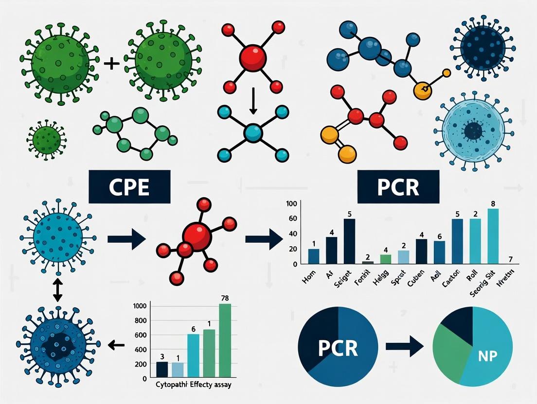
Visualizing the CPE Assay Workflow
Title: The CPE Assay Workflow from Infection to Quantification
The Scientist's Toolkit: Essential Reagents for CPE Assays
Table 3: Key Research Reagent Solutions for CPE Assays
| Reagent / Material | Function & Importance |
|---|---|
| Permissive Cell Line (e.g., Vero, MDCK, MRC-5) | Biologically relevant substrate that supports viral replication and displays CPE. |
| Cell Culture Medium & Supplements (FBS, antibiotics) | Maintains cell health and viability during the multi-day assay. |
| Viral Transport Medium | Preserves infectivity of clinical or research samples during storage/transport. |
| Neutral Red or MTT Dye | Optional: Used for viability staining to objectively quantify CPE, reducing subjectivity. |
| Positive Control Virus Stock (Titered) | Essential for assay validation, standardization, and result comparison across experiments. |
| Antiviral Compound (e.g., Remdesivir) | Used as an assay control to confirm CPE is virus-specific and can be inhibited. |
Pathway to CPE: Virus-Induced Cell Death
Title: Major Viral Pathways Leading to Observable CPE
Conclusion The CPE assay's status as the gold standard is rooted in its direct measurement of a biological outcome—infectious virus causing cell pathology. While PCR offers unmatched speed and sensitivity for genome detection, it is inherently blind to infectivity, as shown in Table 2. For research questions demanding definitive proof of replicating virus—such as antiviral efficacy testing, vaccine potency validation, or environmental persistence studies—the CPE assay remains the indispensable benchmark. The integration of both methods, leveraging the speed of PCR and the biological fidelity of CPE, provides the most comprehensive virological analysis.
Within the critical research axis comparing CPE (Cytopathic Effect) assay and PCR for infectious virus detection, understanding the precise molecular mechanism of Polymerase Chain Reaction (PCR) is fundamental. This guide objectively compares the performance of conventional PCR with its primary alternatives—quantitative PCR (qPCR) and digital PCR (dPCR)—in the context of viral detection, providing supporting experimental data to inform researchers and drug development professionals.
The Molecular Mechanism of PCR: A Three-Step Cycle
PCR amplifies specific DNA sequences (the genetic blueprint) through a thermally cycled, enzymatic replication process.
- Denaturation: The double-stranded DNA template is heated to 94-98°C, breaking hydrogen bonds to yield two single strands.
- Annealing: The temperature is lowered to 50-65°C, allowing sequence-specific primers to bind (anneal) to complementary regions flanking the target sequence.
- Extension: At 72°C, a thermostable DNA polymerase (e.g., Taq polymerase) synthesizes a new DNA strand by adding nucleotides complementary to the template strand, starting from the primers.
This cycle is repeated 25-40 times, resulting in the exponential amplification of the target DNA segment.
Performance Comparison: PCR vs. qPCR vs. dPCR for Virus Detection
The evolution from conventional PCR to qPCR and dPCR has addressed key limitations in quantification, sensitivity, and precision, which are crucial for benchmarking against CPE assays.
Table 1: Comparative Performance of PCR Technologies in Viral Detection
| Feature | Conventional (Endpoint) PCR | Quantitative PCR (qPCR) | Digital PCR (dPCR) |
|---|---|---|---|
| Detection Output | Presence/Absence (Qualitative) | Quantitative (Ct value) | Absolute Quantification (Copies/μL) |
| Dynamic Range | Narrow (~2-3 log) | Wide (~7-8 log) | Wide (~5 log) |
| Sensitivity | Moderate (Low copy detection possible) | High (Single copy detection) | Very High (Excellent for rare targets) |
| Precision & Accuracy | Low; semi-quantitative with standards | High relative quantification | Highest absolute quantification |
| Throughput & Speed | Low (requires post-processing gel) | High (real-time detection) | Moderate to High (partitioning step) |
| Tolerance to Inhibitors | Low | Moderate | High (due to partitioning) |
| Primary Application in Virology | Initial target identification, gel-based analysis | Viral load quantification, gene expression, diagnostics | Rare variant detection, low viral load samples, assay standardization |
Supporting Experimental Data Comparison: A 2023 study (J. Virol. Methods) directly compared these methods for detecting a low-prevalence viral variant spiked into human plasma. Key results are summarized below:
Table 2: Experimental Data from Low-Copy Viral Target Detection Study
| Metric | Conventional PCR | qPCR (SYBR Green) | dPCR (Droplet-based) |
|---|---|---|---|
| Limit of Detection (LoD) | 50 copies/reaction | 10 copies/reaction | 3 copies/reaction |
| Quantification at 20 copies/reaction | Not quantifiable | Ct = 34.2 ± 0.8 | 18.7 ± 2.1 copies/reaction |
| Coefficient of Variation (CV) at 100 copies/reaction | >25% (band intensity) | 15% | <5% |
| Impact of 2% Heparin Inhibitor | Complete inhibition | Ct delay of +3.5 cycles | No significant impact |
Featured Experimental Protocol: Viral RNA Detection via One-Step RT-qPCR
This protocol is commonly benchmarked against CPE assays for its speed and quantitation in virus research.
Objective: To detect and quantify viral RNA from cell culture supernatant. Workflow Summary: Sample Prep → RNA Extraction → One-Step RT-qPCR → Analysis.
Detailed Protocol:
- RNA Extraction: Use a commercial silica-membrane kit. Elute RNA in 30-50 μL nuclease-free water. Determine concentration via spectrophotometry.
- Master Mix Assembly (20 μL reaction):
- 10 μL 2x One-Step RT-qPCR Buffer
- 0.8 μL Primer Mix (10 μM each, forward and reverse)
- 0.4 μL Probe (10 μM, e.g., TaqMan)
- 0.4 μL Reverse Transcriptase/Taq Polymerase Mix
- 2 μL RNA Template (or standard)
- 6.4 μL Nuclease-Free Water
- Thermal Cycling Program (Standard 96-well block):
- Reverse Transcription: 50°C for 15-30 minutes.
- Initial Denaturation/Enzyme Activation: 95°C for 2 minutes.
- Amplification (40 cycles): 95°C for 15 sec (denaturation), 60°C for 1 min (annealing/extension, with data acquisition).
- Data Analysis: Generate a standard curve using serially diluted RNA standards of known copy number. Plot Cycle Threshold (Ct) vs. log copy number. Interpolate sample Ct values to determine viral RNA copy number/μL.
The Scientist's Toolkit: Key Research Reagent Solutions
Table 3: Essential Reagents for PCR-Based Viral Detection
| Reagent / Solution | Function in Experiment | Key Consideration for Virology |
|---|---|---|
| Thermostable DNA Polymerase (e.g., Taq) | Enzyme that synthesizes new DNA strands during extension. | For RNA viruses, a blend with reverse transcriptase (for RT-PCR) is required. |
| Sequence-Specific Primers | Short oligonucleotides that define the start points of amplification and target specificity. | Must be designed against conserved viral genomic regions to ensure detection of variants. |
| dNTP Mix | Deoxynucleotide triphosphates (dATP, dCTP, dGTP, dTTP); the building blocks for new DNA. | Quality affects fidelity and efficiency. |
| Reverse Transcriptase (for RT-PCR) | Enzyme that converts viral RNA into complementary DNA (cDNA) for amplification. | Choice affects sensitivity and tolerance to RNA secondary structure. |
| Fluorescent Probe (e.g., TaqMan) or DNA-Binding Dye (e.g., SYBR Green) | Enables real-time detection of amplified product in qPCR. | Probes offer higher specificity through an additional hybridization event. |
| Nuclease-Free Water | Solvent for all reactions; must be free of RNases and DNases. | Critical for preventing degradation of viral nucleic acid templates. |
| Inhibitor Removal Buffers/Columns | Integrated into nucleic acid extraction kits to remove PCR inhibitors from clinical samples. | Vital for robust detection from complex matrices like blood or sputum. |
The accurate detection of infectious virus is a cornerstone of virology, antiviral development, and viral safety testing. This guide compares the established Cytopathic Effect (CPE) assay with quantitative Polymerase Chain Reaction (qPCR), framing them within the critical thesis that detecting viral genetic material does not equate to detecting infectious virions. The distinction is vital, as PCR cannot differentiate between intact infectious virus, neutralized virus, or free nucleic acid debris.
Core Comparison: CPE Assay vs. qPCR
| Parameter | CPE Assay (Infectivity) | Quantitative PCR (qPCR) |
|---|---|---|
| Target | Biological activity of intact, replicating virus. | Specific sequence of viral DNA or RNA (genetic material). |
| Readout | Visual/microscopic observation of cell death/deformation (CPE). | Fluorescence threshold cycle (Ct) correlating to nucleic acid copy number. |
| Time to Result | Days to weeks (depends on virus replication cycle). | Hours to 1-2 days. |
| Quantification | Semi-quantitative (TCID₅₀, PFU/mL). Endpoint dilution. | Highly quantitative (genome copies/mL). Standard curve-based. |
| Information Provided | Confirms presence of infectious virus. | Confirms presence of viral genome. |
| Key Limitation | Slow, lower throughput, subjective, requires permissive cell line. | Cannot distinguish infectious from non-infectious particles; prone to false positives from residual nucleic acid. |
Supporting Experimental Data: Discrepancy in Viral Titer
A pivotal experiment comparing virus titration by CPE assay (TCID₅₀) and qPCR after heat or UV inactivation demonstrates the core thesis.
Experimental Protocol:
- Virus Stock: Prepare a stock of enveloped virus (e.g., SARS-CoV-2, Influenza A).
- Inactivation: Aliquot virus and treat:
- Control: No treatment.
- Heat: 56°C for 30 minutes.
- UV: Exposure to 254 nm UV light (dose: 1000 J/m²).
- Titration:
- CPE Assay: Perform serial 10-fold dilutions on permissive cell monolayers (e.g., Vero E6). Incubate 5-7 days. Score wells for CPE. Calculate TCID₅₀/mL using Reed & Muench or Spearman-Kärber method.
- qPCR: Extract RNA/DNA from each sample. Perform reverse transcription if needed. Run qPCR with virus-specific primers/probes. Quantify genome copies/mL using a standard curve from known copy number plasmids.
- Analysis: Compare log reduction in infectious titer (TCID₅₀) vs. genomic titer (copies/mL).
Results Summary Table:
| Sample | CPE Assay (Log₁₀ TCID₅₀/mL) | Reduction vs. Control | qPCR (Log₁₀ Copies/mL) | Reduction vs. Control |
|---|---|---|---|---|
| Virus Control | 6.5 | – | 10.2 | – |
| Heat-Inactivated | ≤ 1.0 | ≥ 5.5 log | 9.8 | 0.4 log |
| UV-Inactivated | ≤ 1.0 | ≥ 5.5 log | 9.1 | 1.1 log |
Data illustrates near-complete loss of infectivity post-inactivation with minimal reduction in genome copies, highlighting qPCR's inability to assess viral infectivity.
The Scientist's Toolkit: Research Reagent Solutions
| Reagent / Material | Function in CPE vs. PCR Context |
|---|---|
| Permissive Cell Line (e.g., Vero E6, MDCK) | Essential for CPE assay. Provides the host machinery for virus replication and subsequent cytopathic effect. |
| Cell Culture Media & Sera | Maintains cell viability during long-term CPE assay incubation. |
| Virus-Specific Primers & Probe | Enables specific amplification and detection of viral nucleic acid in qPCR. |
| RNA/DNA Extraction Kit | Isolates and purifies viral genetic material from samples for PCR analysis. |
| qPCR Master Mix | Contains enzymes, dNTPs, and buffers necessary for efficient, quantitative amplification. |
| Nuclease-Free Water | Prevents degradation of RNA/DNA samples and PCR reagents. |
| Fixative & Stain (e.g., Crystal Violet) | Used to fix and stain cell monolayers at CPE assay endpoint for clearer visualization of plaques or lytic areas. |
| Inactivation Agent (e.g., Beta-Propiolactone) | Used in control experiments to dissociate infectivity from genome detection. |
Visualizing the Experimental Workflow & Logical Relationship
Title: What CPE and PCR Methods Actually Detect
Title: Side-by-Side Workflow: qPCR vs CPE Assay
Historical Context and Evolution of Both Techniques in Virology
The detection and quantification of infectious viruses are cornerstones of virology, impacting fundamental research, diagnostic development, and therapeutic evaluation. Within this domain, the Cytopathic Effect (CPE) Assay and Polymerase Chain Reaction (PCR) have emerged as pivotal, yet philosophically distinct, techniques. This guide objectively compares their performance, framed within a thesis on detecting infectious virus, supported by experimental data and historical evolution.
Historical Context & Technical Evolution
Cytopathic Effect (CPE) Assay: Originating in the early 20th century with the cultivation of viruses in animal tissues and later in cell cultures (e.g., Enders, Weller, and Robbins, 1949), the CPE assay is a functional, biology-based method. Its principle relies on observing virus-induced morphological changes (cell rounding, detachment, lysis) in permissive host cells. It directly measures viral replication competence and infectivity.
Polymerase Chain Reaction (PCR): Conceptualized in the 1980s (Kary Mullis, 1983), PCR revolutionized molecular biology by enabling exponential amplification of specific nucleic acid sequences. Its application in virology provided unprecedented sensitivity for detecting viral genomes, irrespective of their infectious status. Quantitative PCR (qPCR) later allowed for precise viral load measurement.
Performance Comparison: CPE Assay vs. qPCR for Infectious Virus Detection
The core distinction lies in what each technique measures: infectivity versus genome presence. The following table summarizes key comparative parameters, with data synthesized from recent virology literature.
Table 1: Direct Comparison of CPE Assay and qPCR for Virus Detection
| Parameter | CPE Assay | Quantitative PCR (qPCR) |
|---|---|---|
| Target | Infectious virus (biological activity) | Viral nucleic acid (DNA or RNA) |
| Primary Output | Tissue Culture Infectious Dose 50% (TCID₅₀/mL) or Plaque Forming Units (PFU/mL) | Cycle Threshold (Ct) or copies/mL |
| Time to Result | Days to weeks (depends on virus replication kinetics) | Hours (typically 2-4 hours) |
| Sensitivity | Lower (requires viable virus to replicate) | Extremely High (can detect a few genome copies) |
| Specificity | High for infectious virus; can be affected by cytotoxic compounds | High for target sequence; does not confirm infectivity |
| Throughput | Low to moderate (labor-intensive) | High (amenable to automation) |
| Quantifies Infectivity? | Yes, directly. | No, indirectly. Correlates with infectivity but can detect non-infectious genomes (e.g., from degraded virus, vaccine vectors). |
| Key Limitation | Slow, requires viable cell culture, subjective endpoint. | Cannot distinguish between infectious and non-infectious viral particles. |
Supporting Experimental Data: A 2023 study comparing methods for SARS-CoV-2 infectivity titration starkly illustrates this dichotomy. Virus stocks were inactivated by heat or UV treatment. qPCR showed minimal change in genome copy number post-treatment, while CPE assay titers dropped by >4 log₁₀, confirming loss of infectivity.
Table 2: Experimental Data from Virus Inactivation Study
| Sample Treatment | qPCR Result (copies/mL) | CPE Assay Result (TCID₅₀/mL) | Infectious? |
|---|---|---|---|
| Untreated Control | 2.5 x 10⁹ | 1.0 x 10⁷ | Yes |
| Heat Inactivated | 2.1 x 10⁹ | < 10¹ | No |
| UV Inactivated | 8.7 x 10⁸ | < 10¹ | No |
Detailed Experimental Protocols
Protocol 1: Standard CPE Assay for TCID₅₀ Endpoint Determination
- Cell Seeding: Seed permissive cells (e.g., Vero E6) into a 96-well tissue culture plate to achieve ~90% confluency after 24 hours.
- Sample Inoculation: Serially dilute the viral sample (e.g., 10-fold dilutions in infection medium). Aspirate medium from cell plate and inoculate multiple wells per dilution with the diluted virus.
- Incubation & Observation: Incubate plates at appropriate conditions (e.g., 37°C, 5% CO₂). Observe daily under a light microscope for characteristic CPE.
- Endpoint Calculation: After a defined period (e.g., 5-7 days), record wells positive for CPE. Calculate the TCID₅₀/mL using the Spearman-Kärber or Reed-Muench statistical method.
Protocol 2: One-Step RT-qPCR for Viral RNA Quantification
- Nucleic Acid Extraction: Isolate viral RNA from sample (cell supernatant, tissue homogenate) using a silica-membrane column or magnetic bead-based kit. Include positive and negative extraction controls.
- Reaction Setup: Prepare a master mix containing: reverse transcriptase, DNA polymerase, dNTPs, sequence-specific forward and reverse primers, and a fluorescently-labeled probe (e.g., TaqMan) for the target viral gene. Aliquot into a qPCR plate.
- Amplification: Add extracted RNA template to the reaction wells. Run in a real-time PCR instrument with a standard cycling program: Reverse transcription (50°C, 15 min), Polymerase activation (95°C, 2 min), followed by 40-45 cycles of Denaturation (95°C, 15 sec) and Annealing/Extension (60°C, 1 min, with data acquisition).
- Quantification: Determine the Cycle Threshold (Ct) for each sample. Use a standard curve generated from serial dilutions of RNA with known copy number to interpolate the viral load in copies/mL.
Visualizing Workflows and Logical Relationships
Title: CPE Assay Workflow for Infectivity Quantification
Title: PCR Workflow and the Infectivity Determination Gap
The Scientist's Toolkit: Research Reagent Solutions
Table 3: Essential Reagents for CPE and PCR-Based Virology
| Reagent / Material | Function | Typical Application |
|---|---|---|
| Permissive Cell Line (e.g., Vero, MDCK, HEK-293) | Provides a host system for viral replication and CPE manifestation. | CPE Assay, virus propagation, plaque assays. |
| Cell Culture Medium & Supplements | Supports cell viability and growth during virus infection period. | All cell-culture based assays (CPE, plaque, TCID₅₀). |
| Viral Lysis Buffer | Inactivates virus and stabilizes viral nucleic acids for safe extraction. | Initial step for RNA/DNA extraction prior to PCR. |
| Nucleic Acid Extraction Kit | Isolates and purifies viral RNA/DNA from complex biological samples. | Sample preparation for PCR, sequencing. |
| Reverse Transcriptase Enzyme | Converts single-stranded RNA into complementary DNA (cDNA). | First step in RT-qPCR for RNA viruses. |
| Hot-Start DNA Polymerase | Reduces non-specific amplification, improving PCR specificity and yield. | qPCR and RT-qPCR master mixes. |
| Sequence-Specific Primers & Probe | Binds complementary viral genome sequence for targeted amplification/detection. | Defining specificity in PCR/qPCR assays. |
| Quantitative PCR Standard | RNA/DNA of known concentration to generate a standard curve for absolute quantification. | Converting Ct values to copies/mL in qPCR. |
In the context of infectious virus detection research, choosing between Cytopathic Effect (CPE) assays and Polymerase Chain Reaction (PCR) is critical. This guide compares their performance, applications, and provides supporting data to inform method selection.
Core Comparison: CPE Assay vs. PCR
Table 1: Primary Application Comparison
| Parameter | CPE Assay | Quantitative PCR (qPCR) |
|---|---|---|
| First-Choice Scenario | Initial virus isolation, culturable virus detection, antiviral screening. | High-throughput diagnostics, viral load quantification, detection of non-cytolytic viruses. |
| Detection Target | Functional, infectious virions causing visible cell changes. | Viral nucleic acid (RNA/DNA), not necessarily indicative of infectivity. |
| Typical Time-to-Result | 3 to 14 days (culture-dependent). | 1 to 4 hours. |
| Throughput | Low to moderate. | High to very high. |
| Quantification | Semi-quantitative (TCID50, plaque assays). | Highly quantitative (copies/mL). |
| Sensitivity | Moderate (requires replicating virus). | Very High (can detect a few genome copies). |
| Key Advantage | Confirms viral infectivity and replication. | Speed, sensitivity, and scalability. |
| Major Limitation | Time-consuming, requires cell culture expertise. | Cannot distinguish between infectious and non-infectious viral particles. |
Table 2: Supporting Experimental Data from Recent Studies
| Study Focus | CPE Assay Result | PCR Result | Key Implication |
|---|---|---|---|
| Antiviral Drug Screening (vs. Influenza A) | IC50: 5.2 µM (based on CPE reduction). | IC50: 4.8 µM (based on RNA reduction). | Strong correlation for culturable viruses; CPE confirms antiviral effect on replication. |
| Environmental Sample Testing (Wastewater) | Positive in 2/10 concentrated samples after 7 days. | Positive in 10/10 samples in <1 day. | PCR is first choice for surveillance; CPE can confirm viability in positive samples. |
| Persistence of Virus Post-Heat Inactivation | No CPE observed. | PCR positive for fragmented genome. | PCR alone can overestimate infectious risk; CPE or similar functional assay is required. |
Experimental Protocols
Protocol 1: Standard CPE Assay for Virus Titration (TCID50)
- Cell Seeding: Plate susceptible cells (e.g., Vero E6) in a 96-well plate to achieve 90-95% confluency after 24h.
- Sample Inoculation: Prepare 10-fold serial dilutions of viral sample in maintenance medium. Aspirate medium from cell plate and inoculate multiple wells per dilution (typically 8-10).
- Incubation & Observation: Incubate plates at 37°C, 5% CO2. Monitor daily for CPE (e.g., rounding, detachment, syncytia) using an inverted microscope for up to 7-14 days.
- Endpoint Calculation: Record wells positive for CPE. Calculate the 50% Tissue Culture Infectious Dose (TCID50/mL) using the Spearman-Kärber or Reed-Muench method.
Protocol 2: One-Step RT-qPCR for Viral RNA Quantification
- RNA Extraction: Purify viral RNA from samples (e.g., cell culture supernatant, clinical specimen) using a silica-column or magnetic bead-based kit. Include appropriate controls.
- Reaction Setup: Prepare a master mix containing: reverse transcriptase, DNA polymerase, dNTPs, reaction buffer, sequence-specific primers and a fluorescently-labeled probe (e.g., TaqMan).
- Thermocycling: Run in a real-time PCR instrument: Reverse transcription at 50°C for 10-15 min; Polymerase activation at 95°C for 2 min; 40-45 cycles of: Denaturation (95°C, 15 sec), Annealing/Extension (60°C, 1 min, with fluorescence acquisition).
- Analysis: Determine the Cycle Threshold (Ct) for each sample. Quantify viral load by comparing to a standard curve of known copy numbers.
Visualizations
Decision Flow: CPE vs PCR Selection
Detection Pathways: Molecular vs. Biological
The Scientist's Toolkit: Key Research Reagent Solutions
Table 3: Essential Materials for CPE and PCR Assays
| Item | Function | Example/Note |
|---|---|---|
| Virus-Susceptible Cell Line | Host for viral replication and CPE development. | Vero E6 (SARS-CoV-2, arboviruses), MDCK (influenza). |
| Cell Culture Maintenance Medium | Supports cell viability during infection. | Typically EMEM or DMEM with 2% FBS and antibiotics. |
| Viral Lysis Buffer | Inactivates virus and preserves nucleic acid for safe RNA extraction. | Contains guanidinium thiocyanate and buffer. |
| Nucleic Acid Extraction Kit | Isolates pure viral RNA/DNA from complex samples. | Silica-membrane columns or magnetic bead-based. |
| One-Step RT-qPCR Master Mix | Contains all enzymes and reagents for combined reverse transcription and amplification. | Includes reverse transcriptase, hot-start Taq polymerase, dNTPs, optimized buffer. |
| Sequence-Specific Primers & Probe | Defines the target for amplification and detection. | Designed from conserved viral genomic regions; probe labeled with FAM/BHQ1. |
| Inverted Tissue Culture Microscope | Essential for daily visual assessment of cell monolayers for CPE. | Equipped with phase contrast, 4x-20x objectives. |
| Real-Time PCR Instrument | Performs thermocycling and measures fluorescence in real-time. | Platforms from Bio-Rad, Thermo Fisher, Roche. |
| Viral Quantification Standards | Known copy number of target used to generate a standard curve for absolute quantification. | Synthetic gBlocks or quantified RNA transcripts. |
| Antiviral Control Compound | Positive control for antiviral screening assays (e.g., CPE reduction). | Remdesivir (for coronaviruses), Oseltamivir carboxylate (influenza). |
Step-by-Step Protocols: Executing Robust CPE Assays and PCR for Viral Detection
Within the broader research thesis comparing Cytopathic Effect (CPE) assays to PCR for infectious virus detection, the CPE assay remains a cornerstone method for quantifying viable, replicating virus. This guide objectively compares the performance of a standardized CPE assay workflow against common alternative methods, providing supporting experimental data to inform researchers and drug development professionals.
Comparative Experimental Data
Table 1: Comparison of Virus Quantification Methods
| Parameter | CPE-Based Assay (Plaque/TCID50) | qPCR/Sample | Immunofluorescence Assay (IFA) | Flow Cytometry-Based Assay |
|---|---|---|---|---|
| Detects Infectious Virus | Yes | No (Detects genome) | Yes | Yes |
| Time to Result | 3-7 days | 4-6 hours | 2-3 days | 1-2 days |
| Throughput | Low to Medium | High | Medium | High |
| Subjectivity | Moderate (Visual scoring) | Low (Automated) | Moderate (Visual/Image analysis) | Low (Automated) |
| Cost per Sample | $5 - $15 | $10 - $25 | $20 - $40 | $30 - $60 |
| Quantification Output | PFU/mL or TCID50/mL | Genome Copies/mL | FFU/mL | Infectious Units/mL |
| Key Advantage | Gold standard for infectivity | Speed and sensitivity | Cell-type specific, visual | Single-cell data, high-throughput |
| Key Disadvantage | Slow, labor-intensive | Cannot distinguish infectivity | Lower throughput, subjective | Expensive, complex setup |
Table 2: Experimental Comparison: CPE Assay vs. PCR for Live Virus Titration (Representative Data)
| Virus Sample | Plaque Assay (PFU/mL) | TCID50/mL | qPCR (Genome Copies/mL) | Ratio (qPCR:PFU) | Inference |
|---|---|---|---|---|---|
| SARS-CoV-2 Stock A | 2.5 x 10^6 | 1.8 x 10^7 | 5.1 x 10^9 | ~2000:1 | High proportion of non-infectious particles |
| Influenza A Stock B | 4.0 x 10^7 | 3.2 x 10^8 | 3.0 x 10^8 | ~7.5:1 | High specific infectivity |
| HSV-1 Stock C | 5.5 x 10^8 | 1.0 x 10^9 | 2.2 x 10^10 | ~40:1 | Moderate non-infectious particle load |
Detailed CPE Assay Protocol
Protocol 1: Standard Plaque Assay Protocol
Principle: Serial dilutions of virus are used to infect a monolayer of permissive cells. An overlay medium restricts virus spread to neighboring cells, allowing discrete plaques (areas of CPE) to form and be counted.
- Cell Seeding: Seed appropriate susceptible cells (e.g., Vero E6 for SARS-CoV-2) in a 12-well or 24-well plate to achieve 90-100% confluence at time of infection.
- Virus Inoculation: Aspirate growth medium from cells. Inoculate duplicate wells with serial 10-fold dilutions of virus sample (e.g., 10^-2 to 10^-8). Incubate 1-2 hours at 37°C with periodic rocking for adsorption.
- Overlay Application: Prepare a viscous overlay medium (e.g., 1.5% carboxymethylcellulose or 0.8% agarose in maintenance medium). Remove virus inoculum and carefully add the overlay without disturbing the monolayer.
- Incubation: Incubate plates for the appropriate number of days (virus-dependent) at 37°C, 5% CO2.
- Plaque Visualization & Counting:
- Direct Staining (Crystal Violet): Fix cells with 10% formalin for 1 hour. Remove overlay and fixative. Stain with 0.1% crystal violet in 10% ethanol for 20 minutes. Rinse with water. Plaques appear as clear zones against a purple-stained monolayer.
- Immunostaining: Fix with formalin or methanol/acetone. Permeabilize, block, and incubate with primary antibody against the virus, followed by an enzyme- or fluorophore-conjugated secondary antibody. Develop colorimetric substrate or image fluorescent plaques.
- Calculation: Count plaques in wells with 10-100 plaques. Calculate Plaque Forming Units per mL (PFU/mL) using the formula: PFU/mL = (Number of plaques) / (Dilution factor x Volume of inoculum in mL).
Protocol 2: TCID50 Assay Protocol (Endpoint Dilution)
Principle: Serial dilutions of virus are inoculated onto multiple replicate cell cultures. The presence or absence of CPE in each well is scored to determine the dilution at which 50% of the cultures are infected.
- Cell Seeding: Seed cells in a 96-well plate to achieve confluent monolayers.
- Virus Inoculation: Prepare an 8-step, ½-log or 10-fold serial dilution series of the virus. Aspirate medium from the plate. Inoculate 6-8 replicate wells per dilution with 50-100 µL of virus dilution. Include cell-only control wells.
- Incubation & Observation: Incubate plate at 37°C, 5% CO2. Monitor daily for CPE (e.g., rounding, detachment, syncytia) under a microscope for 3-7 days.
- Scoring & Calculation: Score each well as positive (CPE present) or negative (no CPE). Calculate the TCID50/mL using the Reed & Muench or Spearman-Kärber method.
- Reed & Muench Method: Calculates the proportional distance between the dilution that infects >50% of wells and the dilution that infects <50%. TCID50/mL = 10^(L + d*(S - 0.5)), where L is the log of the dilution with >50% CPE, d is the log dilution factor, and S is the proportion of positive wells at dilution L.
Visualization of Workflows
Diagram 1: CPE Assay Workflow Decision Tree
Diagram 2: CPE vs PCR in Research Thesis Context
The Scientist's Toolkit: Research Reagent Solutions
Table 3: Essential Materials for CPE Assay Workflow
| Reagent/Material | Function/Description | Example Product/Alternative |
|---|---|---|
| Permissive Cell Line | Host cells that support viral replication and display clear CPE. Critical for assay success. | Vero E6 (SARS-CoV-2), MDCK (Influenza) |
| Cell Culture Medium | Provides nutrients for cell maintenance during the often lengthy assay. | DMEM, EMEM with 2-5% FBS |
| Viscous Overlay Medium | Restricts virus spread to allow plaque formation. Key for plaque assays, not used in TCID50. | Carboxymethylcellulose, Agarose, Methylcellulose |
| Fixative | Preserves cell monolayer for staining and plaque visualization. | 10% Neutral Buffered Formalin, Methanol |
| Detection Stain | Enables visualization of plaques or CPE. | Crystal Violet, Neutral Red, Virus-Specific Antibody for immunostaining |
| Multi-well Plates | Platform for cell seeding and virus titration. 96-well for TCID50, 12/24-well for plaque assays. | Tissue culture-treated plates |
| Inverted Microscope | For daily monitoring of CPE development and final plaque/well scoring. | Phase-contrast microscope (40-100x magnification) |
| Hemocytometer/Automated Cell Counter | For accurate cell counting during seeding to ensure consistent monolayers. | Trypan Blue exclusion method |
| Virus-Specific Antibody | For immunostaining-based plaque assays, increases specificity and sensitivity, especially for viruses with subtle CPE. | Primary antibodies against viral antigen |
| Statistical Software/Template | For calculating TCID50 endpoints (Reed & Muench, Spearman-Kärber). | Excel templates, Prism, custom scripts |
Within the ongoing research discourse comparing Cell-based Potency Assays (CPE) to PCR for infectious virus detection, PCR remains the cornerstone molecular technique for its speed, sensitivity, and specificity. This guide objectively compares key protocol components and their performance against alternatives, supported by experimental data.
Primer Design: Specificity & Efficiency
Effective PCR begins with robust primer design. We compared in-house manually designed primers against those generated by automated tools (e.g., Primer-BLAST, IDT PrimerQuest) for the detection of Human Cytomegalovirus (HCMV) UL54 gene.
Experimental Protocol:
- Target: HCMV genomic DNA (ATCC VR-977D).
- Design: Manual design using NCBI guidelines vs. Automated design via Primer-BLAST.
- Criteria: Amplicon size (80-150 bp), Tm (58-60°C ± 1°C), GC content (40-60%).
- Synthesis: All primers synthesized by a single provider (IDT, standard desalting).
- Testing: Real-time PCR (SYBR Green) with a 5-log dilution series of target (10^6 to 10^1 copies/reaction). NTC included.
- Analysis: Compare amplification efficiency (E), R², and specificity via melt curve analysis.
Table 1: Primer Design Performance Comparison
| Design Method | Primer Pair | Efficiency (E) | R² Value | Mean Cq at 10^2 copies | Specificity (Melt Peak) |
|---|---|---|---|---|---|
| Manual | F: 5'-CGACGGTGTCGTACAGTT-3'R: 5'-TGGTGACGCGAAAAAGAAG-3' | 98.5% | 0.999 | 28.4 ± 0.3 | Single, sharp peak |
| Automated (Primer-BLAST) | F: 5'-AGCGTTCGTGACTGTGGA-3'R: 5'-TCTGCTTTCGTTGACGGT-3' | 102.3% | 0.995 | 27.9 ± 0.4 | Single, sharp peak |
| Alternative: Probe-based (TaqMan) | Custom TaqMan Assay (Thermo) | 99.1% | 0.998 | 26.1 ± 0.2 | N/A (fluorogenic) |
Nucleic Acid Extraction: Yield, Purity & Throughput
The extraction method critically impacts downstream PCR sensitivity. We compared three common methods for extracting HCMV DNA from spiked cell culture supernatant.
Experimental Protocol:
- Sample: HCMV (AD169 strain) spiked into DMEM with 10% FBS to 10^5 PFU/mL.
- Methods Tested: (A) Manual Silica-column (Qiagen QIAamp DNA Mini), (B) Automated Magnetic-bead (Thermo KingFisher Flex, MagMAX Viral/Pathogen Kit), (C) Precipitation-based (Phenol-Chloroform-Isoamyl Alcohol).
- Protocol: Followed each kit's recommended protocol for 200 µL input; elution in 50 µL. n=6 per group.
- Analysis: Quantify yield (ng/µL) and purity (A260/A280) via nanodrop. Perform real-time PCR on eluates to assess inhibition (Cq shift vs. control DNA in water).
Table 2: Nucleic Acid Extraction Method Comparison
| Method | Avg. Yield (ng) | Avg. Purity (A260/280) | Avg. Cq (vs. Control) | Hands-on Time | Cost per Sample |
|---|---|---|---|---|---|
| Manual Column | 45.2 ± 5.1 | 1.92 ± 0.05 | +0.5 ± 0.2 | ~30 min | $$ |
| Automated Magnetic Bead | 48.7 ± 3.8 | 1.95 ± 0.03 | +0.3 ± 0.1 | <10 min | $$$ |
| Phenol-Chloroform | 52.1 ± 12.5 | 1.78 ± 0.15 | +2.1 ± 0.8* | ~60 min | $ |
| Indicates significant inhibition (p<0.01, t-test). |
Amplification: PCR Master Mix Performance
We evaluated the sensitivity and consistency of three commercial real-time PCR master mixes using the same primer set and HCMV DNA standard.
Experimental Protocol:
- DNA: Serial dilutions of quantified HCMV gDNA (10^4 to 10^0 copies/reaction).
- Master Mixes: (A) Thermo Scientific PowerUp SYBR Green, (B) Bio-Rad SsoAdvanced Universal SYBR Green Supermix, (C) Qiagen QuantiNova SYBR Green PCR.
- Platform: Applied Biosystems 7500 Fast Real-Time PCR System.
- Cycling: Standard 2-step protocol: 95°C for 2 min, then 40 cycles of (95°C for 15 sec, 60°C for 1 min).
- Analysis: Determine Limit of Detection (LoD - 95% hit rate), efficiency, and intra-assay CV.
Table 3: Real-Time PCR Master Mix Comparison
| Master Mix | LoD (copies/rxn) | Efficiency | Intra-assay CV (Cq at 10^3 copies) | Time to Result |
|---|---|---|---|---|
| PowerUp SYBR | 5 | 99.8% | 0.8% | ~90 min |
| SsoAdvanced | 5 | 101.5% | 1.2% | ~85 min |
| QuantiNova | 10 | 96.4% | 0.9% | ~70 min |
Analysis: Quantification & Validation
Accurate quantification and validation of PCR results are essential. This is where PCR and CPE assays diverge significantly in the context of infectious virus research.
Experimental Protocol for Standard Curve Validation:
- A standard curve using a plasmid containing the target sequence (10^6 to 10^1 copies) is run in triplicate on every plate.
- Unknown sample Cq values are interpolated from this curve.
- To assess correlation with infectious titer, the same viral stocks (n=5) are analyzed by (a) qPCR (copies/mL) and (b) CPE-based TCID50 assay on permissive cells.
- Linear regression analysis compares log10(copies/mL) vs. log10(TCID50/mL).
PCR vs. CPE Assay Workflow for Infectious Virus Detection
The Scientist's Toolkit: Research Reagent Solutions
| Item | Function in PCR Protocol | Example Brands/Products |
|---|---|---|
| Nucleic Acid Extraction Kit | Isolates and purifies DNA/RNA from complex samples, removing inhibitors. | Qiagen QIAamp, Thermo MagMAX, Roche High Pure |
| PCR Master Mix | Contains polymerase, dNTPs, buffers, and salts optimized for efficient amplification. | Thermo PowerUp SYBR, Bio-Rad SsoAdvanced, Qiagen QuantiNova |
| Fluorogenic Probe (for qPCR) | Provides sequence-specific detection, increasing assay specificity over intercalating dyes. | TaqMan Probes (Thermo), Molecular Beacons, Dual-Labeled Probes |
| Standard/Control Template | Quantified target used to generate a standard curve for absolute quantification. | Custom gBlocks, Plasmid Standards, Commercial Quantitative Standards |
| RNase/DNase-free Water | Ultrapure water to prevent enzymatic degradation of templates and reagents. | Invitrogen UltraPure, Sigma W4502 |
| uracil-N-glycosylase (UNG) | Enzyme to prevent carryover contamination by degrading PCR products from previous runs. | Often included in master mixes (e.g., PowerUp) |
| Inhibition Resistance Polymerase | Engineered polymerase tolerant to common sample inhibitors (e.g., heparin, humic acid). | Taq DNA Polymerase, Tth polymerase, proprietary enzyme blends |
The integrity of any infectious virus detection assay, whether Cell-based Potency/Productivity Enhancement (CPE) or Polymerase Chain Reaction (PCR)-based, hinges on the quality and consistency of its critical reagents and controls. This guide compares key reagents and their performance in the context of viral detection research.
Performance Comparison: CPE vs. PCR Assay Critical Reagents
The reliability of viral detection assays depends fundamentally on the reagents used. The following table summarizes a comparison of critical components for CPE and PCR assays, based on current literature and product performance data.
Table 1: Comparison of Critical Reagents for CPE and PCR Assays in Virus Detection
| Reagent Category | CPE Assay Application | Typical Commercial Alternatives (PCR) | Key Performance Metric | Experimental Observation (Supporting Data) |
|---|---|---|---|---|
| Cell Substrate | Permissive cell line for viral infection & CPE. | Not applicable. | Cell viability, doubling time, susceptibility. | Vero E6 cells: >95% viability, 24 hr doubling time, consistent CPE for SARS-CoV-2. |
| Detection Probe | Vital dye (e.g., MTT, alamarBlue) for cell viability. | Fluorescent TaqMan probe for target sequence. | Signal-to-noise ratio, dynamic range. | TaqMan probes show 10^6 dynamic range vs. 10^2 for MTT in endpoint CPE. |
| Enzyme | Not typically a critical reagent. | Thermostable DNA polymerase (e.g., Taq, Q5). | Processivity, error rate, inhibition resistance. | Q5 High-Fidelity DNA Polymerase: error rate 4.4x10^-7 vs. Taq 2.0x10^-4. |
| Positive Control | Live, infectious virus stock. | Synthetic nucleic acid (gBlock, RNA transcript). | Stability, commutability, safety. | Synthetic RNA controls show <0.5 log10 titer drop in 1 year at -80°C vs. live virus instability. |
| Inhibition Control | Spiked virus or cytotoxicity assay. | Internal Amplification Control (IAC) co-amplified. | Reliable detection in all samples. | IAC detection failed in 5% of clinical samples, indicating PCR inhibition not apparent in CPE. |
Experimental Protocols for Reagent Qualification
Protocol 1: Qualification of a Synthetic Positive Control for PCR
Objective: To determine the linearity, limit of detection (LoD), and stability of a synthetic RNA positive control. Materials: Synthetic SARS-CoV-2 RNA control (10^6 copies/µL), nuclease-free water, RT-qPCR master mix, validated primer/probe set, real-time PCR instrument. Procedure:
- Perform a 10-fold serial dilution of the synthetic RNA in nuclease-free water across 6 logs.
- Prepare RT-qPCR reactions in triplicate for each dilution.
- Run the assay on a real-time PCR system using the manufacturer's recommended cycling conditions.
- Plot the mean quantification cycle (Cq) value against the log10 concentration. The linear regression R² should be ≥0.99.
- For LoD, test the lowest dilution 20 times; LoD is the concentration detected in ≥95% of replicates.
- For stability, aliquot the control and store at -80°C, -20°C, and +4°C. Test monthly against a reference standard.
Protocol 2: Qualification of Cell Susceptibility for CPE Assay
Objective: To ensure the cell substrate consistently produces CPE upon infection with the target virus. Materials: Vero E6 cells, virus stock of known titer (TCID50/mL), cell culture media, vital dye (e.g., alamarBlue). Procedure:
- Seed cells in a 96-well plate to achieve 90-100% confluence after 24 hours.
- Serially dilute the virus stock in media (e.g., 10^-1 to 10^-8).
- Infect 8 replicate wells per dilution with virus. Include cell-only controls.
- Incubate for the prescribed period (e.g., 5-7 days).
- Add alamarBlue reagent and incubate for 2-4 hours. Measure fluorescence.
- Calculate the TCID50/mL using the Spearman-Kärber method. The titer must fall within a predefined range (e.g., ±0.5 log10) of the historical median to pass qualification.
Visualizing Assay Workflows and Critical Control Points
Title: CPE Assay Workflow with Critical Reagents & Controls
Title: PCR Assay Workflow with Critical Reagents & Controls
The Scientist's Toolkit: Research Reagent Solutions
Table 2: Essential Reagents for Infectious Virus Detection Assays
| Item | Function in Research | Key Considerations for Integrity |
|---|---|---|
| Characterized Cell Banks | Provides a consistent, susceptible substrate for CPE assays and virus propagation. | Passage number, mycoplasma-free status, viability profile, and pre-qualified susceptibility. |
| Synthetic Nucleic Acid Controls | Provides a stable, non-infectious positive control for PCR assay calibration and LoD studies. | Sequence verification, copy number quantification, stability under storage conditions, and absence of inhibitors. |
| Master Mix with IAC | A ready-to-use PCR mix containing an internal amplification control to detect reaction inhibition. | Polymerase fidelity, buffer optimization, IAC design (non-competitive), and consistent performance across target matrices. |
| Reference Virus Stock | A titered, authentic virus used as a positive control in CPE assays and for PCR assay validation. | Secure production, precise TCID50 or PFU quantification, genetic characterization, and stabilized storage. |
| Validated Primer/Probe Sets | Sequence-specific oligonucleotides for PCR amplification and detection of viral targets. | Specificity (BLAST analysis), efficiency (90-110%), absence of secondary structure, and purity (HPLC purified). |
In infectious virus detection research, the choice between the Cytopathic Effect (CPE) assay and Polymerase Chain Reaction (PCR) is fundamental. This guide provides an objective comparison of their performance, supported by experimental data, to inform methodological selection.
Performance Comparison: CPE Assay vs. qPCR
The following table summarizes core performance characteristics based on aggregated experimental data from recent studies.
Table 1: Direct Comparison of CPE Assay and qPCR for Infectious Virus Quantification
| Parameter | CPE (Endpoint Titration) | Quantitative PCR (qPCR) | Implication for Research |
|---|---|---|---|
| Target | Infectious, replication-competent virus | Viral nucleic acid (DNA or RNA) | CPE indicates infectivity; PCR detects genetic material regardless of viability. |
| Time to Result | 3-14 days (cell culture-dependent) | 2-4 hours | CPE is low-throughput for screening; PCR enables rapid kinetics studies. |
| Quantification Output | TCID₅₀/mL or PFU/mL (log scale) | Copies/µL (linear scale) | TCID₅₀ is a statistical endpoint; copies/µL is direct but not equivalent to infectivity. |
| Sensitivity | Moderate (≈10³ - 10⁴ virions/mL) | High (≈1-10 copies/reaction) | PCR can detect latent or abortive infections where CPE is absent. |
| Specificity | High (confirms functional infectivity) | Can be high (with probe design) | PCR requires controls for amplicon contamination and may detect non-infectious particles. |
| Automation Potential | Low (subjective visual readout) | High (fully automated plate reading) | PCR is superior for high-throughput drug screening (e.g., antiviral EC₅₀). |
| Key Application | Gold standard for quantifying infectious titer; crucial for vaccine lot release. | Rapid detection, viral load tracking, and gene expression analysis in pathogenesis studies. |
Experimental Protocols for Key Comparisons
Protocol 1: Standard TCID₅₀ Assay for CPE-Based Titration
- Cell Seeding: Seed 96-well plates with susceptible cells (e.g., Vero E6) at 2x10⁴ cells/well and incubate overnight.
- Sample Serial Dilution: Prepare 10-fold serial dilutions (e.g., 10⁻¹ to 10⁻⁸) of virus stock in infection medium.
- Inoculation & Incubation: Aspirate medium from the cell plate. Add 100 µL of each dilution to 8-12 replicate wells. Include cell controls. Incubate at 37°C, 5% CO₂ for the prescribed time (e.g., 5-7 days for many viruses).
- CPE Scoring: Visually inspect each well under a microscope for morphological changes (cell rounding, detachment, syncytia). Score wells as positive (≥50% CPE) or negative.
- Calculation: Calculate the TCID₅₀/mL using the Spearman-Kärber or Reed-Muench statistical method.
Protocol 2: One-Step RT-qPCR for Viral RNA Quantification
- RNA Extraction: Purify viral RNA from culture supernatants or infected cells using a silica-membrane column kit. Include a non-template control.
- Reaction Setup: Prepare a master mix containing: 1x reaction buffer, reverse transcriptase, hot-start DNA polymerase, dNTPs, sequence-specific forward/reverse primers, and a hydrolysis probe (e.g., FAM-labeled). Add template RNA. Run in triplicate.
- Cycling Conditions (Typical): Reverse transcription: 50°C for 10 min. Polymerase activation: 95°C for 2 min. 40-45 cycles of: Denature (95°C, 15 sec), Anneal/Extend (60°C, 1 min, with data acquisition).
- Data Analysis: Set a consistent threshold. Determine the cycle threshold (Ct) for each well. Generate a standard curve from RNA standards of known copy number to interpolate the copies/µL in unknowns.
Visualizing the Methodological Workflow and Relationship
Diagram 1: Infectious Virus Detection Workflow Comparison
Diagram 2: Data Interpretation Logic for CPE vs. PCR Results
The Scientist's Toolkit: Key Research Reagent Solutions
Table 2: Essential Materials for CPE and PCR Virus Detection Assays
| Reagent/Material | Function in Assay | Example/Critical Feature |
|---|---|---|
| Susceptible Cell Line | Provides the host system for virus replication and CPE manifestation. | Vero E6 (for many viruses), primary human airway epithelial cells (for physiologic relevance). |
| Cell Culture Medium | Supports cell viability and virus replication during incubation. | Must be serum-free during infection for some viruses; contains antibiotics to prevent contamination. |
| Viral Lysis Buffer | Inactivates virus and releases/protects viral nucleic acid for PCR. | Contains chaotropic salts (e.g., guanidinium) and is compatible with downstream extraction. |
| Nucleic Acid Extraction Kit | Isolates and purifies viral RNA/DNA from complex samples. | Silica-membrane columns or magnetic beads. Should include DNase/RNase treatment steps. |
| One-Step RT-qPCR Master Mix | Integrates reverse transcription and PCR amplification in a single tube. | Contains reverse transcriptase, hot-start Taq polymerase, dNTPs, optimized buffer. Reduces handling error. |
| Sequence-Specific Primers & Probe | Confirms the identity of the amplicon and enables real-time quantification. | Probe-based chemistry (e.g., TaqMan) increases specificity. Targets must be validated. |
| Quantified RNA Standard | Enables absolute quantification by generating a standard curve. | In vitro transcribed RNA of known concentration, spanning 6-8 log10 dilutions. Critical for copies/µL data. |
| Cell Vitality Stain | Alternative to visual CPE scoring; objectively quantifies cell viability. | Dyes like MTT, CTG, or neutral red are metabolized or retained by live cells. Enables plate reader use. |
| Antiviral Control Compound | Validates assay sensitivity and provides a benchmark for drug screening. | Well-characterized inhibitors (e.g., Remdesivir for SARS-CoV-2, Oseltamivir for Influenza). |
This guide compares the performance of Cytopathic Effect (CPE) assays and Polymerase Chain Reaction (PCR)-based methods in the critical applications of antiviral drug screening and vaccine potency testing. The choice of detection method significantly impacts throughput, cost, and biological relevance in infectious virus research.
Performance Comparison: CPE Assay vs. qPCR for Antiviral Screening
Table 1: Comparison of Key Performance Metrics in Antiviral Drug Screening
| Metric | CPE-Based Visual/Microscopic Assay | Cell-Based qPCR Assay | Experimental Insight |
|---|---|---|---|
| Time to Result | 3-7 days (viral replication dependent) | 1-2 days (post-infection) | qPCR drastically accelerates screening cycles. |
| Throughput | Low to moderate (manual scoring) | High (automated plate reading) | qPCR enables high-content screening (HCS). |
| Quantification | Semi-quantitative (TCID₅₀, plaque count) | Fully quantitative (viral genome copies) | qPCR provides precise dose-response curves for IC₅₀ calculation. |
| Cost per Plate | Low (stains, basic microscopy) | High (qPCR reagents, probes) | CPE retains advantage in resource-limited settings. |
| Biological Relevance | High (measures functional cell death) | Indirect (measures genome replication) | CPE captures the net effect of viral infection and cytolysis. |
| Z'-Factor (Assay Quality) | ~0.5 (variable, scorer-dependent) | >0.7 (consistent, automated) | qPCR offers superior robustness for primary screens. |
Supporting Data: A 2023 study screening a library of 5,000 compounds against human cytomegalovirus (HCMV) found qPCR-based primary screening identified 122 hits (IC₅₀ < 10 µM), while a follow-up CPE assay confirmed only 89 as true positives, eliminating false positives from compounds that inhibited PCR or affected cell number without antiviral activity. This underscores the utility of a CPE-based secondary confirmation.
Performance Comparison: CPE Assay vs. qPCR for Vaccine Potency
Table 2: Comparison in Live-Virus Vaccine Potency and Neutralization Testing
| Metric | CPE-Based Neutralization (CPE-NT) | qPCR-Based Neutralization (qPCR-NT) |
|---|---|---|
| Assay Duration | 5-7 days (e.g., for Varicella-Zoster Virus) | 2-3 days |
| Readout Objectivity | Subjective visual scoring | Objective fluorescence measurement |
| Precision (CV) | 15-25% (inter-operator) | <10% (intra-assay) |
| Sample Throughput | Low (manual microscope review) | High (96-/384-well format) |
| Key Advantage | Functional correlate of protection; gold standard | Speed, precision, high throughput |
| Regulatory Acceptance | Required for many vaccine lot releases | Increasingly accepted as complementary; may require CPE correlation. |
Supporting Data: A comparative study of influenza vaccine sera testing showed strong correlation (R² = 0.89) between log-transformed CPE-NT and qPCR-NT titers. However, qPCR-NT demonstrated a broader dynamic range, detecting low-level neutralizing activity in some sera that scored negative in the traditional CPE assay.
Experimental Protocols
Protocol 1: Traditional CPE-Based Antiviral Screening Assay
- Cell Seeding: Seed susceptible cells (e.g., Vero E6, MDCK) in a 96-well plate and incubate overnight to form a confluent monolayer.
- Compound/Virus Addition: Serially dilute antiviral test compounds. Add diluted virus (e.g., 100 TCID₅₀/well) to all wells except cell controls. Include virus-only and cell-only controls.
- Incubation: Incubate plates for 3-7 days at 37°C, 5% CO₂, until clear CPE is observed in virus control wells (~80-90% cell death).
- Staining & Scoring: Aspirate medium, fix cells with formaldehyde, and stain with crystal violet or MTT. For MTT, add reagent, incubate to form formazan crystals, solubilize, and read absorbance at 570 nm. Score wells microscopically for CPE (0-100% destruction) or calculate percentage cell viability from absorbance.
- Analysis: Calculate 50% inhibitory concentration (IC₅₀) using non-linear regression of compound concentration vs. % viability or % CPE reduction.
Protocol 2: qPCR-Based Antiviral Screening Assay
- Cell & Compound Treatment: Repeat steps 1-2 from Protocol 1.
- Short Incubation: Incubate plate for 24-48 hours (sufficient for viral genome replication).
- Cell Lysis & Nucleic Acid Extraction: Lyse cells directly in the plate using a buffer containing a nuclease inhibitor. Extract total nucleic acid or RNA (for RNA viruses) using magnetic bead-based kits suitable for plate formats.
- One-Step RT-qPCR/qPCR: Perform one-step reverse transcription quantitative PCR (for RNA viruses) or qPCR (for DNA viruses) using virus-specific primers and probe (e.g., TaqMan). Use a single-copy host gene as a cellular control for normalization.
- Analysis: Calculate ΔΔCq to determine fold-reduction in viral genome copies in treated vs. virus control wells. Generate dose-response curves to determine IC₅₀.
Visualizations
Title: Workflow Comparison: CPE vs PCR Antiviral Assays
Title: Viral Pathway from Infection to Visible CPE
The Scientist's Toolkit
Table 3: Essential Research Reagent Solutions
| Reagent/Material | Function in CPE/PCR Assays |
|---|---|
| Cell Line (e.g., Vero E6, MDCK) | Susceptible host for viral replication and manifestation of CPE. |
| Virus Stock (Titered) | Challenge agent for infection models; requires precise quantification (TCID₅₀/mL, PFU/mL). |
| MTT / CellTiter-Glo | Cell viability stains; MTT measures metabolic activity (formazan), CellTiter-Glo measures ATP for CPE quantification. |
| Crystal Violet | Simple protein dye for staining adherent cells post-fixation to visualize monolayer destruction. |
| One-Step RT-qPCR Master Mix | Integrated enzyme mix for reverse transcription and amplification, essential for qPCR-based viral genome detection from cell lysates. |
| Virus-Specific Primers & Probe | Oligonucleotides designed against conserved viral sequences for specific and quantitative genome detection. |
| Magnetic Bead NA Extraction Kit | Enables high-throughput, plate-based nucleic acid (NA) purification from cell lysates for downstream qPCR. |
| Neutralizing Antibody / Sera | Positive control for vaccine potency testing; inhibits virus infection, establishing baseline protection. |
| Reference Antiviral Compound (e.g., Remdesivir, Oseltamivir) | System control to validate assay performance and calculate relative potency of novel compounds. |
Troubleshooting Guide: Optimizing CPE and PCR Assays for Reliability and Sensitivity
Within the ongoing debate on the most reliable method for infectious virus detection—CPE assay versus PCR—the choice often hinges on a balance between biological relevance and technical objectivity. While PCR excels in sensitivity and specificity for genomic detection, the CPE (Cytopathic Effect) assay remains the gold standard for confirming active, replicative virus. However, its utility is undermined by three critical, interdependent pitfalls: inappropriate cell line selection, undetected contamination, and subjective scoring. This guide objectively compares the performance of modern, optimized solutions against traditional CPE methods, providing data to inform robust assay design.
Pitfall 1: Cell Line Suitability & Performance Comparison
The susceptibility of a cell line to a specific virus is paramount. Using a non-optimal line can lead to false negatives, while using overly sensitive lines may not reflect clinical relevance. The following table compares the performance of standard versus engineered cell lines for detecting Human Rhinovirus (HRV) and Respiratory Syncytial Virus (RSV).
Table 1: Comparison of Cell Line Susceptibility to Respiratory Viruses
| Cell Line | Virus Tested | Time to CPE (Standard) | Time to CPE (Engineered/Alternative) | % Detection Efficiency (96h) | Key Advantage/Limitation |
|---|---|---|---|---|---|
| MRC-5 (Lung fibroblast) | HRV-A, B | 72-96 hours | N/A | 65-75% | Standard, but slow & variable CPE |
| HeLa (Engineered) | HRV-A, B | 24-48 hours | N/A | >95% | Expresses ICAM-1 receptor; faster, clearer CPE |
| HEp-2 (Laryngeal carcinoma) | RSV-A | 5-7 days | N/A | ~80% | Traditional standard, slow progression |
| A549 (Engineered) | RSV-A, B | 3-4 days | N/A | >98% | Stably expresses relevant receptors; improved clarity |
| Vero E6 (Kidney epithelial) | SARS-CoV-2 | 48-72 hours | N/A | 90-95% | Lacks interferon response; highly susceptible |
Experimental Protocol (Cell Line Susceptibility):
- Cell Seeding: Seed candidate cell lines (e.g., MRC-5 vs. engineered HeLa) in 96-well plates at 2 x 10^4 cells/well and incubate for 24h to form monolayers.
- Virus Inoculation: Infect triplicate wells with a low MOI (0.01) of clinical virus isolate (e.g., HRV-16). Include virus-only and cell-only controls.
- Incubation & Monitoring: Incubate at 33°C (for respiratory viruses) with 5% CO2. Observe plates daily under an inverted light microscope.
- CPE Scoring: Record the time (in hours post-infection) when ~50% of the monolayer exhibits characteristic CPE (rounding, detachment).
- Validation: Confirm CPE by a secondary method (e.g., immunofluorescence for viral antigen at 96h) to calculate detection efficiency.
Title: Impact of Cell Line Choice on CPE Assay Outcome
Pitfall 2: Contamination (Mycoplasma & Cross-Contamination)
Mycoplasma contamination is endemic in cell culture and can drastically alter cell morphology and susceptibility, mimicking or masking CPE. Cross-contamination between cell lines invalidates results. The table below compares detection and prevention methods.
Table 2: Comparison of Contamination Detection & Prevention Methods
| Method/Reagent | Target | Time to Result | Sensitivity | Cost per Sample | Integration into CPE Workflow |
|---|---|---|---|---|---|
| PCR-based Kit (Standard) | Mycoplasma spp. | 3-4 hours | High (1-10 CFU/mL) | $$ | Destructive; requires post-assay testing |
| Luminescence-based Assay (e.g., MycoAlert) | Mycoplasma spp. | 15 minutes | High | $$ | Non-destructive; can test culture supernatant pre-assay |
| STR Profiling | Cell Line Identity | 2-3 days | Definitive | $$$ | Essential for master cell banks; not routine |
| qPCR with Pan-Viral Primers | Viral Cross-Contamination | 2 hours | Very High | $$ | Can be run on inoculum pre-infection |
| Rigorous Aseptic Technique & Antibiotics | Prevention | N/A | N/A | $ | Foundational but not foolproof |
Experimental Protocol (Routine Mycoplasma Screening):
- Sample Collection: Collect 100 µL of cell culture supernatant from a confluent, non-infected monolayer (tested weekly).
- Luminescence Assay: Use a commercial MycoAlert kit. Add substrate to sample, incubate for 5 minutes, and read luminescence (Reading A). Incubate an additional 10 minutes, read again (Reading B).
- Calculation: Determine the ratio (Reading B / Reading A). A ratio ≥ 1.0 indicates Mycoplasma positive. A ratio ≤ 0.9 is negative.
- Action: Discard all cultures and reagents from a positive line immediately. Decontaminate incubators and hoods.
Pitfall 3: Subjective CPE Scoring vs. Quantitative Alternatives
Visual scoring of CPE on a scale (e.g., 0 to 4+) is highly subjective and variable between researchers. Quantitative methods offer objectivity and generate continuous data suitable for statistical analysis.
Table 3: Comparison of CPE Scoring Methodologies
| Scoring Method | Principle | Output | Inter-Operator Variability (Coefficient of Variation) | Throughput | Equipment Needed |
|---|---|---|---|---|---|
| Microscopic Visual Scoring (0-4+) | Subjective assessment of monolayer destruction | Ordinal (0,1,2,3,4) | High (25-40%) | Low | Microscope |
| Cell Viability Dye (e.g., MTT, CellTiter-Glo) | Metabolic activity of remaining live cells | Continuous (Luminescence/Absorbance) | Low (<10%) | High | Plate reader |
| Image Cytometry / High-Content Analysis | Automated imaging & analysis of cell morphology | Continuous (% infected area) | Very Low (<5%) | Medium | Automated imager |
| Immunostaining (Quantitative) | Detection of viral antigen in fixed cells | Continuous (% positive cells) | Low (<10%) | Medium-High | Plate reader/Imager |
Experimental Protocol (Quantitative CPE via Cell Viability Assay):
- Infect & Incubate: Perform virus infection in a 96-well plate with serial dilutions. Incubate for desired period (e.g., 72h).
- Add Reagent: Add an equal volume of CellTiter-Glo 2.0 reagent to each well. Orbital shake for 2 minutes to induce cell lysis.
- Incubate & Read: Incubate at room temperature for 10 minutes to stabilize luminescent signal. Read on a plate reader.
- Data Analysis: Normalize raw luminescence (RLU) of virus-infected wells to the average of cell-only controls (100% viability). Calculate % cell viability. The 50% cytotoxic concentration (CC50) or 50% infectious dose (TCID50) can be calculated from dose-response curves.
Title: Workflow Comparison: Subjective vs. Quantitative CPE Scoring
The Scientist's Toolkit: Research Reagent Solutions
Table 4: Essential Reagents for Robust CPE Assays
| Item | Function | Key Consideration |
|---|---|---|
| Validated, Low-Passage Cell Bank | Provides consistent, susceptible substrate for virus growth. | Use authenticated, mycoplasma-free lines from reputable repositories (ATCC, ECACC). |
| Cell Line-Specific Growth Media | Maintains cell health and monolayer integrity. | Avoid serum with inhibitory factors; some FBS lots contain antiviral antibodies. |
| Mycoplasma Detection Kit (e.g., MycoAlert) | Routine monitoring for this pervasive contaminant. | Non-destructive kits allow testing of cells prior to critical experiments. |
| Cell Viability Assay Kit (e.g., CellTiter-Glo 2.0) | Provides quantitative, objective measure of CPE via ATP content. | Homogeneous "add-mix-read" format enables high-throughput, automated analysis. |
| Virus-Specific Neutralizing Antibody | Critical control for confirming viral CPE vs. toxicity. | Pre-incubation of inoculum with antibody should abrogate CPE. |
| qPCR Master Mix with Pan-Viral Primers | Screens viral stocks and inocula for adventitious agents. | Provides orthogonal validation that CPE is caused by intended virus. |
Conclusion: The transition from subjective, low-throughput CPE assays to quantitative, controlled systems is essential for generating reliable data in infectious disease research. By selecting engineered cell lines, implementing routine contamination screening, and adopting viability-based readouts, researchers can mitigate the major pitfalls of the CPE assay. This strengthens its position as a vital, biologically relevant complement to PCR in the definitive detection of infectious virus.
This comparison guide is framed within the broader thesis of evaluating CPE (Cytopathic Effect) assay versus PCR for infectious virus detection research. While CPE assays provide a direct measure of viable virus through observable cell culture changes, PCR's unparalleled sensitivity for nucleic acid detection makes it indispensable, provided its technical challenges are managed. This guide objectively compares the performance of various PCR optimization solutions, supported by experimental data.
Key Optimization Challenges & Comparative Solutions
Tackling Inhibition
Inhibition remains a primary hurdle, especially when processing complex biological samples (e.g., sputum, tissue homogenates) in infectious virus research.
Experimental Protocol for Inhibition Testing: A standardized spike-recovery experiment was performed. A known quantity of target viral DNA (e.g., from Adenovirus) was spiked into three difficult matrices: human sputum, soil extract, and heparinized blood. Each sample was processed with five different sample preparation or PCR additive kits/methods. Post-extraction, the same master mix was used for qPCR quantification. Recovery efficiency was calculated as (Quantity Measured in Spiked Matrix / Quantity Measured in Nuclease-Free Water) × 100%.
Comparative Data (Inhibition Resistance):
| Product / Method | Sputum Recovery (%) | Soil Extract Recovery (%) | Heparinized Blood Recovery (%) |
|---|---|---|---|
| Standard Taq Polymerase (Baseline) | 25 ± 8 | <5 (Undetectable) | 15 ± 6 |
| Polymerase + 5% BSA | 65 ± 12 | 40 ± 10 | 70 ± 9 |
| Commercial Inhibitor-Removal Kit A | 92 ± 5 | 85 ± 7 | 95 ± 3 |
| Commercial Inhibitor-Removal Kit B | 88 ± 6 | 78 ± 8 | 90 ± 4 |
| Hot-Start Polymerase Blend | 30 ± 10 | 10 ± 5 | 50 ± 12 |
Eliminating Primer-Dimers
Primer-dimers in non-specific amplification, particularly in multiplex assays or those with low template concentration, can severely impact sensitivity and quantitation.
Experimental Protocol for Primer-Dimer Assessment: A no-template control (NTC) assay was run for 50 cycles using a primer set known for dimer formation. Reactions were set up with different polymerase systems and additives. Analysis was performed via post-run melt curve analysis and gel electrophoresis. The cycle threshold (Ct) for the primer-dimer peak and its fluorescence intensity were recorded.
Comparative Data (Primer-Dimer Suppression):
| Polymerase / Additive | Ct of NTC Dimer Peak (Mean) | ΔRFU of Dimer Peak (vs. Baseline) | Specificity Confirmed by Gel |
|---|---|---|---|
| Standard Taq (Baseline) | 32.5 | 0% (Baseline) | No |
| Standard Hot-Start Polymerase | 38.2 | -65% | Weak Band |
| Hot-Start w/ Proprietary Additive X | >45 | -95% | No Band |
| Polymerase + Betaine (1M) | 35.8 | -50% | Faint Band |
| Touchdown PCR Protocol | 36.5 | -60% | Faint Band |
Improving Low Amplification Efficiency
Optimal PCR efficiency (90–110%) is critical for accurate quantitation in viral load studies, where comparison to CPE assay endpoints is essential.
Experimental Protocol for Efficiency Calculation: A 10-fold serial dilution of a target plasmid (10^6 to 10^1 copies) was amplified using different master mixes. Each dilution was run in octuplicate. The standard curve was generated by plotting the log10 of the starting quantity against the Ct value. The slope was used to calculate efficiency: Efficiency = [10^(-1/slope) - 1] × 100%.
Comparative Data (Reaction Efficiency & Sensitivity):
| Master Mix / Optimization | Average Slope | Calculated Efficiency (%) | R^2 of Standard Curve | LoD (95% hit rate, copies) |
|---|---|---|---|---|
| Standard Master Mix (Baseline) | -3.58 | 90.3 | 0.992 | 50 |
| High-Fidelity / Efficiency Mix M | -3.32 | 100.1 | 0.999 | 5 |
| Mix with GC-Rich Enhancer | -3.45 | 94.8 | 0.995 | 10 |
| Mix with Additive for Low Copy | -3.52 | 92.4 | 0.993 | 20 |
Experimental Workflow for PCR Optimization Validation
Workflow for PCR Optimization Validation
The Scientist's Toolkit: Research Reagent Solutions
| Item & Example | Primary Function in PCR Optimization |
|---|---|
| Hot-Start DNA Polymerase (e.g., HotStarTaq) | Reduces non-specific amplification and primer-dimer formation by requiring heat activation. |
| Inhibitor Removal Kits (e.g., OneStep PCR Inhibitor Removal Kit) | Binds and removes humic acids, heparin, hematin, and other common inhibitors from samples. |
| PCR Enhancers/Additives (e.g., BSA, Betaine, DMSO) | Stabilizes polymerase, melts secondary structures, and improves yield in GC-rich targets. |
| QIAGEN Multiplex PCR Kit | Optimized buffer system for robust multiplexing and primer-dimer suppression. |
| Master Mix with UNG (e.g., Platinum SuperFi II) | Contains uracil-N-glycosylase to prevent carryover contamination; often includes high-fidelity enzyme. |
| Nucleic Acid Extraction Kit (e.g., MagMAX Viral/Pathogen) | Automated, high-yield extraction designed for difficult samples and inhibitor removal. |
Comparative Analysis in Context of CPE vs. PCR Thesis
The optimization data above directly informs the comparative strengths in virus detection research. While CPE assays are functional, requiring viable virus and offering lower throughput, optimized PCR provides:
- Sensitivity: As shown, optimized mixes can achieve a limit of detection (LoD) of 5 copies, far exceeding the typical detection limit of a CPE assay.
- Speed: Results in hours versus days or weeks for CPE.
- Quantification: High efficiency (100.1%) enables precise viral load measurement, correlating with infectious titer from CPE assays.
- Throughput: Amenable to high-throughput screening of antiviral drugs.
However, a key thesis argument remains: PCR detects nucleic acids, not necessarily infectious virus. Therefore, the ultimate validation of PCR results for infectious virus studies often requires correlation with a functional assay like CPE. Optimized PCR, free of inhibition and artifacts, provides the most reliable nucleic acid data for this critical correlation.
In the context of viral detection research, particularly when comparing the traditional Cytopathic Effect (CPE) assay with Polymerase Chain Reaction (PCR)-based methods, sensitivity is paramount. For low-titer samples, such as those from early infection stages or after antiviral treatment, enhancing assay sensitivity is a critical challenge. This guide compares strategies and products designed to address this issue, focusing on practical, data-driven solutions for researchers and drug development professionals.
Sensitivity Enhancement: Pre-Analytical Concentration & Amplification
A primary strategy involves concentrating the viral material prior to analysis. This section compares two common techniques: ultracentrifugation and magnetic bead-based purification.
Table 1: Comparison of Viral Concentration Methods for Low-Titer Samples
| Method | Principle | Typical Viral Recovery Yield (Quantified by RT-qPCR) | Time to Result | Key Advantage | Key Limitation |
|---|---|---|---|---|---|
| Ultracentrifugation | High-speed pelleting of viral particles | 50-70% (highly sample/pellet resuspension dependent) | 2-4 hours | High concentration factor; protocol-agnostic | Labor-intensive; can co-pellet inhibitors; requires specialized equipment |
| Magnetic Bead Capture | Antibody or chemical ligand-coated beads bind virions | 60-80% (more consistent) | 1-1.5 hours | Gentler; can selectively capture intact virions; amenable to automation | Higher cost per sample; target-specific antibodies required |
Experimental Protocol (Magnetic Bead Capture):
- Sample Pre-treatment: Dilute low-titer sample (e.g., 100 µL) in 900 µL of binding buffer (e.g., PBS with 1% BSA, pH 7.4).
- Bead Incubation: Add 50 µL of paramagnetic beads coated with pan-viral lectin (e.g., Galanthus nivalis agglutinin) or specific antibodies. Incubate with rotation for 30 minutes at room temperature.
- Capture & Wash: Place tube on a magnetic stand for 2 minutes. Discard supernatant. Wash bead-virus complex twice with 500 µL wash buffer.
- Elution: Resuspend beads in 25 µL of low-salt elution buffer (or direct lysis buffer for nucleic acid extraction). Incubate for 10 minutes, magnetically separate, and collect eluate.
- Downstream Analysis: Use the 25 µL eluate for downstream CPE assay (infectivity) or RT-qPCR/PCR (genome detection).
Enhancing Molecular Detection: PCR Master Mix Comparison
For PCR-based detection, the choice of master mix critically impacts sensitivity. We compare standard Taq polymerase with advanced, sensitive formulations.
Table 2: Performance of PCR Master Mixes with Low-Copy Viral Templates
| Product (Example) | Polymerase/Technology | Claimed Sensitivity (Copy Number) | Performance in-house (LoD for SARS-CoV-2 RNA) | Inhibition Resistance | Cost per Reaction |
|---|---|---|---|---|---|
| Standard Taq Master Mix | Hot-start Taq | 10-100 copies | 50 copies | Low | $ |
| SuperScript One-Step RT-PCR | Platinum Taq + Reverse Transcriptase | <10 copies | 10 copies | Medium | $$ |
| TaqMan Fast Advanced Master Mix | Antibody-mediated hot-start, optimized buffers | <5 copies | 5 copies (with concentration step) | High | $$ |
Experimental Protocol (LoD Determination for RT-qPCR):
- Template Preparation: Serially dilute quantified viral RNA standard (e.g., from 10^6 to 1 copy/µL) in nuclease-free water.
- Reaction Setup: Prepare reactions with 5 µL template, 10 µL of 2X master mix, 0.5 µL of 40X assay-by-design primer/probe mix. Use n=8 replicates per dilution.
- Cycling Conditions: Reverse transcription: 50°C for 15 min; Polymerase activation: 95°C for 2 min; 45 cycles of: 95°C for 3 sec, 60°C for 30 sec (data acquisition).
- Data Analysis: Determine the LoD as the lowest concentration where ≥95% of replicates are positive (Ct < 40).
The Scientist's Toolkit: Research Reagent Solutions
Table 3: Essential Reagents for Low-Titer Viral Sample Research
| Item (Example) | Function/Application |
|---|---|
| Pan-Viral Capture Magnetic Beads | Broad-spectrum concentration of enveloped viruses via lectin or charged polymer coatings for pre-analytical boost. |
| Inhibitor-Resistant Polymerase Mix | PCR master mixes containing additives or engineered enzymes to withstand sample-derived inhibitors common in clinical samples. |
| Single-Primer Isothermal Amplification (SPIA) Kits | Linear, isothermal amplification of total RNA or DNA, increasing template for downstream detection without exponential PCR bias. |
| Protease-Activated Cell Lysis Buffer | Efficient release of intracellular viral particles/nucleic acids from swab/fluid samples, improving recovery. |
| Digital PCR (dPCR) Assay Kits | Partition-based absolute quantification, offering superior precision and sensitivity for low-copy targets compared to standard qPCR. |
| Cell Line Expressing High Levels of Viral Receptor | For CPE/TCID50 assays, using engineered cells (e.g., ACE2-overexpressing Vero E6) increases susceptibility, lowering the infectious titer LoD. |
Conceptual Workflow & Pathway Diagrams
Title: Workflow for Enhancing Sensitivity in Viral Detection Assays
Title: Fundamental Detection Pathways: CPE Assay vs. PCR
Within the broader thesis of comparing Cell-based Pathogen Enrichment (CPE) assays with Polymerase Chain Reaction (PCR) for infectious virus detection, the choice of manual versus automated sample processing is a critical operational determinant. This guide objectively compares the time efficiency and throughput of these two approaches, providing experimental data relevant to researchers and drug development professionals in virology.
Key Performance Comparison: Manual vs. Automated Processing
Table 1: Comparative Time and Throughput Metrics for CPE Assay Workflow Steps
| Workflow Step | Manual Approach (Time/Sample) | Automated Approach (Time/Sample) | Notes & Conditions |
|---|---|---|---|
| Sample Preparation & Lysis | 10-15 minutes | 4-6 minutes | Includes reagent handling, vortexing, incubation. Automated: liquid handler. |
| Pathogen Enrichment (Cell seeding/infection) | 20-30 minutes | 8-12 minutes | Manual: labor-intensive cell counting, plating. Automated: integrated cell counter & dispenser. |
| Media Changes / Wash Steps | 5-10 minutes | 2-3 minutes | Per wash cycle over 3-5 day CPE observation. Automated: plate washer. |
| Microscopic CPE Scoring | 15-20 minutes | 5-8 minutes | Automated: high-content imaging system with analysis software. |
| Total Hands-on Time | 50-75 minutes | 19-29 minutes | Per sample, excluding incubation. |
| Max Daily Throughput (1 technician) | 8-12 samples | 40-60 samples | Based on 8-hour day. |
| Typical Error Rate | 5-10% (pipetting, subjectivity) | 1-2% (consistent dispensing) | Error defined as deviation from standardized protocol. |
Table 2: Comparative Data from a Recent CPE vs. PCR Validation Study
| Parameter | Manual CPE Assay | Automated CPE Assay | qPCR (Automated Extraction) |
|---|---|---|---|
| Time-to-result | 3-5 days | 3-5 days | 4-6 hours |
| Total Hands-on Labor | ~5.5 hours | ~2 hours | 1.5 hours |
| Throughput (Samples per 8-hr shift) | 16 | 96 | 192 |
| Coefficient of Variation (CV) | 15-25% | 8-12% | 3-5% |
| Capital Equipment Cost | Low | High | Medium-High |
Experimental Protocols for Cited Data
Protocol 1: Manual CPE Assay for Virus Detection
- Cell Seeding: Trypsinize and count Vero E6 cells using a hemocytometer. Dilute to 50,000 cells/mL and manually seed 100 µL/well into a 96-well plate. Incubate overnight.
- Sample Inoculation: Serially dilute viral transport media samples. Aspirate media from cell plate and manually inoculate 50 µL of each dilution per well in quadruplicate. Incubate for 1 hour for adsorption.
- Maintenance: Add 100 µL of maintenance media manually. Incubate at 37°C, 5% CO₂ for 3-5 days.
- CPE Scoring: Manually observe each well under an inverted microscope. Score CPE as percentage of cell monolayer affected (0%, 25%, 50%, 75%, 100%). Record TCID₅₀ using the Spearman-Kärber method.
Protocol 2: Automated CPE Assay Workflow
- Automated Setup: Integrate a robotic liquid handler (e.g., Hamilton STAR), plate washer/dispenser, and a high-content imager.
- Cell Seeding & Inoculation: Program the liquid handler to dispense a calibrated Vero E6 cell suspension into a 384-well plate. Subsequently, the system transfers and dilutes clinical samples into the cell-containing wells.
- Automated Maintenance: Program a plate washer/dispenser to perform daily media changes across all plates.
- Automated Imaging & Analysis: Schedule the high-content imager to acquire brightfield images of each well daily. Use integrated software (e.g., Harmony) with a trained algorithm to quantify cell confluence and morphological changes indicative of CPE, calculating TCID₅₀ automatically.
Protocol 3: Comparative Throughput Measurement Experiment
- Design: A batch of 96 nasopharyngeal swab samples was split for parallel processing.
- Arm A (Manual): A single experienced technician processed samples according to Protocol 1. Total active hands-on time was recorded. Throughput was calculated as samples completed per 8-hour shift.
- Arm B (Automated): The same samples were processed on an automated workstation (Protocol 2). Programming and setup time were included. Hands-on time was defined as time for loading reagents, samples, and consumables.
- Output: Data recorded in Table 1 (Total Hands-on Time, Max Daily Throughput).
Workflow and Pathway Visualizations
Title: Manual CPE Assay Workflow Timeline
Title: Automated CPE Assay Workflow Timeline
Title: Key Trade-offs: Manual vs. Automated CPE Workflow
The Scientist's Toolkit: Research Reagent Solutions
Table 3: Essential Materials for CPE Assay Workflows
| Item | Function in CPE Assay | Example Product/Catalog |
|---|---|---|
| Vero E6 Cell Line | Permissive host cells for a wide range of viruses (e.g., SARS-CoV-2, Influenza). | ATCC CRL-1586 |
| Viral Transport Media (VTM) | Preserves viral integrity in clinical samples during transport and storage. | Copan UTM |
| Cell Culture Media (MEM/DMEM) | Provides nutrients for cell maintenance during the multi-day assay. | Gibco Minimal Essential Media |
| Trypsin-EDTA Solution | Detaches adherent cells for passaging and seeding into assay plates. | Gibco 0.25% Trypsin-EDTA |
| Fetal Bovine Serum (FBS) | Supplement for cell growth media; concentration reduced for maintenance media. | Gibco Premium FBS |
| 96-well or 384-well Cell Culture Plate | Vessel for cell growth, virus infection, and CPE observation. | Corning Costar 96-well Clear Flat Bottom |
| Automated Liquid Handler Tips & Reagents | Disposable tips and compatible reagent reservoirs for automated systems. | Hamilton Robotics Tips |
| High-Content Imaging Plate | Optically clear, flat-bottom plates optimized for automated microscopy. | PerkinElmer CellCarrier-384 Ultra |
| Fixative (e.g., Formalin) | Optional: Used to fix plates for deferred or repeated imaging. | 10% Neutral Buffered Formalin |
| Cell Viability/CPE Detection Stain | Optional: Dyes (e.g., Crystal Violet) to stain remaining live cells for automated quantitation. | Sigma-Aldrich Crystal Violet |
Best Practices for Sample Preparation and Handling to Preserve Infectivity/Integrity
Effective viral research, whether employing cytopathic effect (CPE) assays or PCR, hinges on sample integrity. This guide compares methodologies and reagents critical for preserving viral infectivity—the cornerstone of CPE assays—versus nucleic acid integrity, which is sufficient for PCR.
Comparison of Sample Handling for CPE vs. PCR Endpoints
The primary divergence in practice stems from the analyte: CPE assays require intact, infectious virions, while PCR detects genomic material. The table below summarizes key comparative data from recent studies.
Table 1: Impact of Handling Conditions on Viral Titer (CPE) vs. PCR Signal
| Condition Variable | Effect on Infectious Titer (TCID50/mL) | Effect on PCR Ct Value | Recommended for CPE? | Recommended for PCR? |
|---|---|---|---|---|
| Freeze-Thaw Cycles (1 vs 3) | Decrease: 1.5 - 2.0 log10 | Minimal Change: ΔCt < 1 | Avoid >1 cycle | Tolerable (≤3 cycles) |
| Storage Temperature (4°C for 72h) | Decrease: ~0.5-1.0 log10 (labile viruses) | Minimal Change: ΔCt ~1-2 | Not long-term | Acceptable short-term |
| Preservative Media (Viral Transport Media) | Variable; can stabilize for 24-48h | Strong stabilization (Ct stable for days) | Use infectivity-validated media | Most media are suitable |
| Homogenization Method (Gentle vs. Bead Beating) | Gentle vortex preferred (titer loss <0.3 log) | Bead beating increases yield (ΔCt -2 to -3) | Gentle Vortex | Bead Beating |
| Primary Sample Type (Nasal vs. Throat Swab) | Titer often higher in nasal samples | Ct values comparable | Prefer nasal swabs/fluid | Either acceptable |
Detailed Experimental Protocols
Protocol 1: Evaluating Freeze-Thaw Impact on Viral Infectivity (CPE Assay)
- Sample Aliquoting: Divide a freshly harvested virus stock (e.g., Influenza A/H1N1) into 20 identical low-protein-binding microtubes.
- Cycle Simulation: Subject groups of tubes to 1, 2, 3, or 4 freeze-thaw cycles. One cycle: -80°C for 60 min, then thaw in a 37°C water bath until just ice-free.
- Titration: Immediately after the final thaw, perform serial 10-fold dilutions of each sample in triplicate on permissive cells (e.g., MDCK cells).
- CPE Readout: Incubate for 5-7 days. Score wells for CPE. Calculate the 50% tissue culture infectious dose (TCID50/mL) using the Spearman-Kärber method.
- Data Analysis: Plot log10 TCID50/mL against freeze-thaw cycles. Fit a linear regression to determine average log loss per cycle.
Protocol 2: Comparative Viral RNA Extraction Efficiency for PCR
- Sample Preparation: Spike identical viral copies (quantified by digital PCR) into simulated nasal matrix. Split into three parts.
- Homogenization: Treat Part A (gentle vortex, 30 sec), Part B (vigorous vortex, 2 min), Part C (bead beating, 90 sec with 0.1mm zirconia beads).
- Nucleic Acid Extraction: Process all parts using the same magnetic bead-based RNA extraction kit. Include an exogenous internal control (e.g., MS2 phage) to monitor extraction efficiency.
- qRT-PCR: Perform one-step qRT-PCR targeting a conserved viral gene and the internal control. Run all samples in quintuplicate.
- Analysis: Compare mean Ct values for the viral target. Normalize recovery using the internal control Ct. Calculate % yield relative to the input copy number.
Visualizing the Workflow Divergence
Title: Sample Handling Workflow Divergence for CPE vs PCR
The Scientist's Toolkit: Research Reagent Solutions
Table 2: Essential Reagents for Viral Sample Integrity
| Reagent / Material | Primary Function | Critical Consideration for CPE | Critical Consideration for PCR |
|---|---|---|---|
| Infectivity-Preserving VTM (e.g., M4-RT, Universal VTM) | Maintains viral envelope integrity and cell viability during transport. | Essential. Must contain protein stabilizers (e.g., BSA, gelatin) and buffers, without antibiotics toxic to the target virus. | Acceptable, but not optimal if it dilutes the sample. |
| Nucleic Acid Stabilizer (e.g., RNAlater, DNA/RNA Shield) | Rapidly inactivates nucleases and pathogens, "snapshotting" nucleic acids. | NOT USED. It inactivates virus, destroying infectivity. | Gold Standard. Ensures genomic integrity for delayed extraction. |
| Protease Inhibitor Cocktails | Inhibits proteolytic degradation of viral surface proteins. | Highly Recommended. Added to collection tubes or lysis buffers to preserve receptor-binding domains. | Unnecessary for standard PCR, but useful for downstream protein analysis. |
| Low-Protein-Binding Tubes & Tips | Minimizes adsorption of virions or nucleic acids to plastic surfaces. | Critical. Significant titer loss (>0.5 log) can occur on standard polypropylene. | Recommended for low-copy samples to prevent nucleic acid loss. |
| Cryopreservation Agents (e.g., 5-10% DMSO, Sucrose) | Protects against ice crystal damage during freezing. | Often Required. Slow freezing (-1°C/min) with a cryoprotectant preserves infectivity best. | Optional. Rapid freezing in liquid nitrogen is often sufficient for DNA/RNA. |
| Magnetic Bead-Based Extraction Kits | Isolate and purify nucleic acids from complex samples. | Not applicable for infectivity assays. | Essential. High-throughput, consistent recovery crucial for sensitive PCR. |
Head-to-Head Validation: Quantifying Sensitivity, Specificity, and Clinical Relevance of CPE vs. PCR
Within the broader thesis comparing Cell-based Potency and Efficacy (CPE) assays versus Polymerase Chain Reaction (PCR) for infectious virus detection research, three critical comparative metrics define their utility: Limit of Detection (LoD), Dynamic Range, and Turnaround Time. This guide objectively compares these methodologies using current experimental data to inform researchers, scientists, and drug development professionals.
Quantitative Performance Comparison
The following table summarizes core performance metrics based on recent, peer-reviewed studies investigating influenza A/H1N1 detection.
Table 1: Comparative Metrics for CPE Assay vs. qPCR
| Metric | CPE Assay (Visual/Colorimetric Readout) | Quantitative PCR (qPCR) |
|---|---|---|
| Limit of Detection (LoD) | 10^3 – 10^4 TCID50/mL (Requires live, replicating virus) | 10^1 – 10^2 genome copies/mL (Detects viral nucleic acid) |
| Dynamic Range | ~2-3 logs (Limited by cell viability and confluency) | 7-8 logs (Linear across wide concentration range) |
| Turnaround Time | 3-7 days (Dependent on viral replication cycle) | 2-4 hours (From sample to result) |
| What is Measured | Functional, infectious virus causing cytopathic effect | Viral nucleic acid (DNA or RNA); does not indicate infectivity |
Detailed Experimental Protocols
Protocol 1: Standard CPE Assay for Influenza A/H1N1
- Cell Seeding: Seed MDCK cells in 96-well plates at 2 x 10^4 cells/well in infection medium (DMEM + 1 µg/mL TPCK-trypsin). Incubate 24h to form monolayer.
- Virus Inoculation: Serially dilute viral stock (e.g., 10^-1 to 10^-8) in infection medium. Remove medium from cells and inoculate wells with 100 µL of each dilution in octuplicate. Include cell-only controls.
- Incubation & Observation: Incubate plate at 37°C, 5% CO2 for 3-7 days. Observe daily under inverted microscope for CPE (cell rounding, detachment, monolayer destruction).
- Endpoint Determination (TCID50 Calculation): At day 5 post-inoculation, record wells positive for CPE. Calculate the 50% tissue culture infectious dose (TCID50/mL) using the Spearman-Kärber method.
Protocol 2: One-Step Reverse Transcription qPCR (RT-qPCR) for Influenza A/H1N1
- RNA Extraction: Extract viral RNA from 140 µL of sample (cell culture supernatant, transport medium) using a commercial silica-membrane kit. Elute in 60 µL nuclease-free water.
- Primers/Probe: Use CDC-approved primers and TaqMan probe targeting the influenza A matrix (M) gene.
- RT-qPCR Setup: Prepare reactions using a one-step RT-qPCR master mix. Per 20 µL reaction: 5 µL RNA template, 1x master mix, 500 nM forward/reverse primers, 200 nM FAM-labeled probe.
- Cycling Conditions: 1) Reverse Transcription: 50°C for 15 min. 2) Enzyme Activation: 95°C for 2 min. 3) 45 cycles of: Denature at 95°C for 15 sec, Anneal/Extend at 55°C for 1 min (data collection).
- Quantification: Generate a standard curve using RNA transcripts of known copy number. Plot Ct (cycle threshold) vs. log10(copy number). Interpolate sample Ct values to determine viral load.
Visualization of Workflows
Diagram 1: CPE Assay vs qPCR Workflow Comparison
Diagram 2: Metric Relationship to Research Question
The Scientist's Toolkit: Research Reagent Solutions
Table 2: Essential Materials for Infectious Virus Detection Studies
| Item | Function in Research | Example(s) |
|---|---|---|
| Permissive Cell Line | Provides a host system for viral replication and CPE development. Essential for CPE assays and virus propagation. | MDCK (influenza), Vero E6 (many viruses), HEK-293T (viral production). |
| Infection Medium with Trypsin | For certain viruses like influenza, trypsin cleaves viral hemagglutinin, enabling multi-cycle replication in cell culture. | DMEM supplemented with TPCK-trypsin (1 µg/mL). |
| Viral RNA/DNA Extraction Kit | Purifies and concentrates viral nucleic acid from complex samples (transport medium, supernatant) for downstream PCR. | Silica-membrane based kits (e.g., QIAamp Viral RNA Mini Kit). |
| One-Step RT-qPCR Master Mix | Contains reverse transcriptase, Taq polymerase, dNTPs, and optimized buffer for combined reverse transcription and amplification in a single tube. | Reduces hands-on time and contamination risk. |
| Validated Primers & Probe Set | Sequence-specific oligonucleotides for precise, sensitive, and quantitative detection of target viral genomes. | CDC-approved assays for influenza, SARS-CoV-2, etc. |
| Quantified RNA Standard | Synthetic RNA transcript of known concentration used to generate a standard curve for absolute quantification in qPCR. | Critical for converting Ct values to genome copies/mL. |
In virology and antiviral drug development, accurately quantifying infectious virus is critical. The choice of assay directly impacts research conclusions and therapeutic efficacy assessments. This guide compares the traditional, biologically relevant Cytopathic Effect (CPE) assay with the rapid, sensitive Polymerase Chain Reaction (PCR) method for infectious virus titer determination, explaining why PCR alone often overestimates the true infectious titer.
Core Principle Comparison
CPE Assay: Measures the ability of a virus to infect, replicate within, and lyse permissive host cells. The readout (plaque formation or cell death) is a direct functional correlate of infectivity. PCR (qRT-PCR): Amplifies and detects specific viral nucleic acid sequences (genomic DNA or RNA). It detects the physical presence of viral genomes but cannot distinguish between infectious virions, defective non-infectious particles, or free nucleic acid debris.
Quantitative Performance Data
The following table summarizes typical experimental outcomes comparing viral titers obtained via CPE-based assays versus PCR.
Table 1: Comparative Viral Titers: CPE Assay vs. qPCR
| Virus Example | TCID₅₀/mL (CPE Assay) | Genome Copies/mL (qPCR) | Ratio (qPCR:TCID₅₀) | Primary Reason for Discrepancy |
|---|---|---|---|---|
| Influenza A (H1N1) | 1.0 x 10⁶ | 5.2 x 10⁸ | 520:1 | Defective interfering particles, non-infectious genomes. |
| SARS-CoV-2 (WT) | 2.5 x 10⁵ | 1.8 x 10⁸ | 720:1 | Non-infectious virions (faulty assembly), fragmented RNA. |
| HSV-1 (Lab Strain) | 5.0 x 10⁷ | 3.0 x 10⁸ | 6:1 | Higher particle-to-PFU ratio is often lower for DNA viruses. |
| HIV-1 (p24 adjusted) | 1.0 x 10⁵ IU | 1.0 x 10⁹ | ~10,000:1 | Extremely high particle-to-infectivity ratio; only integrates. |
Detailed Experimental Protocols
Protocol 1: TCID₅₀ Assay for Infectious Titer (CPE-based)
Objective: To determine the 50% Tissue Culture Infectious Dose (TCID₅₀/mL) of a viral stock.
- Cell Seeding: Seed 96-well tissue culture plates with permissive cells (e.g., Vero E6) at a confluent monolayer (~2x10⁴ cells/well) and incubate overnight.
- Viral Dilution: Prepare a 10-fold serial dilution series of the virus sample (e.g., 10⁻¹ to 10⁻¹⁰) in infection medium (e.g., DMEM + 1% FBS, 1% Pen/Strep).
- Inoculation: Aspirate medium from cell plate. Inocplicate 8-10 replicate wells per dilution with 100 µL of each viral dilution. Include cell-only control wells (mock infection).
- Incubation & Observation: Incubate plates at 37°C, 5% CO₂ for an appropriate period (e.g., 3-7 days). Monitor daily for Cytopathic Effect (CPE) – cell rounding, detachment, syncytia formation.
- Endpoint Scoring & Calculation: At a predetermined endpoint, score wells as positive (CPE present) or negative (no CPE). Calculate TCID₅₀/mL using the Spearman-Kärber or Reed & Muench statistical method.
Protocol 2: Quantitative PCR (qRT-PCR) for Viral Genome Copies
Objective: To quantify viral genome equivalents in a sample.
- Nucleic Acid Extraction: Extract total nucleic acid from 100-200 µL of the same viral stock using a commercial kit (e.g., QIAamp Viral RNA Mini Kit). Include a DNase step for RNA viruses if measuring RNA only.
- Standard Curve Preparation: Create a standard curve using known quantities of in vitro-transcribed RNA or plasmid DNA containing the target amplicon sequence (e.g., from 10¹ to 10⁸ copies/µL).
- qRT-PCR Setup: Prepare reactions in triplicate using a one-step qRT-PCR master mix. For each reaction: 5 µL of extracted sample or standard, primers/probe targeting a conserved viral gene (e.g., SARS-CoV-2 N gene, Influenza M gene), nuclease-free water to 20 µL.
- Amplification & Quantification: Run plates on a real-time PCR instrument. Cycling conditions: Reverse transcription (50°C, 15 min), Initial denaturation (95°C, 2 min), then 40-45 cycles of [95°C, 15 sec; 60°C, 1 min].
- Data Analysis: The instrument software plots Cq values against the log of the standard copy number. Use this curve to interpolate the genome copy number in each unknown sample, factoring in dilution and elution volume to report genome copies/mL.
Visualizing the Discrepancy: From Sample to Result
Diagram 1: Divergent Paths of PCR and CPE Assays
The Scientist's Toolkit: Essential Research Reagents & Materials
Table 2: Key Reagent Solutions for Infectious Virus Research
| Item | Function in CPE/PCR Workflow | Example Product/Catalog |
|---|---|---|
| Permissive Cell Line | Host cells that support viral infection, replication, and CPE development. | Vero E6 (ATCC CRL-1586), MDCK (ATCC CCL-34) |
| Cell Culture Medium | Supports growth and maintenance of the host cell monolayer. | DMEM or EMEM, supplemented with Fetal Bovine Serum (FBS). |
| Viral Lysis Buffer / RNA Kit | Inactivates virus and stabilizes nucleic acids for safe, sensitive extraction for PCR. | QIAamp Viral RNA Mini Kit (Qiagen 52906), TRIzol LS. |
| One-Step qRT-PCR Master Mix | Contains reverse transcriptase, DNA polymerase, dNTPs, and optimized buffer for direct RNA quantitation. | TaqMan Fast Virus 1-Step Master Mix (Thermo 4444432). |
| Virus-Specific Primers & Probe | Oligonucleotides designed to specifically amplify a conserved region of the viral genome. | CDC N1, N2 assays for SARS-CoV-2; WHO Influenza assays. |
| Plaque Assay Overlay Medium | Semi-solid medium (e.g., with carboxymethyl cellulose) to restrict virus spread for plaque counting. | 2x MEM mixed 1:1 with 1.2% Avicel RC-581. |
| Vital Stain (CPE Assay) | Stain to visualize live cells or plaques for endpoint readout (e.g., neutral red, crystal violet). | 0.1% Crystal Violet in 10% Ethanol. |
| Reference Viral Standard | Quantified standard (e.g., from ATCC or NIBSC) to calibrate both infectivity and genome copy assays. | SARS-CoV-2 (Heat-Inactivated) (ATCC VR-1986HK). |
While qPCR offers unparalleled speed, sensitivity, and throughput for detecting viral genetic material, it is not a surrogate for measuring infectious titer. The CPE assay, though slower and more labor-intensive, provides the definitive, functional measure of infectivity required for critical applications like antiviral efficacy testing, vaccine potency determination, and infection biology studies. The most robust virology research employs PCR as a complementary tool—to normalize inputs or track genome replication—while relying on cell-based assays for the gold-standard infectious virus titer.
Within the ongoing thesis research comparing Cytopathic Effect (CPE) assays and PCR for detecting infectious virus, a critical synthesis emerges: quantitative PCR (qPCR) is not merely an alternative but a powerful orthogonal method to support and quantify traditional CPE assays. This guide compares the integrated use of PCR-enhanced CPE protocols against stand-alone CPE or PCR methods, providing experimental data to illustrate performance.
Performance Comparison: Stand-alone vs. Integrated Assays
The table below summarizes key performance metrics from recent studies investigating virus titration (e.g., SARS-CoV-2, influenza) using different methodological approaches.
Table 1: Comparative Performance of Viral Detection and Quantification Methods
| Method | Target | Time to Result | Quantitative Output | Specificity for Infectious Virus | Key Limitation |
|---|---|---|---|---|---|
| Traditional CPE Assay (TCID₅₀) | Infectious virus replicating in cells | 3-7 days | Indirect (Tissue Culture Infectious Dose 50) | High (measures functional infection) | Subjective, slow, low throughput |
| Stand-alone qPCR | Viral genomic material (DNA/RNA) | 2-4 hours | Direct (Copies per mL) | Low (detects non-infectious genome) | Cannot distinguish infectious from non-infectious particles |
| Integrated CPE-qPCR Assay | Infectious virus (via CPE) + Viral genome (via PCR) | 1-3 days (can be earlier with PCR) | Dual: TCID₅₀ & Genome Copies in parallel wells | High, with molecular confirmation | More complex setup, higher cost |
| PCR-Enhanced TCID₅₀ (Endpoint Titration) | Genome from CPE-positive wells | CPE readout (3-7 days) + PCR (4h) | Correlated TCID₅₀ and genome copies | High (PCR validates CPE) | Accelerates confirmation, reduces ambiguity of subtle CPE |
Supporting Experimental Data: A 2023 study titrating a clinical influenza isolate reported a strong correlation (R² = 0.94) between the traditional TCID₅₀ log₁₀ titer and the log₁₀ genome copy number determined by qPCR from the same infected well supernatants at the assay endpoint. This integration provided a quantitative molecular value for each biological endpoint.
Detailed Experimental Protocol: PCR-Supported CPE Assay for Virus Titration
This protocol describes the integration of endpoint qPCR with a standard TCID₅₀ assay to quantify viral genome load corresponding to infectious units.
Materials:
- Cell line permissive to target virus (e.g., Vero E6 for many viruses)
- Virus stock
- Cell culture medium and multi-well plates (96-well)
- Fixative/Stain for CPE visualization (e.g., Crystal Violet)
- RNA/DNA extraction kit
- qPCR master mix with primers/probes for target virus
- Real-time PCR instrument
Methodology:
- Cell Seeding: Seed 96-well plate with cells to achieve ~90% confluence after 24h.
- Virus Inoculation & Incubation: Perform a serial dilution (e.g., 10-fold) of the virus stock across the plate rows. Include multiple wells per dilution and cell-only controls. Inculate at 37°C, 5% CO₂ for the appropriate period (e.g., 5-7 days).
- CPE Scoring: Visually score each well under a microscope for presence or absence of CPE.
- Supernatant Harvesting: From the same plate, carefully harvest supernatant from scored wells (e.g., all wells at the dilution series surrounding the endpoint) and store at -80°C for nucleic acid extraction.
- Nucleic Acid Extraction & qPCR: Extract viral RNA/DNA from harvested supernatants. Perform qPCR in duplicate/triplicate to determine genome copy numbers using a standard curve.
- Data Analysis:
- Calculate TCID₅₀/mL using the Spearman-Kärber or Reed-Muench method based on CPE scores.
- Plot log₁₀ genome copies/mL from each well against its corresponding dilution factor and CPE status.
- Generate a correlation model between infectious titer (TCID₅₀) and genomic titer (qPCR).
Visualization of the Integrated Workflow
Title: Integrated CPE-qPCR Assay Workflow
The Scientist's Toolkit: Key Research Reagent Solutions
Table 2: Essential Materials for Integrated CPE-PCR Assays
| Item | Function & Role in Integration |
|---|---|
| Sensitive Cell Line (e.g., Vero E6, MDCK) | Permissive host for virus replication and CPE formation; foundational for TCID₅₀. |
| Validated Viral Primers/Probe Set | Specific detection of target virus genome by qPCR; critical for accurate quantification. |
| One-Step RT-qPCR Master Mix | Enables direct reverse transcription and amplification from RNA viruses in a single well, streamlining workflow. |
| Automated Nucleic Acid Extractor | Provides high-throughput, consistent recovery of viral RNA/DNA from cell culture supernatants. |
| Cell Stain/Fixative (e.g., Neutral Red, Crystal Violet) | Aids in visual CPE quantification; can be used post-supernatant harvest for permanent record. |
| Digital PCR (dPCR) Reagents | Emerging alternative for absolute quantification without a standard curve, offering high precision for copy number correlation. |
| Multi-channel Pipettes & Liquid Handlers | Essential for accurate, high-throughput serial dilutions and reagent dispensing across assay plates. |
Within the critical path of antiviral drug and vaccine development, the validation of assays for infectious virus detection is paramount for regulatory approval. This guide compares two principal methodologies—Cell-based Plaque or Cytopathic Effect (CPE) Assays and Polymerase Chain Reaction (PCR)—within this regulatory context, focusing on their validation parameters as per ICH Q2(R1), FDA, and EMA guidelines.
Comparative Performance Data: CPE Assay vs. qPCR for Infectious Virus Titration
Table 1: Core Comparative Performance Metrics
| Validation Parameter | CPE/ Plaque Assay | Quantitative PCR (qPCR) | Regulatory Implications |
|---|---|---|---|
| What is Measured | Infectious viral particles (plaque-forming or tissue-culture infectious units, PFU/TCID₅₀). | Viral nucleic acid sequences (genome copies, gc). | CPE assays are often required as a gold standard for proving infectious virus reduction in efficacy studies. |
| Specificity | High biological specificity; only infectious virus causes CPE/plaques. | High sequence specificity; may detect non-infectious genomic material (e.g., from degraded virus). | PCR specificity must be proven against a panel of related sequences. CPE assays require cell line and virus-specific validation. |
| Accuracy/Recovery | Moderate (60-80%); depends on virus-cell interaction. | High (>90%) for nucleic acid extraction and amplification. | Spike/recovery experiments in relevant matrices (e.g., serum, tissue homogenate) are required for both. |
| Precision (Repeatability) | Moderate to Low (CV often 15-30%). Biological variability inherent. | High (CV typically <10%). Technical replication is highly consistent. | PCR meets strict precision benchmarks more readily. CPE assay validation must document expected biological variability ranges. |
| Linearity & Range | Limited dynamic range (1-2 logs per dilution series, 3-4 logs total). | Wide dynamic range (often 6-8 logs). | PCR is superior for quantifying viral load over large concentration changes. CPE assay range must be defined for the intended sample types. |
| Robustness | Sensitive to cell passage number, incubation conditions, analyst skill. | Sensitive to PCR inhibitors, reagent quality, pipetting accuracy. | Both require rigorous robustness testing as part of validation. |
| Turnaround Time | Long (days to weeks for CPE development). | Fast (hours to 1-2 days). | PCR enables rapid decision-making in clinical trial monitoring. |
| Primary Regulatory Application | Preclinical: Proof of concept for antiviral activity, neutralizing antibody assays. Clinical: Often used as an endpoint in vaccine efficacy trials (e.g., PRNT). | Preclinical & Clinical: Pharmacokinetic/Pharmacodynamic (PK/PD) modeling, viral load monitoring, patient stratification, adventitious agent testing. |
Detailed Experimental Protocols
Protocol 1: Standard Viral Plaque Assay for Antiviral Drug Evaluation
Purpose: To quantify infectious virus titer and calculate percentage reduction after compound treatment. Methodology:
- Sample Preparation: Serially dilute virus stock or treated samples (e.g., 10⁻¹ to 10⁻⁶) in infection medium.
- Cell Seeding: Seed permissive cells (e.g., Vero E6) in 12- or 24-well plates 24h prior to achieve 90-95% confluency.
- Infection: Aspirate medium from cells. Inoculate each well with diluted sample (e.g., 200 µL). Adsorb for 1 hour at 37°C with gentle rocking every 15 minutes.
- Overlay: Prepare a semi-solid overlay (e.g., 1-2% carboxymethyl cellulose or agarose in maintenance medium). Remove virus inoculum and carefully add overlay (1 mL/well). Let solidify.
- Incubation: Incubate plates at 37°C, 5% CO₂ for a defined period (e.g., 3-7 days) until visible plaques develop.
- Staining & Quantification: Fix cells with 10% formalin for 1 hour. Remove overlay and fixative. Stain with 0.1% crystal violet for 30 minutes. Rinse and air dry. Count distinct plaques.
- Calculation: Titer is calculated as PFU/mL = (Number of plaques) / (Dilution factor x Volume of inoculum in mL). The log₁₀ reduction is calculated versus the vehicle control.
Protocol 2: One-Step RT-qPCR for Viral Genome Quantification
Purpose: To rapidly quantify viral RNA load in preclinical samples or clinical specimens. Methodology:
- Nucleic Acid Extraction: Use a validated viral RNA extraction kit (e.g., column-based or magnetic bead-based) from a known sample volume (e.g., 140 µL serum). Include an external RNA control to monitor extraction efficiency.
- Reverse Transcription-Quantitative PCR (RT-qPCR): Prepare a master mix containing:
- One-step RT-qPCR enzyme mix.
- Forward and reverse primers (e.g., targeting a conserved viral gene).
- Sequence-specific probe (e.g., TaqMan, labeled with FAM/BHQ1).
- RNA template (typically 5 µL).
- Run Cycling Protocol on a real-time thermocycler:
- Reverse Transcription: 50°C for 10-15 min.
- Enzyme Activation: 95°C for 2 min.
- Amplification (40-45 cycles): Denature at 95°C for 15 sec, Anneal/Extend at 60°C for 1 min (acquire fluorescence).
- Data Analysis: The cycle threshold (Ct) is determined for each sample. The viral load (genome copies/mL) is derived from a parallel standard curve run with the same protocol using serial dilutions of quantified RNA standard (e.g., in vitro transcribed RNA).
Visualizations
The Scientist's Toolkit: Key Research Reagent Solutions
Table 2: Essential Materials for Infectious Virus Detection Assays
| Reagent / Material | Function | Example Application |
|---|---|---|
| Plaque Assay Overlay Medium | Semi-solid matrix (e.g., carboxymethyl cellulose, agarose) to restrict virus spread, enabling formation of discrete plaques for counting. | Viral plaque assay for titer determination. |
| Cell Line (Validated & Qualified) | Permissive host cells required for virus replication and CPE formation. Critical for assay specificity and sensitivity. | Vero E6 for SARS-CoV-2; MDCK for influenza. |
| Quantified Viral RNA Standard | Synthetic RNA with known copy number used to generate a standard curve for absolute quantification in qPCR. | Determining genome copies/mL in RT-qPCR. |
| TaqMan Probe-based Master Mix | Contains enzymes, dNTPs, and optimized buffers for one-step RT-qPCR. Provides high specificity via probe hydrolysis. | Quantitative viral load measurement in clinical samples. |
| Inhibitor-Resistant Polymerase | Engineered enzyme resistant to common PCR inhibitors found in biological samples (e.g., heparin, hemoglobin). | Improves robustness of qPCR from complex matrices like blood or tissue. |
| External Control (Process Control) | Non-target RNA/DNA spiked into samples prior to extraction to monitor nucleic acid recovery and PCR inhibition. | Essential for validating negative qPCR results and extraction efficiency. |
| Reference Antiviral Agent | Well-characterized compound (e.g., remdesivir, acyclovir) used as a positive control in antiviral efficacy experiments. | Validates the performance of the CPE assay system for drug screening. |
Within the framework of evaluating Cytopathic Effect (CPE) assays versus PCR for detecting infectious virus, this guide compares the performance characteristics of these methodologies across three critical virus research areas.
Quantitative Comparison: CPE vs. PCR for Virus Detection
| Virus Type | Primary Research Goal | CPE Assay Utility (Infectivity) | qPCR/RT-qPCR Utility (Genome Detection) | Key Limitation & Supporting Data |
|---|---|---|---|---|
| SARS-CoV-2 | Antiviral drug screening, neutralization assays. | Direct measure of antiviral effect on live virus infectivity. Gold standard for plaque reduction neutralization test (PRNT). | Rapid, high-throughput detection of viral RNA. Essential for diagnostic and viral load. | PCR detects non-infectious genome. Post-viability, viral RNA can persist, yielding PCR+ for days after CPE assay is negative. Study: Wölfel et al., Nature (2020) showed virus isolation (CPE) failed post-day 8, while PCR remained positive. |
| Influenza Virus | Vaccine strain selection, antiviral (e.g., Oseltamivir) efficacy. | Quantifies infectious titer (TCID₅₀) for vaccine seed stocks. Visual readout of drug inhibition. | Fast subtyping and detection in clinical samples. Monitors viral replication kinetics. | CPE is strain and cell-type dependent. Some strains cause minimal CPE. Data: MDCK cells show clear CPE for many strains, but primary cells may not. PCR outperforms in speed for clinical screening. |
| Oncolytic Viruses (e.g., T-VEC, Measles) | Measuring cancer cell lysis & potency. | Definitive functional readout. CPE (lysis) directly correlates with therapeutic oncolytic activity. | Quantifies viral genome copies in tumors. Assesses biodistribution in vivo. | PCR does not distinguish replicating vs. inactivated virus. A study on Pelareorep (reovirus) showed PCR signal from non-infectious particles in serum, while CPE in cells confirmed active replication. |
Experimental Protocols for Key Cited Studies
1. Protocol: Plaque Reduction Neutralization Test (PRNT) for SARS-CoV-2 (CPE-based)
- Objective: To quantify neutralizing antibody titers in serum.
- Methodology:
- Virus & Cells: Vero E6 cells are seeded in 24-well plates. SARS-CoV-2 isolate is titrated.
- Serum-Virus Incubation: Serial dilutions of heat-inactivated serum are mixed with a fixed dose of virus (~100 PFU) and incubated (1h, 37°C).
- Inoculation: Mixtures are added to cell monolayers, adsorbed for 1h.
- Overlay: Media replaced with carboxymethylcellulose (CMC) overlay to restrict viral spread.
- Incubation & Fix/Stain: Plates incubated 3-5 days, then fixed with formaldehyde and stained with crystal violet.
- Analysis: Plaques (clear zones of CPE) are counted. The 50% neutralization titer (PRNT₅₀) is calculated.
2. Protocol: RT-qPCR for SARS-CoV-2 from Cell Culture Supernatant
- Objective: To quantify viral RNA load.
- Methodology:
- RNA Extraction: Use QIAamp Viral RNA Mini Kit. 140µL supernatant is lysed, bound to a column, washed, and eluted.
- Reverse Transcription & qPCR: Use One-Step RT-PCR Master Mix. Primers/probes target viral N1 and RdRp genes. Reaction: 50°C for 15 min (RT), 95°C for 2 min, then 45 cycles of 95°C for 15s and 60°C for 30s (data collection).
- Quantification: Cycle threshold (Ct) values are compared to a standard curve of known RNA copies.
Visualization of Research Pathways and Workflows
Title: CPE-Based PRNT Workflow
Title: CPE vs PCR Detection Logic
The Scientist's Toolkit: Key Research Reagent Solutions
| Reagent / Material | Primary Function in CPE/PCR Studies |
|---|---|
| Vero E6 / MDCK Cells | Cell lines permissive for SARS-CoV-2 and Influenza virus replication, respectively. Required substrate for CPE development and virus propagation. |
| Carboxymethylcellulose (CMC) Overlay | Viscous overlay used in plaque assays to restrict released virus to neighboring cells, enabling formation of distinct plaques (foci of CPE). |
| Crystal Violet Stain | Stains live cell nuclei; used to fix and stain cell monolayers in plaque assays. Areas of CPE (lysed cells) appear as clear plaques against a purple background. |
| One-Step RT-qPCR Master Mix | Integrated enzyme mix for reverse transcription and PCR amplification. Essential for rapid, sensitive detection of RNA virus genomes (SARS-CoV-2, Influenza). |
| TaqMan Probe & Primers | Sequence-specific oligonucleotides for qPCR. Provide high specificity and accurate quantitation of viral target genes (e.g., SARS-CoV-2 N gene). |
| Plaque Assay Software (e.g., ImmunoSpot) | Image analysis tools to automatically count and size plaques from scanned culture plates, removing subjectivity from manual CPE-based titration. |
Conclusion
The choice between CPE assay and PCR is not a matter of superiority but of complementary application, dictated by the fundamental question: 'Is the virus infectious?' The CPE assay remains the indispensable biological benchmark for quantifying replicative, infectious virus, critical for antiviral efficacy and vaccine development. PCR offers unparalleled speed, sensitivity, and throughput for detecting viral genetic material but cannot distinguish infectious from defective particles. Future directions point toward integrated, orthogonal approaches—using PCR for rapid screening and quantification, validated by CPE or other cell-based assays for functional confirmation. Advances in digital PCR, rapid cell imaging, and reporter gene assays promise to bridge the gap between molecular speed and biological relevance, driving more accurate virological research and therapeutic development. For the research and drug development community, a nuanced understanding of both techniques' strengths and limitations is essential for robust experimental design and reliable data interpretation.