CRISPR-Cas in Viral Diagnostics: From Basic Mechanisms to Next-Generation POC Applications
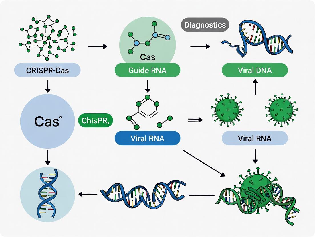
This article provides a comprehensive review of the CRISPR-Cas system's transformative role in viral diagnostics, tailored for researchers and drug development professionals.
CRISPR-Cas in Viral Diagnostics: From Basic Mechanisms to Next-Generation POC Applications
Abstract
This article provides a comprehensive review of the CRISPR-Cas system's transformative role in viral diagnostics, tailored for researchers and drug development professionals. We begin by exploring the foundational principles of CRISPR-Cas biology and its natural antiviral function, establishing the basis for its diagnostic repurposing. The core methodological section details leading platforms like SHERLOCK, DETECTR, and HOLMES, with step-by-step workflows for detecting RNA and DNA viruses. We then address critical troubleshooting and optimization challenges, including sensitivity limits, off-target effects, and sample preparation. Finally, we present a rigorous comparative analysis validating CRISPR diagnostics against gold-standard methods (qPCR, LAMP) and emerging technologies, evaluating performance metrics, cost, and scalability. The conclusion synthesizes the path from lab bench to point-of-care use and future clinical integration.
CRISPR Basics: Understanding the Molecular Scissors and Their Natural Antiviral Role
Within the context of viral diagnostics research, understanding the native function of CRISPR-Cas systems in prokaryotes provides a foundational blueprint. These systems confer adaptive immunity against invasive genetic elements, such as viruses and plasmids, through a genetically encoded memory of past infections. This memory is leveraged in diagnostics via engineered guide RNAs and Cas proteins for sensitive nucleic acid detection.
Core Functional Stages: An Application-Oriented Perspective
The process can be delineated into three distinct stages, each offering unique molecular tools for diagnostics development.
Adaptation: Memory Acquisition
During this initial stage, the Cas1-Cas2 integrase complex captures short fragments (~30-40 bp) of invasive DNA, known as protospacers, and integrates them as new spacers into the CRISPR array at the leader sequence end. This process requires a Protospacer Adjacent Motif (PAM) in the invader DNA, a critical feature exploited in diagnostics to ensure target specificity.
Table 1: Key Quantitative Features of the Adaptation Stage
| Parameter | Typical Range/Value | Significance for Diagnostics |
|---|---|---|
| Protospacer Length | 30-40 base pairs | Determines guide RNA length and specificity. |
| New Spacer Integration Site | Adjacent to the leader sequence | Preserves chronological infection record. |
| PAM Requirement (Type II-A) | 5'-NGG-3' (S. pyogenes Cas9) | Defines targetable sequences; used for assay design. |
| Adaptation Efficiency | Highly variable; often <1 spacer/viral genome | Highlights need for engineered, high-efficiency systems. |
Protocol 1.1: In Vitro Assay for Cas1-Cas2 Integration Activity
- Purpose: To study spacer acquisition kinetics or engineer novel integrases.
- Materials:
- Purified Cas1-Cas2 complex (from E. coli or recombinant).
- Linearized plasmid DNA containing a CRISPR array with leader sequence.
- Fluorescently labeled protospacer DNA duplex (e.g., FAM-labeled 33-bp dsDNA with 5'-TTG-3' PAM).
- Reaction Buffer: 20 mM HEPES (pH 7.5), 150 mM KCl, 10 mM MnCl₂, 1 mM DTT.
- Stop Solution: 50 mM EDTA, 0.1% SDS.
- Agarose gel electrophoresis system.
- Procedure:
- Assemble a 20 µL reaction: 50 nM CRISPR array plasmid, 100 nM protospacer DNA, 200 nM Cas1-Cas2 in Reaction Buffer.
- Incubate at 37°C for 60 minutes.
- Terminate reaction with 5 µL Stop Solution.
- Resolve products on a 1.2% agarose gel. Successful integration results in a higher molecular weight band shift.
Diagram 1: CRISPR Adaptive Immunity - Spacer Acquisition
Expression & Processing: crRNA Biogenesis
The CRISPR array is transcribed into a long pre-crRNA, which is processed into mature CRISPR RNAs (crRNAs). In Class 2 systems (e.g., Type II Cas9), a trans-activating crRNA (tracrRNA) is essential for processing by RNase III and subsequent activity.
Table 2: crRNA Processing Across Major System Types
| System Type | Processing Machinery | Mature crRNA Component | Diagnostic Utility |
|---|---|---|---|
| Type I (Class 1) | Cas6 endoribonuclease | crRNA with repeat handle | Used in multi-Cas complex detection (e.g., Cascade). |
| Type II (Class 2) | RNase III + tracrRNA | crRNA:tracrRNA duplex or sgRNA | Simplified, single-protein system (Cas9, Cas12, Cas13). |
| Type V (Class 2) | Cas12 protein itself | crRNA with minimal handle | Enables "collateral cleavage" used in SHERLOCK/DETECTR. |
Protocol 2.1: Generating sgRNA for Cas9 Diagnostic Assays
- Purpose: To produce single guide RNAs (sgRNAs) combining crRNA and tracrRNA for streamlined detection assays.
- Materials:
- DNA template (PCR product or plasmid with T7 promoter, 20-nt guide sequence, and sgRNA scaffold).
- T7 RNA Polymerase Kit (with NTPs, buffer).
- DNase I (RNase-free).
- RNA Clean-up Kit (e.g., silica column-based).
- Nuclease-free water.
- Procedure:
- Perform in vitro transcription (IVT): Mix 1 µg DNA template with T7 polymerase, NTPs, and buffer per kit instructions.
- Incubate 37°C, 4 hours.
- Add 1 µL DNase I, incubate 15 min at 37°C.
- Purify RNA using Clean-up Kit. Elute in 30 µL nuclease-free water.
- Quantify by Nanodrop (260/280 ratio ~2.0).
Interference: Target Degradation
Mature crRNA guides the Cas effector complex to complementary nucleic acid sequences. Upon PAM-dependent target recognition, Cas nucleases are activated to cleave the invader. Notably, some Cas enzymes (e.g., Cas12a, Cas13a) exhibit trans-cleavage activity post-activation, indiscriminately degrading nearby reporter molecules—the core mechanism of many CRISPR-diagnostic platforms.
Table 3: Interference Mechanisms of Common Cas Effectors
| Cas Protein | PAM Requirement | Cleavage Target | Key Diagnostic Feature |
|---|---|---|---|
| Cas9 (Type II) | 5'-NGG-3' (SpCas9) | dsDNA (blunt ends) | High-fidelity target binding; used for precise detection. |
| Cas12a (Type V) | 5'-TTTV-3' | dsDNA (staggered ends) | Collateral ssDNA cleavage; enables amplified signal. |
| Cas13a (Type VI) | Non-specific (ssRNA) | ssRNA | Collateral ssRNA cleavage; ideal for RNA virus detection. |
Protocol 3.1: Cas12a-based Fluorescent Detection of Viral DNA (DETECTR Assay)
- Purpose: Rapid, isothermal detection of target viral DNA sequence.
- Materials:
- Recombinant LbCas12a protein.
- crRNA designed against target viral sequence (e.g., HPV16 E6/E7).
- Isothermal Amplification Mix (e.g., RPA or LAMP reagents).
- Fluorescent Reporter: ssDNA oligo labeled with 5'-FAM, 3'-BHQ1.
- Reaction Buffer: 20 mM HEPES (pH 6.8), 100 mM KCl, 5 mM MgCl₂, 5% PEG-8000.
- Real-time or endpoint fluorescence reader.
- Procedure:
- Pre-amplification: Amplify sample DNA using isothermal (RPA/LAMP) method for 15-20 min at 37-42°C.
- Detection Reaction: In a fresh tube, mix 50 nM Cas12a, 60 nM crRNA, 500 nM ssDNA reporter in Reaction Buffer.
- Add 2 µL of pre-amplified product (or target control) to a final volume of 20 µL.
- Incubate at 37°C for 15-30 minutes.
- Readout: Measure fluorescence increase (Ex/Em: 485/535 nm) in real-time or at endpoint. A significant increase over no-target control indicates positive detection.
Diagram 2: Cas12a-based Detection via Collateral Cleavage
The Scientist's Toolkit: Key Reagent Solutions for CRISPR Diagnostics Research
Table 4: Essential Research Reagents & Materials
| Item | Function in Research | Example/Notes |
|---|---|---|
| Recombinant Cas Proteins | Core effector enzyme for interference. | Purified SpCas9, LbCas12a, LwaCas13a. Commercial vendors: NEB, IDT, Thermo Fisher. |
| crRNA/sgRNA Synthesis Kits | Produce guide RNAs for target recognition. | In vitro transcription kits or synthetic custom RNA oligos. |
| Isothermal Amplification Kits | Pre-amplify target for sensitive detection without thermal cyclers. | RPA (TwistDx), LAMP (Eiken), NASBA kits. |
| Fluorescent/Colorimetric Reporters | Signal generation upon Cas collateral activity. | ssDNA reporters (FAM/BHQ1) for Cas12; ssRNA (FAM/Quencher) for Cas13; lateral flow strips. |
| Positive Control Target DNA/RNA | Validate assay performance and establish limits of detection (LoD). | Synthetic gBlocks, PCR amplicons, or whole viral genomes. |
| Nuclease-free Buffers & Water | Prevent degradation of sensitive RNA components and reagents. | Essential for all reaction assembly steps. |
| Lateral Flow Strips | Portable, visual readout for point-of-care applications. | Detect labeled cleavage products (e.g., FAM/biotin reporters). |
| Programmable Nucleic Acid Enzymes | For orthogonal signal amplification (e.g., with CRISPR). | Used in assays like CASEXPAR or CARMEN for multiplexing. |
This Application Note delineates the translation of CRISPR-Cas adaptive immune systems into next-generation diagnostic tools, framed within a thesis focused on advancing viral detection methodologies. The intrinsic programmability of Cas nucleases, particularly Cas12 and Cas13, allows for precise targeting of nucleic acid sequences, transforming these systems from cellular defenders into instruments for sensitive and specific pathogen identification. The following sections provide structured data, detailed protocols, and essential toolkits to facilitate implementation in research and development settings.
Data Presentation: Key Performance Metrics of CRISPR-Cas Diagnostics
Table 1: Comparison of Major CRISPR-Cas Systems for Viral Diagnostics
| System | Target Molecule | Collateral Activity | Reporters Used | Typical Detection Limit (copies/µL) | Time-to-Result (min) | Key Viral Application Example |
|---|---|---|---|---|---|---|
| Cas12a (e.g., LbCas12a) | dsDNA/ssDNA | ssDNA trans-cleavage | FQ-reporters, Lateral Flow | 1 - 10 | 30 - 60 | SARS-CoV-2, HPV |
| Cas13a (e.g., LwCas13a) | ssRNA | ssRNA trans-cleavage | FQ-reporters, Lateral Flow | 0.1 - 1 | 20 - 40 | SARS-CoV-2, Dengue, Zika |
| Cas9 | dsDNA | None (nickase) | Fluorescence, Electrochemical | 10 - 100 | 60 - 120 | HBV, HIV |
| Cas14/Cas12f | ssDNA | ssDNA trans-cleavage | FQ-reporters | 0.1 - 1 | < 30 | SNP detection, SARS-CoV-2 variants |
Table 2: Pre-amplification vs. Amplification-Free CRISPR-Dx Approaches
| Parameter | Pre-amplification (e.g., RPA, RT-RPA) | Isothermal Amplification-Coupled (e.g., SHERLOCK, DETECTR) | Amplification-Free (Direct Detection) |
|---|---|---|---|
| Sensitivity | High (aM - fM) | Extremely High (single digit copies) | Moderate to Low (pM - nM) |
| Speed | ~30-90 min | ~60 min | < 30 min |
| Complexity | Medium (2-step process) | Medium (integrated workflow) | Low (single pot) |
| Risk of Contamination | High | High | Very Low |
| Ideal Use Case | Clinical lab, low viral load | Ultrasensitive field detection | Point-of-care, high viral load screening |
Experimental Protocols
Protocol 1: SHERLOCKv2 for SARS-CoV-2 RNA Detection (Adapted from Gootenberg et al., 2017, 2018) Principle: Reverse Transcription Recombinase Polymerase Amplification (RT-RPA) followed by Cas13-mediated collateral cleavage of an RNA reporter. Materials:
- Sample: Nasopharyngeal swab RNA extract.
- Enzymes: LwCas13a, T7 RNA polymerase, Reverse Transcriptase.
- Amplification: RT-RPA primers for SARS-CoV-2 N gene, RT-RPA kit.
- Reporter: Quenched Fluorescent RNA Reporter (e.g., FAM-rU-rU-rU-BHQ1).
- Buffer: NEBuffer 2.1 or equivalent. Procedure:
- Isothermal Amplification: Prepare a 10 µL RT-RPA mix containing 5 µL RNA sample, primers (400 nM final), and RT-RPA master mix. Incubate at 42°C for 20 min.
- T7 Transcription: Dilute 2 µL of RPA product in 8 µL of T7 transcription mix (T7 polymerase, NTPs). Incubate at 37°C for 15 min.
- Cas13 Detection: Prepare a 20 µL detection mix containing: 1 µL LwCas13a (100 nM), 2 µL crRNA (100 nM), 1 µL RNA reporter (1 µM), 5 µL T7 transcription product, and NEBuffer 2.1. Mix gently.
- Fluorescence Measurement: Load into a real-time PCR instrument or plate reader. Incubate at 37°C and measure fluorescence (FAM channel) every 30 seconds for 30 minutes.
- Analysis: A positive sample shows an exponential increase in fluorescence over time. Determine the time-to-positive (TTP) or endpoint fluorescence.
Protocol 2: HUDSON-DETECTR for Direct Detection of Viral DNA from Serum (Adapted from Myhrvold et al., 2018) Principle: Heating Unextracted Diagnostic Samples to Obliterate Nucleases (HUDSON) for sample prep, followed by RPA and Cas12a detection via lateral flow. Materials:
- Sample: Human serum or plasma.
- Enzymes: LbCas12a.
- Amplification: RPA primers for target viral DNA (e.g., HPV-16 E6), RPA kit.
- Reporter: FAM-TTATT-Biotin ssDNA reporter.
- Lateral Flow Strips: Compatible with FAM/Biotin (e.g., Milenia HybriDetect). Procedure:
- HUDSON Sample Prep: Mix 5 µL serum with 5 µL HUDSON buffer (EDTA, TCEP). Heat at 95°C for 5 min, then 4°C for 5 min. This inactivates nucleases and liberates viral nucleic acids.
- RPA Amplification: Use 2 µL of the treated sample in a 50 µL RPA reaction per manufacturer's instructions. Incubate at 39°C for 20 min.
- Cas12a Lateral Flow Detection: a. Prepare a 20 µL cleavage mix: 5 µL RPA product, 300 nM LbCas12a, 300 nM crRNA, 100 nM FAM/Biotin reporter, in Cas12 reaction buffer. b. Incubate at 37°C for 10 min. c. Apply 75 µL of running buffer to a lateral flow strip well. Pipette 10 µL of the cleavage reaction into the same well. d. Allow to run for 2-5 minutes.
- Interpretation: Positive: Both test (T) and control (C) lines appear. Negative: Only the control (C) line appears.
Mandatory Visualization
Title: Generic CRISPR-Cas Diagnostic Workflow
Title: Cas13 RNA Targeting & Trans-Cleavage Mechanism
The Scientist's Toolkit: Essential Research Reagent Solutions
Table 3: Key Reagents for CRISPR-Cas Viral Diagnostic Development
| Reagent Category | Specific Example(s) | Function & Rationale |
|---|---|---|
| CRISPR Nuclease | Purified LbCas12a, LwCas13a | The core effector enzyme. Commercial high-purity, high-activity grades ensure consistent collateral cleavage kinetics. |
| Synthetic crRNA | HPLC-purified crRNA targeting conserved viral sequences (e.g., SARS-CoV-2 ORF1ab). | Provides target specificity. Chemical synthesis allows for rapid prototyping of guide RNAs against emerging variants. |
| Isothermal Amplification Mix | RT-RPA kit (e.g., TwistAmp), LAMP kit. | Enables rapid, instrument-free nucleic acid amplification to boost sensitivity prior to CRISPR detection. |
| Fluorescent Reporters | ssDNA-FQ (for Cas12), ssRNA-FQ (for Cas13) reporters (e.g., FAM-TTATT-BHQ1). | The cleavable substrate that generates a quantitative signal upon Cas activation. Dual-labeled quenchers are critical for low background. |
| Lateral Flow Reporters | FAM/Biotin-labeled ssDNA reporters, compatible lateral flow strips (e.g., HybriDetect). | Enables visual, instrument-free readout ideal for point-of-care applications. |
| Sample Prep Reagents | HUDSON buffers (EDTA/TCEP), magnetic silica beads for RNA/DNA extraction. | Prepares complex clinical samples (serum, saliva) by inactivating inhibitors and releasing nucleic acids. |
| Positive Control Template | Synthetic gBlocks, plasmid clones, or in vitro transcribed RNA of the target viral sequence. | Essential for assay validation, optimization of crRNA efficiency, and as a run control. |
| Nuclease-Free Buffers & Water | Certified nuclease-free water, optimized Cas reaction buffers (e.g., NEBuffer). | Prevents degradation of sensitive RNA/DNA components and ensures optimal enzyme activity. |
Within the broader thesis on developing robust CRISPR-Cas systems for decentralized viral diagnostics, the selection of the effector enzyme is paramount. Cas12, Cas13, and engineered Cas9 variants represent the leading platforms, each with distinct mechanisms, strengths, and limitations. This application note provides a comparative analysis and detailed protocols for their implementation in diagnostic assays.
Enzyme Mechanisms & Comparative Properties
Table 1: Core Characteristics of Key Diagnostic CRISPR-Cas Enzymes
| Feature | Cas12 (e.g., LbCas12a) | Cas13 (e.g., LwaCas13a) | Cas9 Variants (e.g., dCas9, enCas12a) |
|---|---|---|---|
| Target Nucleic Acid | DNA (ss/ds) | RNA (ss) | DNA or RNA (depends on variant) |
| Collateral Activity | ssDNA cleavage (trans) | ssRNA cleavage (trans) | Typically none; engineered for signal transduction (e.g., cleavage of reporter) |
| Guide RNA | crRNA only (shorter) | crRNA only | crRNA + tracrRNA (or sgRNA) |
| PAM/PFS Requirement | PAM Required (e.g., TTTV for LbCas12a) | PFS Required (non-G for LwaCas13a) | PAM Required (e.g., NGG for SpCas9) |
| Primary Diagnostic Use | DNA virus detection, SNP genotyping | RNA virus detection, gene expression | Fused to reporters (e.g., dCas9-fluorescent protein) for visualization without cleavage |
| Key Assay Names | DETECTR, HOLMES | SHERLOCK, CARMEN | CASFISH, REPAIR |
| Typical LOD | ~aM to low fM (10-18 - 10-15 M) | ~aM to low fM (10-18 - 10-15 M) | Varies; often less sensitive than collateral-effect systems |
Application Protocols
Protocol A: Cas12a-based DNA Detection (DETECTR Workflow)
Objective: Detect a double-stranded DNA viral target (e.g., HPV16) using LbCas12a collateral ssDNase activity. Workflow Diagram Title: Cas12 DETECTR Assay Workflow
Materials & Reagents:
- Target DNA: Purified or crude lysate containing viral DNA.
- RPA Reagents: TwistAmp Basic kit (enzymes, rehydration buffer, primers).
- LbCas12a Protein: Commercially sourced recombinant enzyme.
- Target-specific crRNA: Designed with complementarity to the target amplicon and cognate of the TTTV PAM sequence.
- ssDNA FQ Reporter: Oligo with fluorophore (e.g., FAM) and quencher (e.g., BHQ1).
- Reaction Buffer: NEBuffer 2.1 or equivalent.
- Fluorimeter or Plate Reader: For endpoint or real-time detection.
Procedure:
- Isothermal Amplification: Perform RPA per manufacturer’s instructions. Use primers amplifying an 80-200 bp region of the target virus. Incubate at 37-42°C for 15-20 min.
- CRISPR Reaction Setup: Prepare a master mix containing:
- 50 nM LbCas12a
- 50 nM crRNA
- 100 nM ssDNA FQ Reporter
- 1X Reaction Buffer
- Detection: Combine 5 µL of the RPA amplicon with 15 µL of the CRISPR master mix. Incubate at 37°C. Monitor fluorescence (λex/λem ~485/535 nm) in real-time or measure endpoint signal after 30 min.
Protocol B: Cas13-based RNA Detection (SHERLOCK Workflow)
Objective: Detect an RNA viral target (e.g., SARS-CoV-2 genomic RNA) using LwaCas13a collateral RNase activity. Workflow Diagram Title: Cas13 SHERLOCK Assay Workflow
Materials & Reagents:
- Target RNA: Purified viral RNA.
- RT-RPA/RTT Reagents: TwistAmp Basic kit plus reverse transcriptase.
- T7 RNA Polymerase: For in vitro transcription.
- LwaCas13a Protein: Commercially sourced recombinant enzyme.
- Target-specific crRNA: Designed to target the transcribed RNA, avoiding a 5' G (PFS requirement for LwaCas13a).
- ssRNA FQ Reporter: RNA oligo with fluorophore and quencher (e.g., FAM/UU/BBQ).
- Reaction Buffer: Specific Cas13 buffer (e.g., 40 mM HEPES, 60 mM NaCl, 6 mM MgCl2, pH 6.8).
Procedure:
- Target Amplification & Conversion: Perform RT-RPA using primers that embed a T7 promoter sequence in the amplicon. Incubate at 42°C for 30 min.
- T7 Transcription: Use 2 µL of the RPA product in a 10 µL T7 transcription reaction at 37°C for 30-60 min to generate target RNA.
- CRISPR Detection: Prepare a master mix containing:
- 50 nM LwaCas13a
- 50 nM crRNA
- 100 nM ssRNA FQ Reporter
- 1X Reaction Buffer
- Combine 2 µL of the transcription reaction with 18 µL of the CRISPR master mix. Incubate at 37°C and measure fluorescence over time.
Protocol C:dCas9-FP for Visual FluorescentIn SituDetection (CASFISH)
Objective: Visually localize viral DNA sequences in fixed cells using catalytically dead Cas9 (dCas9) fused to a fluorescent protein. Workflow Diagram Title: dCas9-FP FISH Assay Workflow
Materials & Reagents:
- Fixed Cells: Cell culture infected with virus (e.g., AAV), fixed with paraformaldehyde and permeabilized.
- dCas9-FP Protein: Purified dCas9 (D10A, H840A mutants for SpCas9) fused to eGFP or mCherry.
- sgRNA: Target-specific, complexed with dCas9-FP to form ribonucleoprotein (RNP).
- Permeabilization Buffer: PBS with 0.1-0.5% Triton X-100.
- Hybridization Buffer: Containing dextran sulfate and formamide to enhance RNP binding.
- Wash Buffer: SSC buffer with varying stringency.
Procedure:
- Sample Preparation: Fix and permeabilize cells. Treat with RNase-free DNase to expose ssDNA target regions if necessary.
- RNP Complex Formation: Pre-complex 50 nM dCas9-FP with 150 nM sgRNA in hybridization buffer for 15 min at room temperature.
- Hybridization: Apply the RNP complex to the fixed cells. Incubate in a dark, humidified chamber at 37°C for 60 min.
- Washing: Wash cells 3x with wash buffer to reduce background.
- Imaging: Mount samples and visualize using a fluorescence microscope with appropriate filter sets for the FP tag.
The Scientist's Toolkit: Essential Research Reagents
Table 2: Key Reagents for CRISPR Diagnostic Development
| Reagent | Function in Assays | Example Vendor/Product |
|---|---|---|
| Recombinant Cas Proteins | Core enzyme for target recognition and signal generation (collateral cleavage or reporter fusion). | IDT (Alt-R S.p. Cas9, AapCas12b), NEB (LbCas12a, LwCas13a), Mammoth Biosciences. |
| Custom crRNA/sgRNA | Guides the Cas protein to the specific target sequence. Critical for specificity. | Synthesized by IDT, Sigma-Aldrich, or in vitro transcribed. |
| Fluorophore-Quencher (FQ) Reporters | ssDNA or ssRNA oligos that yield fluorescence upon collateral cleavage. Key signal transducer. | Custom ordered from IDT or Biosearch Technologies (e.g., Black Hole Quencher dyes). |
| Isothermal Amplification Kits | Amplifies target to detectable levels without thermocyclers (enables point-of-care use). | TwistAmp (RPA) from TwistDx, or LAMP kits from NEB. |
| Fluorescence Plate Reader / Lateral Flow Strips | Detection modalities. Plate readers offer quantitative data; lateral flow enables visual, binary readouts. | BioTek instruments, or Milenia HybriDetect strips. |
| dCas9-Fusion Constructs | For visualization or enrichment without cleavage (e.g., dCas9-GFP, dCas9-APEX2). | Addgene plasmids for expression and purification. |
Application Notes
This document details the prototypical workflow for utilizing CRISPR-Cas systems, specifically Cas12a and Cas13, for viral nucleic acid diagnostics. The approach leverages the collateral (trans) cleavage activity upon target recognition, enabling highly sensitive and specific detection. This is framed within the broader thesis of developing rapid, deployable, and sequence-specific diagnostics for emerging viral threats.
Core Principle: A CRISPR RNA (crRNA) guides the Cas enzyme to a complementary viral DNA or RNA target. Upon binding and cis-cleavage of the target, the enzyme undergoes a conformational change, activating its non-specific collateral nuclease activity. This activity cleaves nearby reporter molecules (e.g., fluorescent quenched probes), generating a detectable signal.
Key Advantages:
- Specificity: Programmable via crRNA to distinguish single-nucleotide polymorphisms.
- Sensitivity: Can achieve attomolar detection through signal amplification via collateral cleavage.
- Speed: Results can be obtained in 30-60 minutes.
- Instrument Flexibility: Can be adapted to fluorescence plate readers, lateral flow strips, or portable readers.
Table 1: Comparison of Common Cas Enzymes for Viral Diagnostics
| Parameter | Cas12a (e.g., LbCas12a) | Cas13a (e.g., LwaCas13a) | Cas13d (e.g., RfxCas13d) |
|---|---|---|---|
| Target Nucleic Acid | Single-stranded DNA (ssDNA) | Single-stranded RNA (ssRNA) | Single-stranded RNA (ssRNA) |
| Prototypical PAM Requirement | TTTV (V = A, C, G) | Non-G for LwaCas13a | None (minimal constraints) |
| Collateral Substrate | ssDNA reporters | ssRNA reporters | ssRNA reporters |
| Reported Limit of Detection (LoD) | ~aM - fM (for SARS-CoV-2) | ~aM - fM (for SARS-CoV-2) | Low fM (for influenza) |
| Optimal Temperature | 37°C | 37°C | 37-42°C |
| Key Diagnostic Platforms | DETECTR, HOLMES | SHERLOCK, CARMEN | SHERLOCKv2 |
Table 2: Example crRNA Design Parameters for Viral Targets
| Design Factor | Optimal Recommendation | Rationale |
|---|---|---|
| crRNA Spacer Length | 20-24 nucleotides (nt) for Cas12a; 28-30 nt for Cas13 | Balances specificity and binding efficiency. |
| Target Region | Conserved genomic region (e.g., viral polymerase gene) | Ensures detection across viral variants. |
| GC Content | 40-60% | Prevents secondary structure, improves hybridization. |
| Avoidance | Homology to human genome, self-complementarity | Minimizes off-target effects and crRNA misfolding. |
Experimental Protocols
Protocol 1: Design and Preparation of crRNAs for Viral Detection
Objective: To design and synthesize crRNAs targeting a conserved region of a viral genome (e.g., SARS-CoV-2 ORF1ab gene).
- Target Identification: Use databases (NCBI Virus, GISAID) to align sequences of the target virus. Identify a highly conserved region (≥ 95% identity across strains).
- PAM Identification (for Cas12a): Scan the conserved region for the presence of a 5'-TTTV-3' PAM sequence on the opposite strand of your target DNA. The target sequence will be the 20-24 nt preceding the PAM on the PAM-containing strand.
- Spacer Design: Extract the 20-24 nt target sequence directly adjacent to the PAM. Verify specificity using BLAST against the human genome.
- crRNA Synthesis: Order the crRNA as a synthetic, chemically modified RNA oligo with the format:
- Cas12a: 5'-[20-24 nt spacer]-UUUU-3' (direct repeat).
- Cas13: 5'-[28-30 nt spacer embedded in direct repeat]-3' (use published repeat sequences, e.g., for LwaCas13a).
- Resuspension: Resuspend the lyophilized crRNA in nuclease-free TE buffer or water to a stock concentration of 100 µM. Store at -80°C.
Protocol 2: Formation of RNP Complex and Fluorescent Detection Assay
Objective: To assemble the Cas-crRNA ribonucleoprotein (RNP) and perform a one-pot detection reaction using a fluorescent reporter. Materials: Purified Cas enzyme (Cas12a/Cas13), crRNA (from Protocol 1), target viral DNA/RNA (or amplified product), fluorescent quenched reporter (e.g., ssDNA-FQ for Cas12a), reaction buffer, plate reader. Procedure:
- RNP Complex Assembly:
- Prepare a master mix on ice:
- 50 nM purified Cas enzyme
- 60 nM crRNA
- 1X NEBuffer 2.1 (for Cas12a) or NEBuffer r2.0 (for Cas13)
- Incubate at 25°C for 10 minutes to allow RNP formation.
- Prepare a master mix on ice:
- Detection Reaction Setup:
- To the assembled RNP, add:
- Target nucleic acid (1 µL of sample or amplified product)
- Fluorescent reporter (e.g., 500 nM ssDNA-FQ reporter for Cas12a)
- Nuclease-free water to a final volume of 20 µL.
- Include a no-target control (NTC) with nuclease-free water instead of sample.
- To the assembled RNP, add:
- Signal Measurement:
- Transfer the reaction to a 96-well optical plate.
- Immediately place in a real-time fluorescence plate reader pre-heated to 37°C.
- Measure fluorescence (FAM channel: Ex/Em ~485/535 nm) every 60 seconds for 60 minutes.
- Data Analysis:
- Plot fluorescence vs. time. Positive samples show an exponential increase in fluorescence, while NTC remains flat.
- Set a threshold fluorescence (e.g., 3 standard deviations above the mean NTC) to determine time-to-positive (TTP) or endpoint signal.
Protocol 3: Lateral Flow Readout for Point-of-Care Application
Objective: To adapt the collateral cleavage assay for visual readout on a lateral flow strip. Modification to Protocol 2:
- Reporter Design: Use a dual-labeled reporter (e.g., 5'-FAM, 3'-Biotin for Cas12a; 5'-6-FAM, 3'-Biotin for Cas13).
- Reaction: Perform the RNP and target incubation step as in Protocol 2, using the dual-labeled reporter.
- Strip Development:
- After a 30-minute incubation at 37°C, apply 5-10 µL of the reaction to the sample pad of a lateral flow strip (designed to capture FAM, e.g., with anti-FAM antibodies at the test line).
- Immerse the strip in running buffer.
- Interpretation:
- Positive: Collateral cleavage destroys the reporter, preventing capture at the test (T) line. Only the control (C) line appears.
- Negative: Intact reporter is captured at both the T and C lines.
Visualization: Workflow and Mechanism Diagrams
Title: High-Level Diagnostic Workflow
Title: Cas12a Collateral Cleavage Mechanism
The Scientist's Toolkit: Research Reagent Solutions
Table 3: Essential Materials for CRISPR-based Viral Detection
| Item | Function & Rationale | Example Vendor/Product |
|---|---|---|
| Recombinant Cas Protein | The effector enzyme that executes targeted and collateral cleavage. High purity is critical for low background. | IDT: Alt-R S.p. Cas12a (Cpf1) Nuclease. NEB: LbaCas12a (Cpf1). MCLAB: Purified LwCas13a. |
| Custom crRNA | Provides sequence-specific targeting. Chemically synthesized, often with modifications to enhance stability. | IDT: Alt-R CRISPR-Cas12a crRNA. Synthego: Custom CRISPR RNA. Horizon Discovery: Custom guide RNA. |
| Fluorescent Quenched Reporters | ssDNA or ssRNA oligonucleotides with a fluorophore and quencher. Cleavage separates the pair, generating signal. | IDT: Alt-R Cas12a/Cas13 Reporter (FQ). Biosearch Technologies: Black Hole Quencher probes. |
| Lateral Flow Strips | For visual, instrument-free readout. Strips must be compatible with the reporter label (e.g., anti-FAM test line). | Milenia HybriDetect: 2T strips. Ustar Biotechnologies: CRISPR test strips. |
| Isothermal Amplification Mix | Pre-amplifies target for ultra-high sensitivity (e.g., in SHERLOCK/DETECTR). | NEB: WarmStart LAMP/RT-LAMP Kit. TwistAmp: RPA Basic Kit. |
| Nuclease-Free Buffers & Water | Essential to prevent degradation of RNA guides, reporters, and target. | Thermo Fisher: UltraPure DNase/RNase-Free Water. Ambion: Nuclease-Free Water. |
Application Notes: CRISPR-Cas in Viral Diagnostics
The adaptation of CRISPR-Cas systems from a prokaryotic adaptive immune system to a programmable nucleic acid detection tool represents a paradigm shift in diagnostic technology. Framed within the thesis of developing robust, field-deployable viral diagnostics, this evolution is marked by key transitions: the discovery of the biological function, the engineering of Cas9 for genome editing, the characterization of Cas12 and Cas13 collateral activities, and the subsequent development of sensitive, isothermal diagnostic platforms like SHERLOCK and DETECTR.
Key Quantitative Milestones in CRISPR Diagnostic Development
Table 1: Evolution of CRISPR-Cas Diagnostic Sensitivity and Speed
| System/Platform | Target | Reported Limit of Detection (LoD) | Time to Result | Key Cas Enzyme | Year Demonstrated |
|---|---|---|---|---|---|
| SHERLOCK v1 | Zika Virus, Dengue | 2 aM (attomolar) | 1-2 hours | Cas13a (LwaCas13a) | 2017 |
| DETECTR | HPV16, HPV18 | 1 aM | < 1 hour | Cas12a (LbCas12a) | 2018 |
| SHERLOCK v2 | Multiplexed detection | Low femtomolar | < 2 hours | Cas13, Csm6 | 2018 |
| STOPCovid | SARS-CoV-2 | 100 copies/µL | 30-70 minutes | Cas12b (AapCas12b) | 2020 |
| CRISPR-Micro | SARS-CoV-2 | 10 copies/µL | 30 minutes | Cas12f (Cas14) | 2022 |
| SHINE | SARS-CoV-2 & Variants | 50-250 cp/mL | 50 minutes | Cas13 | 2021 |
Table 2: Comparison of Major CRISPR-Cas Effector Proteins for Diagnostics
| Feature | Cas9 | Cas12 (Type V) | Cas13 (Type VI) | Cas3 (Type I) |
|---|---|---|---|---|
| Primary Activity | dsDNA cleavage | ss/dsDNA cleavage (collateral ssDNase) | ssRNA cleavage (collateral RNase) | dsDNA unfolding & degradation |
| Collateral Activity | No | Yes (ssDNA) | Yes (ssRNA) | No |
| PAM Requirement | Yes (3-5 nt) | Yes (TTTV) | No (protospacer flanking site) | Yes (variable) |
| Guide RNA | crRNA+tracrRNA/sgRNA | crRNA | crRNA | crRNA+Cascade complex |
| Diagnostic Utility | Limited (pre-amplification needed) | High (DETECTR, HOLMES) | High (SHERLOCK, CARMEN) | Emerging |
Experimental Protocols
Protocol 1: SHERLOCK (Specific High-sensitivity Enzymatic Reporter unLOCKing) for Viral RNA Detection
Principle: Following isothermal amplification (RPA or RT-RPA), the Cas13a-crRNA complex binds target viral RNA, activating its collateral RNase activity to cleave a fluorescent RNA reporter, generating a detectable signal.
Materials & Reagents:
- Target Viral RNA Sample
- RT-RPA Reagents (TwistAmp Basic kit, primers)
- Purified LwaCas13a Enzyme
- Custom crRNA (designed against target viral sequence)
- Fluorescent RNA Reporter (e.g., FAM-UU-BHQ1)
- T7 RNA Polymerase (for in vitro transcription if needed)
- Nuclease-free Water & Buffer (NEBuffer 2.1)
Procedure:
- Sample Preparation & Amplification:
- Design RPA primers flanking the target viral region. Perform RT-RPA at 42°C for 20-30 minutes.
- Use 2 µL of extracted viral RNA in a 50 µL RT-RPA reaction.
- CRISPR Detection Reaction Assembly:
- Prepare a 20 µL detection mix containing:
- 1x NEBuffer 2.1
- 50 nM LwaCas13a
- 50 nM crRNA
- 100 nM Fluorescent RNA Reporter
- Nuclease-free water to volume.
- Add 2 µL of the RT-RPA product to the detection mix.
- Prepare a 20 µL detection mix containing:
- Incubation & Detection:
- Incubate the reaction at 37°C for 10-60 minutes.
- Measure fluorescence (FAM channel: Ex 485 nm, Em 520 nm) in real-time or at endpoint using a plate reader or lateral flow strip reader.
- Data Analysis:
- A positive signal is defined as fluorescence exceeding 5 standard deviations above the mean of no-template controls.
Protocol 2: DETECTR (DNA Endonuclease Targeted CRISPR Trans Reporter) for Viral DNA Detection
Principle: Following LAMP or PCR amplification, the Cas12a-crRNA complex binds target viral dsDNA, activating collateral ssDNase activity, cleaving a quenched fluorescent ssDNA reporter.
Materials & Reagents:
- Target Viral DNA Sample
- LAMP Reagents (WarmStart LAMP Kit, specific primers)
- Purified LbCas12a (Cpf1) Enzyme
- Custom crRNA
- Fluorescent ssDNA Reporter (e.g., FAM-TTATT-BHQ1)
- Reaction Buffer (NEBuffer 2.1 or 3.1)
Procedure:
- Isothermal Amplification:
- Perform LAMP at 65°C for 20-30 minutes using 2 µL of extracted viral DNA in a 25 µL reaction.
- CRISPR Detection Setup:
- Prepare a 20 µL DETECTR mix containing:
- 1x Reaction Buffer
- 100 nM LbCas12a
- 120 nM crRNA
- 500 nM ssDNA Reporter
- Add 2 µL of the LAMP amplicon.
- Prepare a 20 µL DETECTR mix containing:
- Signal Generation:
- Incubate at 37°C for 10-30 minutes.
- Detect fluorescence (FAM) or use lateral flow strips: dip the strip into the reaction; cleaved reporter is captured at the test line.
- Validation:
- Include positive (synthetic target) and negative (no template, non-target DNA) controls in each run.
The Scientist's Toolkit: Key Research Reagent Solutions
Table 3: Essential Reagents for CRISPR-Based Viral Diagnostic Development
| Reagent/Material | Supplier Examples | Function in Workflow |
|---|---|---|
| Recombinant Cas Proteins (Cas12a, Cas13a, Cas12b) | Integrated DNA Technologies (IDT), Thermo Fisher, NEB | The core effector enzyme that provides programmable target recognition and collateral cleavage activity. |
| Custom crRNA | IDT, Synthego, Sigma-Aldrich | Guides the Cas protein to the complementary target nucleic acid sequence. Critical for specificity. |
| Isothermal Amplification Kits (RPA, LAMP, NASBA) | TwistDx, NEB, OptiGene | Pre-amplifies target nucleic acid to detectable levels without complex thermocycling. |
| Fluorescent Quenched Reporters (ssDNA for Cas12, RNA for Cas13) | Biosearch Technologies, IDT | Substrate for collateral activity. Cleavage produces a fluorescent signal proportional to target presence. |
| Lateral Flow Strips (e.g., Milenia HybriDetect) | Milenia Biotec, Ustar | For visual, instrument-free readout of cleavage events. |
| Nuclease-free Buffers & Water | Thermo Fisher, NEB | Maintains reaction integrity and prevents degradation of sensitive RNA/DNA components. |
| Synthetic Viral RNA/DNA Controls | BEI Resources, IDT gBlocks | Positive controls for assay development and validation, enabling safe handling without live virus. |
Diagnostic Workflow and Mechanism Diagrams
Title: CRISPR Viral Diagnostic Workflow
Title: Cas13a Collateral Cleavage Mechanism
Building a CRISPR Assay: Step-by-Step Platforms and Workflows for Viral Detection
Within the broader research thesis on CRISPR-Cas systems for viral diagnostics, the SHERLOCK (Specific High-sensitivity Enzymatic Reporter unLOCKing) platform represents a pivotal advancement for RNA virus detection. This Application Note details the use of Cas13a (formerly C2c2), an RNA-guided, RNA-targeting CRISPR enzyme, for the specific, attomolar-level detection of viral RNA from pathogens such as SARS-CoV-2 and Influenza. The system's collateral cleavage activity upon target recognition enables amplified, facile signal readout, positioning it as a transformative tool for point-of-care and laboratory-based surveillance.
Principle of Operation
SHERLOCK detection is a two-step process:
- Pre-amplification: Target viral RNA is isothermally amplified using Recombinase Polymerase Amplification (RPA) or Reverse Transcription-RPA (RT-RPA) to increase copy number.
- Cas13 Detection: The amplified product is incubated with the Cas13-crRNA complex. Upon specific recognition of the target sequence, the collateral RNase activity of Cas13 is activated, cleaving a surrounding quenched fluorescent RNA reporter molecule. This cleavage generates a fluorescent signal detectable by plate readers or lateral flow strips.
Quantitative Performance Data
Table 1: Analytical Sensitivity of SHERLOCK for Key Respiratory Viruses
| Virus Target | Cas Enzyme Variant | Pre-amplification Method | Limit of Detection (LoD) | Time-to-Result | Reference / Assay Name |
|---|---|---|---|---|---|
| SARS-CoV-2 | LwaCas13a | RT-RPA | ~10 copies/µL | <60 minutes | SHERLOCKv1 |
| SARS-CoV-2 | LbuCas13a | RT-LAMP | 42 copies/mL | ~55 minutes | STOPCovid.v2 |
| Influenza A | LwaCas13a | RT-RPA | 2.2 aM (attomolar) | ~90 minutes | - |
| Influenza B | LwaCas13a | RT-RPA | 1.8 aM | ~90 minutes | - |
| RSV | LwaCas13a | RT-RPA | ~100 copies/reaction | <120 minutes | - |
Table 2: Comparison of SHERLOCK with Traditional Methods
| Parameter | SHERLOCK (Cas13) | qRT-PCR | Rapid Antigen Test |
|---|---|---|---|
| Typical LoD | 10-100 copies | 1-10 copies | 10^4-10^5 TCID50/mL |
| Assay Time | 45-90 min | 60-120 min | 15-30 min |
| Equipment Needs | Low (Isothermal) | High (Thermocycler) | Very Low |
| Multiplex Capacity | High (4-plex demonstrated) | Moderate | Low |
| Primary Readout | Fluorescence / Lateral Flow | Fluorescence | Visual Stripe |
Detailed Experimental Protocols
Protocol 4.1: SHERLOCK Assay for SARS-CoV-2 RNA Detection (Fluorometric Readout)
A. Materials & Reagent Preparation
- Sample: Viral RNA extracted from nasopharyngeal swab (e.g., using silica column).
- RT-RPA Master Mix: Contains reverse transcriptase, recombinase, polymerase, and nucleotides.
- Primers: Design specific forward and reverse primers for the SARS-CoV-2 N or E gene.
- Cas13 Detection Mix:
- LwaCas13a or LbuCas13a protein (purified)
- crRNA designed against the amplicon (sequence: 5'-[28-nt spacer]-3')
- Fluorescent Reporter Quencher (FQ) probe (e.g., 5'-[6-FAM]-UUUUU-[BHQ1]-3')
- RNase inhibitor
- Detection buffer (e.g., 20 mM HEPES, 60 mM NaCl, 6 mM MgCl2, pH 6.8)
B. Step-by-Step Procedure
- Pre-amplification (RT-RPA): a. Prepare a 10-µL RT-RPA reaction on ice: 5 µL rehydration buffer, 2.1 µL nuclease-free water, 0.4 µL of each primer (10 µM), 0.5 µL probe, 1 µL template RNA, and 0.5 µL magnesium acetate (280 mM). b. Incubate at 42°C for 25-30 minutes. c. Dilute the amplicon 1:20 in nuclease-free water.
Cas13 Detection Reaction: a. Prepare a 10-µL detection mix: 1.5 µL Cas13 protein (500 nM), 1.5 µL crRNA (500 nM), 0.5 µL FQ reporter (2 µM), 0.2 µL RNase inhibitor, 4.3 µL detection buffer, and 2 µL diluted RPA product. b. Incubate at 37°C for 30-60 minutes in a real-time PCR machine or fluorometer with FAM channel acquisition every 1-2 minutes.
Data Analysis: A positive sample shows a significant increase in fluorescence over time compared to no-template controls.
Protocol 4.2: Multiplex Detection & Lateral Flow Readout
For a lateral flow (LF) readout, replace the FQ reporter with a poly-U sequence labeled with biotin at one end and FAM at the other.
- After the Cas13 detection reaction (37°C, 30 min), dilute the product 1:5 in LF assay buffer.
- Apply 75 µL to a commercial lateral flow strip (e.g., Milenia HybriDetect) with anti-FAM antibodies at the test line and streptavidin control.
- Wait 2-5 minutes. Interpretation: Both control and test lines = positive. Only control line = negative.
Diagrams
Diagram 1: SHERLOCK Assay Workflow from Sample to Result
Diagram 2: Cas13 Detection Mechanism: Binding and Collateral Cleavage
The Scientist's Toolkit: Essential Research Reagent Solutions
Table 3: Key Reagents for SHERLOCK Assay Development
| Reagent / Material | Function in the Assay | Example/Notes |
|---|---|---|
| LwaCas13a or LbuCas13a Protein | RNA-guided RNase enzyme; provides specificity and collateral cleavage activity. | Purified recombinant protein. LbuCas13a often offers higher activity. |
| Synthetic crRNA | Guides Cas13 to the target RNA sequence; defines assay specificity. | 28-nt spacer flanked by a direct repeat sequence. Requires careful design to avoid off-targets. |
| Fluorescent Quenched (FQ) Reporter | Signal-generating molecule; cleavage produces fluorescent signal. | Typically 5-6 Uracil ribonucleotides flanked by a fluorophore (FAM) and quencher (BHQ1). |
| Biotin-FAM Reporter (for Lateral Flow) | Alternative reporter for visual readout on lateral flow strips. | Poly-U RNA with 5' Biotin and 3' FAM. Cleavage separates labels. |
| RT-RPA or RT-LAMP Kit | Isothermal amplification for target RNA pre-amplification. | Commercial kits (TwistAmp, Loopamp) provide robust, single-temperature amplification. |
| RNase Inhibitor | Protects the RNA reporter and target from non-specific degradation. | Essential for maintaining signal-to-noise ratio. |
| Lateral Flow Strips | Device for visual, instrument-free readout. | Strips with anti-FAM at test line, streptavidin control line (e.g., Milenia HybriDetect). |
| Fluorometer / Plate Reader | Quantitative fluorescence measurement. | Enables kinetic monitoring and precise quantification of reaction. |
| Nuclease-free Water & Buffers | Reaction environment preparation. | Critical for preventing RNA degradation and ensuring consistent enzyme activity. |
Application Notes
DETECTR (DNA Endonuclease-Targeted CRISPR Trans Reporter) is a CRISPR-Cas12a-based diagnostic platform for sensitive and specific detection of DNA viruses and DNA-transcribed RNA viruses (e.g., HPV, SARS-CoV-2). Its operation is framed within the broader thesis that CRISPR-Cas systems provide a rapid, programmable, and equipment-lean alternative to PCR for point-of-care viral diagnostics.
Cas12a, upon recognition and cleavage of a target DNA sequence complementary to its guide RNA (crRNA), exhibits promiscuous trans-cleavage activity, indiscriminately degrading single-stranded DNA (ssDNA) reporters. This collateral cleavage generates a fluorescent or lateral flow readout, enabling detection. The system can be coupled with an isothermal pre-amplification step (e.g., RPA, LAMP) for attomolar sensitivity, rivaling qPCR.
A critical application is distinguishing HPV16 and HPV18, high-risk strains responsible for ~70% of cervical cancers. DETECTR can differentiate these from other HPV types in under 90 minutes. For RNA viruses like SARS-CoV-2, a reverse transcriptase step is integrated to convert viral RNA to cDNA prior to RPA amplification and Cas12 detection.
Key Quantitative Performance Data:
Table 1: Performance Metrics of DETECTR for Selected Viruses
| Virus Target | Pre-amplification Method | Limit of Detection (LoD) | Time-to-Result | Specificity | Clinical Sensitivity |
|---|---|---|---|---|---|
| HPV16/18 | RPA | ~1 attomolar (aM) | < 90 min | 100% (no cross-reactivity with 14 other HPV types) | 100% (on contrived samples) |
| SARS-CoV-2 | RT-RPA | 10 copies/µL | 30-45 min | 100% (no cross-reactivity with MERS-CoV, common-cold coronaviruses) | 95% vs. CDC qPCR assay |
| ASFV | LAMP | ~10 copies/µL | 60 min | 100% | 100% (on tissue samples) |
Experimental Protocols
Protocol 1: DETECTR Workflow for DNA Virus (e.g., HPV) from Sample to Result
Principle: Sample DNA is isothermally amplified, then mixed with Cas12-crRNA ribonucleoprotein (RNP) and an ssDNA reporter. Target-specific Cas12 activation triggers reporter cleavage, producing a fluorescent signal.
Materials:
- Purified DNA sample
- RPA kit (TwistAmp Basic)
- Cas12a (e.g., LbCas12a) protein
- Synthetic crRNA targeting viral sequence
- ssDNA FQ-reporter (e.g., 5'-6-FAM-TTATT-3'-BHQ1)
- Fluorescence plate reader or lateral flow strips
- Buffer: 20 mM HEPES, 100 mM NaCl, 5 mM MgCl2, pH 6.8
Procedure:
- Pre-amplification: Perform RPA on extracted DNA per manufacturer's protocol. Use primers designed for the viral target. Incubate at 37-42°C for 15-25 minutes.
- DETECTR Reaction Setup:
- Prepare Cas12-crRNA RNP complex: Pre-incubate 50 nM LbCas12a with 60 nM crRNA in 1x reaction buffer for 10 min at room temperature.
- In a reaction well, combine:
- 5 µL of RPA product (or nuclease-free water for negative control)
- 2 µL of RNP complex
- 1 µL of ssDNA FQ-reporter (500 nM final concentration)
- Adjust to a total volume of 20 µL with 1x reaction buffer.
- Detection:
- Incubate the reaction at 37°C for 10-30 minutes.
- Measure fluorescence (Ex/Em ~485/535 nm) in real-time or at end-point.
- Alternatively, for lateral flow: Use a biotin- and FAM-labeled ssDNA reporter. Post-reaction, apply mix to a strip with anti-FAM antibodies at the test line. Cleavage prevents test line capture; thus, a visible test line indicates negative result, while control line should always appear.
Protocol 2: DETECTR for RNA Virus (e.g., SARS-CoV-2) via RT-RPA
Principle: Viral RNA is reverse transcribed and amplified in a one-step RT-RPA reaction, followed by Cas12 detection.
Procedure:
- One-step RT-RPA: Use a kit (TwistAmp RT) combining reverse transcriptase and RPA enzymes. Add extracted RNA and target-specific primers. Incubate at 42°C for 20 min.
- Cas12 Detection: Follow steps 2-3 from Protocol 1, using a crRNA targeting a conserved region of the SARS-CoV-2 genome (e.g., N gene or E gene).
Diagrams
Title: DETECTR Assay Workflow from Sample to Result
Title: Cas12a Target Recognition and Collateral Cleavage Mechanism
The Scientist's Toolkit
Table 2: Essential Research Reagent Solutions for DETECTR Assays
| Reagent/Material | Function/Description | Example Vendor/Product |
|---|---|---|
| LbCas12a or AsCas12a Protein | The effector nuclease; recognizes T-rich PAM (TTTV) and provides collateral trans-cleavage activity. | IDT, BioLabs, Thermo Fisher |
| Synthetic crRNA | A chimeric guide RNA (typically ~40-44 nt) that programs Cas12a specificity for the target viral sequence. | IDT (custom synthesis), Synthego |
| ssDNA Fluorescent Reporter | A short ssDNA oligo with a fluorophore and quencher. Cleavage separates the pair, generating signal. | 5'-6-FAM-TTATT-BHQ1-3' (common sequence) |
| ssDNA Lateral Flow Reporter | A dual-labeled (e.g., FAM and biotin) ssDNA reporter for visual readout on immunochromatographic strips. | Custom synthesis with 5' FAM, 3' Biotin |
| Isothermal Amplification Kit | Pre-amplifies target to detectable levels. RPA (TwistAmp) is most common; LAMP is also used. | TwistDx RPA kits, NEB LAMP kits |
| Reverse Transcriptase (for RNA viruses) | Converts viral RNA to cDNA for amplification. Often integrated into RT-RPA/RT-LAMP kits. | WarmStart RTx (for LAMP), included in RT-RPA kits |
| Lateral Flow Strips | For visual, equipment-free readout. Test line captures uncleaved reporter. | Milenia HybriDetect, Ustar Biotech |
| Nucleic Acid Extraction Kit | Isolates viral DNA/RNA from clinical matrices (swab, serum, tissue). | Qiagen QIAamp, magnetic bead-based kits |
| Fluorescence Microplate Reader | For quantitative, real-time or end-point fluorescent measurement of reaction. | BioTek Synergy, Thermo Fisher Varioskan |
This protocol details an integrated sample-to-answer workflow for the rapid, sensitive, and specific detection of viral nucleic acids, contextualized within the broader research thesis on developing field-deployable CRISPR-Cas diagnostic systems. The approach leverages the specificity of CRISPR-Cas12a for target recognition and signal amplification, coupled with integrated readout methods suitable for point-of-care use. The system is designed to detect RNA viruses, such as SARS-CoV-2 or Influenza A, from crude sample types, minimizing preprocessing steps.
Key Advantages:
- Speed: Results in 30-50 minutes from raw sample.
- Sensitivity: Achieves detection in the attomolar (aM) range.
- Specificity: Single-nucleotide mismatch discrimination via CRISPR RNA (crRNA) design.
- Portability: Readout compatible with lateral flow strips or compact fluorometers.
Experimental Protocol: Integrated CRISPR-Cas12a Detection
A. Reagents and Materials (The Scientist's Toolkit)
| Research Reagent Solution | Function & Brief Explanation |
|---|---|
| Nucleic Acid Extraction: | |
| Magnetic Silica Beads | Bind nucleic acids under high-salt conditions for purification from lysates. |
| Guanidinium Thiocyanate Lysis Buffer | Chaotropic agent that denatures proteins, inactivates nucleases, and releases nucleic acids. |
| Amplification & Detection: | |
| Reverse Transcriptase Recombinase Polymerase Amplification (RT-RPA) Kit | Isothermal enzyme mix for rapid cDNA synthesis and amplification of target without a thermal cycler. |
| Recombinant LbCas12a Enzyme | CRISPR effector protein; upon target DNA binding, exhibits collateral cleavage of ssDNA reporters. |
| Synthetic crRNA | Guides Cas12a to the specific target amplicon sequence. |
| Fluorescent Quenched ssDNA Reporter (e.g., FAM-TTATT-BHQ1) | Collateral cleavage substrate. Intact reporter is quenched; cleavage yields fluorescent signal. |
| Lateral Flow Strips (FAM/Biotin) | For visual readout. Uses anti-FAM and control lines to capture cleaved reporters. |
| Equipment: | |
| Portable Fluorometer or Heater Block | Maintains constant 37-42°C for isothermal reactions. |
| Blue LED Transilluminator | For visual fluorescence observation if using fluorescent readout. |
B. Step-by-Step Procedure
I. Viral RNA Extraction (Magnetic Bead-Based) Time: 15 minutes
- Lysis: Mix 50 µL of nasopharyngeal swab sample (in viral transport medium) with 100 µL of lysis/binding buffer. Vortex for 10 seconds. Incubate at room temperature for 2 minutes.
- Binding: Add 20 µL of well-resuspended magnetic silica beads. Mix by pipetting. Incubate for 5 minutes at room temperature.
- Washing: Place tube on a magnetic stand. After solution clears, discard supernatant.
- Wash beads twice with 200 µL of 80% ethanol while on the magnet. Discard ethanol.
- Air-dry beads for 2-3 minutes.
- Elution: Remove from magnet. Resuspend beads in 25 µL of nuclease-free water or low-salt elution buffer. Incubate at 65°C for 2 minutes. Place on magnet and transfer 20 µL of clear supernatant containing RNA to a new tube.
II. Isothermal Amplification (RT-RPA) Time: 15-20 minutes at 39°C
- Prepare Master Mix: On ice, combine the following in a 0.2 mL tube:
- Rehydration buffer (from kit): 29.5 µL
- Forward/Reverse Primers (10 µM each): 2.4 µL each
- RNA template: 5 µL
- Nuclease-free water: to 47.5 µL total
- Initiate Reaction: Add 2.5 µL of magnesium acetate (280 mM) to the tube cap. Briefly spin down to mix and start the amplification reaction.
- Incubate: Immediately place tube in a pre-heated block or fluorometer at 39°C for 15 minutes.
III. CRISPR-Cas12a Detection & Readout Time: 10 minutes at 37°C
- Prepare Cas12a-crRNA Detection Mix: For each reaction, combine:
- Nuclease-free water: 8 µL
- Cas12a reaction buffer (10x): 2 µL
- LbCas12a (10 µM): 1 µL
- crRNA (10 µM): 1.5 µL
- Fluorescent ssDNA Reporter (10 µM): 0.5 µL
- Total: 13 µL
- Combine and Incubate: Add 2 µL of the RT-RPA amplicon directly to the detection mix. Mix by pipetting.
- For Fluorescence: Incubate at 37°C for 10 minutes in a portable fluorometer, measuring FAM signal every minute.
- For Lateral Flow: After 10 min incubation, dip a lateral flow strip into 80 µL of running buffer in a tube. Apply 5 µL of the detection reaction to the sample pad. Read results at 5 minutes.
Data Presentation & Performance Metrics
Table 1: Analytical Sensitivity of the Integrated Workflow (SARS-CoV- N Gene Pseudovirus)
| Sample Input (RNA copies/µL) | RT-RPA + Cas12a-Fluorescence (Ct, min) | RT-RPA + Cas12a-Lateral Flow (Band Intensity) | Detection Rate (n=5) |
|---|---|---|---|
| 1000 | 5.2 ± 0.3 | Strong Test Line | 5/5 |
| 100 | 8.1 ± 0.5 | Clear Test Line | 5/5 |
| 10 | 12.4 ± 1.1 | Visible Test Line | 5/5 |
| 1 | 16.8 ± 1.7 | Faint Test Line | 4/5 |
| 0 (NTC) | No Signal ( >30) | No Test Line | 0/5 |
NTC: No Template Control.
Table 2: Comparison of Readout Modalities
| Parameter | Fluorescent Readout (Portable Fluorometer) | Visual Readout (Lateral Flow Strip) |
|---|---|---|
| Time to Result | ~10 min incubation | ~15 min total |
| Quantitative? | Yes (Real-time kinetics) | No (Qualitative: Yes/No) |
| Limit of Detection | ~1 copy/µL | ~10 copies/µL |
| Equipment Needed | Compact fluorometer | None (visual) |
| Best For | Quantification, kinetic studies | Pure field deployment, low-cost screening |
Visualization of Workflows and Mechanisms
Integrated Sample-to-Answer Diagnostic Workflow
Cas12a Detection Mechanism and Readout Integration
Within the broader thesis on developing rapid, sensitive, and field-deployable viral diagnostics using CRISPR-Cas systems, the readout modality is a critical determinant of a test's utility. This application note details three predominant signal detection strategies—fluorescent, lateral flow, and electrochemical—used in conjunction with Cas12/Cas13-based assays. Each modality offers distinct trade-offs in sensitivity, equipment needs, cost, and suitability for point-of-care (POC) applications, directly influencing their adoption for viral pathogen detection.
Fluorescent Signal Detection
Fluorescent readouts are the gold standard for quantitative, high-sensitivity detection in laboratory settings. In CRISPR diagnostics, collateral nuclease activity (e.g., Cas12a, Cas13a) cleaves reporter probes comprising a fluorophore-quencher pair, generating a measurable fluorescent signal proportional to the target nucleic acid concentration.
Research Reagent Solutions & Essential Materials
| Item | Function in Fluorescent Readout |
|---|---|
| Cas12a/Cas13a Enzyme | CRISPR effector protein; provides target-specific recognition and collateral cleavage activity. |
| ssDNA/Fluorescent Reporter Probe | Short oligonucleotide with a fluorophore (e.g., FAM, HEX) and a quencher (e.g., BHQ1); cleavage separates the pair, causing fluorescence. |
| Isothermal Amplification Mix (RPA/LAMP) | For pre-amplification of target viral RNA/DNA to enhance sensitivity. Contains enzymes, primers, NTPs, and buffer. |
| Plate Reader or Real-time PCR Instrument | For quantifying fluorescence signal in a kinetic or endpoint manner. |
| Black/White 96- or 384-well Plates | Low-autofluorescence plates for optimal signal-to-noise ratio during detection. |
Experimental Protocol: Cas12a-based Fluorescent Detection of SARS-CoV-2
Objective: Detect SARS-CoV-2 genomic RNA using recombinase polymerase amplification (RPA) and Cas12a-mediated fluorescent signal generation.
Materials:
- Purified viral RNA sample.
- TwistAmp Basic RPA Kit.
- EnGen Lba Cas12a (NEB).
- Custom-designed crRNA targeting SARS-CoV-2 N gene.
- ssDNA-FQ Reporter (5'-FAM-TTATT-BHQ1-3').
- 1X NEBuffer 2.1.
- Real-time PCR instrument or plate reader.
Procedure:
- RPA Pre-amplification: Prepare a 50 µL RPA reaction per manufacturer's instructions using target-specific primers. Add 5 µL of RNA sample. Incubate at 39°C for 15-20 minutes.
- Cas12a Detection Setup: Prepare a 20 µL detection mix containing:
- 1X NEBuffer 2.1
- 50 nM Lba Cas12a
- 60 nM crRNA
- 500 nM ssDNA-FQ reporter
- 2 µL of the RPA amplicon.
- Signal Measurement: Immediately transfer the reaction to a qPCR instrument. Incubate at 37°C and measure fluorescence (FAM channel) every 30 seconds for 30-60 minutes.
- Data Analysis: Calculate ΔF (fluorescence minus initial background). A positive result is defined by a kinetic curve where ΔF exceeds a threshold (e.g., 5 standard deviations above the mean of no-template controls).
| Parameter | Fluorescent (Cas12a/RPA) | Lateral Flow (Cas12a/RPA) | Electrochemical (Cas13a/RPA) |
|---|---|---|---|
| Typical Limit of Detection (LoD) | 1-10 copies/µL | 10-100 copies/µL | 0.1-1 copies/µL |
| Time-to-Result (post-sample prep) | 30-60 min | 5-15 min | 15-30 min |
| Quantitative Capability | Yes (kinetic/endpoint) | No (yes/no) | Yes (amperometric) |
| Key Equipment Required | Fluorescence reader / qPCR | None (visual) | Portable potentiostat |
| Approx. Cost per Test (Reagents) | $3-5 | $2-4 | $2-5 |
| Best Suited For | Lab-based screening, high-throughput | Point-of-care, low-resource settings | POC with quantitative needs, lab-based |
Lateral Flow Signal Detection
Lateral flow assays (LFA) provide a rapid, instrument-free, visual readout ideal for POC use. The CRISPR collateral cleavage activity is adapted to modulate the accumulation of labeled particles (typically gold nanoparticles) at test and control lines on a nitrocellulose strip.
Research Reagent Solutions & Essential Materials
| Item | Function in Lateral Flow Readout |
|---|---|
| Biotin- & FAM-labeled Reporter | Dual-labeled ssDNA reporter (e.g., Biotin-TTATT-FAM); cleavage by activated Cas prevents test line capture. |
| Anti-FAM Antibody at Test Line | Captures intact FAM-labeled reporters, yielding a signal. Cleaved reporters fail to bind. |
| Streptavidin at Control Line | Captures biotin from any reporter, ensuring proper strip function. |
| Gold-Nanoparticle (AuNP) Anti-FAM Conjugate | Visual label that binds to the FAM moiety on intact reporters. |
| Lateral Flow Strips & Cassette | Nitrocellulose membrane embedded with test/control lines housed in a plastic cassette for sample application. |
Experimental Protocol: DETECTR Lateral Flow for HPV16
Objective: Visually detect HPV16 DNA using Cas12a DETECTR and a lateral flow strip.
Materials:
- Purified human DNA sample.
- Lba Cas12a, crRNA, RPA kit.
- Biotin-FAM-ssDNA Reporter (IDT).
- HybriDetect lateral flow strips (Milenia Biotec).
- Running buffer.
Procedure:
- RPA Amplification: Perform a 50 µL RPA reaction with HPV16-specific primers and sample DNA at 39°C for 15 min.
- Cas12a Cleavage Reaction: Combine 10 µL RPA product with 50 nM Cas12a, 60 nM crRNA, and 500 nM Biotin-FAM reporter in 1X buffer. Incubate at 37°C for 10 min.
- Lateral Flow Readout: Dilute the 20 µL reaction with 80 µL of running buffer. Dip the lateral flow strip into the mixture. Wait 5-10 minutes for capillary flow.
- Result Interpretation:
- Positive: Control line (C) appears; Test line (T) does NOT appear. (Cleaved reporter is not captured).
- Negative: Both Control (C) and Test (T) lines appear. (Intact reporter is captured).
- Invalid: No control line.
Electrochemical Signal Detection
Electrochemical readouts translate CRISPR cleavage events into measurable electrical current changes, offering high sensitivity, quantitative potential, and compatibility with miniaturized, portable devices.
Research Reagent Solutions & Essential Materials
| Item | Function in Electrochemical Readout |
|---|---|
| Methylene Blue (MB)-labeled Reporter | Redox-labeled ssDNA or RNA reporter; cleavage alters its diffusion or binding to the electrode surface, changing current. |
| Screen-Printed Electrodes (SPEs) | Disposable, low-cost electrodes (Working, Reference, Counter) for POC use. |
| Portable Potentiostat | Applies voltage and measures resulting current (amperometry, voltammetry). |
| Self-assembled Monolayer (SAM) | Often used to modify the gold working electrode to control probe immobilization and reduce non-specific binding. |
Experimental Protocol: E-CRISPR for SARS-CoV-2 RNA
Objective: Electrochemically detect SARS-CoV-2 RNA using Cas13a and a methylene blue-labeled reporter.
Materials:
- Viral RNA sample.
- Lwa Cas13a (NEB), crRNA.
- RPA kit.
- MB-labeled ssRNA Reporter (5'-rUrUrArUrU-MB-3').
- Screen-printed gold electrodes (SPGEs).
- Portable potentiostat (e.g., PalmSens).
Procedure:
- RPA Pre-amplification: Amplify target RNA using RT-RPA at 42°C for 20 min.
- Electrode Preparation: Clean SPGEs according to manufacturer protocol (e.g., electrochemical cycling in H₂SO₄).
- Cas13a Reaction: Mix 5 µL RPA product with 50 nM Lwa Cas13a, 75 nM crRNA, and 1 µM MB-reporter in 1X reaction buffer. Incubate at 37°C for 30 min.
- Electrochemical Measurement: Apply 10 µL of the reaction directly onto the SPGE working area. Perform square wave voltammetry (SWV) from -0.5 V to 0 V vs. Ag/AgCl. Measure the reduction peak current of MB (~ -0.25 V).
- Data Analysis: The cleavage of the MB-reporter reduces the electrochemical signal. The percentage decrease in peak current is proportional to the target concentration.
Comparative Visualization of Workflows
CRISPR Diagnostic Readout Modality Workflows
Collateral Cleavage to Signal Pathways
The integration of multiplexing strategies with CRISPR-Cas diagnostics represents a pivotal advancement in the broader thesis of developing robust, field-deployable viral detection systems. The inherent programmability of Cas nucleases, particularly Cas12 and Cas13, allows for the simultaneous targeting of multiple genomic regions. This capability is critical for comprehensive pandemic preparedness, enabling the discrimination of viral variants, co-detection of multiple pathogens, and differentiation between vaccine and wild-type strains in a single, streamlined reaction. This application note details contemporary multiplexing approaches and provides actionable protocols for researchers.
Current Multiplexing Methodologies
Effective multiplexing in CRISPR diagnostics requires strategic coordination of crRNAs, reporters, and reaction conditions to maintain sensitivity and specificity for each target.
Spatial Separation on Solid Supports
Targets are detected in physically distinct locations, such as different lines on a lateral flow strip or wells in a microfluidic chip, each programmed with a unique crRNA.
Orthogonal Enzyme-Reporter Pairs
Utilizing distinct Cas enzyme families (e.g., Cas12a, Cas13a) with their respective non-overlapping reporter substrates (DNA vs. RNA probes) allows for parallel detection in a single pot.
Temporal or Signal-Based Multiplexing
Employing a single reporter with crRNAs designed to activate at different times or intensities based on target abundance, though this is less quantitative.
Barcoded Fluorescence Reporting
Using multiple spectrally distinct fluorescent reporters (e.g., FAM, HEX, Cy5) each linked to the collateral activity of a specific Cas-crRNA complex.
Table 1: Comparison of Primary CRISPR-Cas Multiplexing Strategies
| Strategy | Key Principle | Maxplex* | Advantages | Key Limitation |
|---|---|---|---|---|
| Spatial Separation | Physical partitioning of reactions | High (5-10+) | Minimal cross-talk, compatible with LFA | Increased device complexity |
| Orthogonal Enzymes | Different Cas proteins & reporters | Low (2-3) | True single-pot reaction | Limited orthogonal Cas systems |
| Barcoded Fluorescence | Distinct fluorogenic reporters | Moderate (4-5) | Quantitative, single-pot | Requires fluorimeter, spectral overlap |
| Temporal Sequencing | Sequential cleavage from one reporter | Low (2-3) | Simple reporter system | Semi-quantitative, complex optimization |
*Typical practical multiplexing capacity for viral targets.
Table 2: Performance Metrics of Recent Multiplexed CRISPR Assays (2023-2024)
| Assay Name | Targets | Cas Protein | Multiplex Strategy | LOD (copies/µL) | Time (min) | Reference (Preprint/Journal) |
|---|---|---|---|---|---|---|
| CARMEN-Cas13 | 169 respiratory viruses/subtypes | Cas13a | Microfluidic droplet encoding | 1-10 | ~120 | Nature, 2023 |
| MULTIPLEXDx | SARS-CoV-2, Influenza A/B | Cas12a & Cas13a | Orthogonal enzyme-reporter | 5 (each) | 40 | Sci. Adv., 2024 |
| CRISPR-ARMS | SARS-CoV-2 Variants (4 key mutations) | Cas12b | Allele-specific crRNAs on LFA | 20 | 60 | Cell Rep. Med., 2023 |
| FLASH-CRISPR | HIV-1, HBV, HCV | Cas12a | Barcoded fluorescence (3-plex) | 10-50 | 90 | Nat. Commun., 2024 |
Detailed Experimental Protocol: Orthogonal Cas12a/Cas13a Duplex Assay
This protocol enables the simultaneous detection of two different viral RNA/DNA targets in a single-tube reaction using Cas12a (for DNA) and Cas13a (for RNA).
I. Reagent Preparation
- Nuclease-Free Water
- Reaction Buffer (5X): 100 mM HEPES, 500 mM KCl, 50 mM MgCl2, 5% PEG-8000, pH 6.8.
- Cas12a Enzyme: Lachnospiraceae bacterium Cas12a (LbCas12a), 50 µM stock.
- Cas13a Enzyme: Leptotrichia wadei Cas13a (LwCas13a), 50 µM stock.
- crRNAs: Design crRNA for Cas12a target (DNA virus, e.g., HPV-16) and crRNA for Cas13a target (RNA virus, e.g., SARS-CoV-2). Resuspend to 100 µM.
- Reporter Probes:
- Cas12a Reporter: 5´-6-FAM-TTATT-BHQ1-3´ (ssDNA quenched fluorophore), 10 µM.
- Cas13a Reporter: 5´-HEX-UUAUU-BHQ2-3´ (ssRNA quenched fluorophore), 10 µM.
- Positive Control Templates: Synthetic DNA and RNA targets containing the respective protospacer sequences.
- Equipment: Real-time PCR instrument or plate reader capable of detecting FAM and HEX fluorescence.
II. Assay Workflow
- Assay Setup: In a 0.2 mL PCR tube, combine the following on ice:
- 4 µL 5X Reaction Buffer
- 1 µL LbCas12a (50 µM)
- 1 µL LwCas13a (50 µM)
- 1 µL Cas12a crRNA (100 µM)
- 1 µL Cas13a crRNA (100 µM)
- 1 µL Cas12a Reporter (10 µM)
- 1 µL Cas13a Reporter (10 µM)
- X µL Sample (containing potential DNA & RNA targets)
- Nuclease-free water to a final volume of 20 µL.
- Incubation: Place tube in a real-time PCR machine. Run at 37°C for 60 minutes, with fluorescence readings (FAM and HEX channels) taken every minute.
- Data Analysis: Plot fluorescence (RFU) vs. time. A positive signal is defined as a curve that exceeds the threshold (mean of negative control + 3 standard deviations) within 60 minutes.
III. Key Validation Steps
- Cross-talk Test: Run reactions with only one target present to confirm the corresponding reporter activates without triggering the orthogonal reporter.
- Limit of Detection (LOD): Perform assay with serial dilutions of target templates (e.g., 10^6 to 10^0 copies/µL) in n=8 replicates. LOD is the lowest concentration with ≥95% positivity.
- Inhibitor Testing: Spike targets into relevant clinical matrices (e.g., nasal swab VTM, saliva) to determine tolerance.
Diagram Title: Orthogonal Cas12a/Cas13a Multiplex Assay Workflow
Diagram Title: Orthogonal Cas12 and Cas13 Signaling Pathways
The Scientist's Toolkit: Research Reagent Solutions
Table 3: Essential Reagents for Multiplexed CRISPR-Cas Viral Detection
| Item | Function & Role in Multiplexing | Example Product/Catalog Number (Representative) |
|---|---|---|
| Recombinant Cas12a (LbCas12a) | CRISPR effector for DNA target detection. High collateral activity is essential for sensitivity. | IDT Alt-R LbCas12a (Cpf1) |
| Recombinant Cas13a (LwCas13a) | CRISPR effector for RNA target detection. Provides orthogonal functionality to Cas12a. | IDT Alt-R LwCas13a |
| Custom crRNA Libraries | Target-specific guide RNAs. Must be designed with minimal cross-reactivity for multiplexing. | Synthesized via IDT, Sigma, or Trilink (RNase-free, HPLC purified) |
| Fluorogenic Reporter Probes | ssDNA (for Cas12) or ssRNA (for Cas13) probes with fluorophore/quencher pairs. Different fluorophores enable barcoding. | Biosearch Technologies (FAM/HEX/Cy5 with BHQ-1/2) |
| Isothermal Amplification Mix (RPA/LAMP) | Pre-amplification of target nucleic acids to enhance sensitivity prior to CRISPR detection. | TwistAmp Basic (RPA) or NEB WarmStart (LAMP) |
| Lateral Flow Strips | For spatial multiplexing; different crRNA/cas lines detect different targets. | Milenia HybriDetect, Ustar Biotechnologies |
| Nuclease-Free Buffers | Optimized reaction buffers that support simultaneous activity of multiple Cas proteins. | ThermoFisher Scientific, NEBuffer |
| Synthetic Control Templates | Cloned or gBlock gene fragments and RNA transcripts for assay development and validation. | IDT gBlocks, Twist Synthetic Genes |
Application Notes
CRISPR-Cas systems have transitioned from a gene-editing tool to a cornerstone of next-generation viral diagnostics. This application note details their deployment against four critical viral targets—HIV, HPV, Dengue, and emerging pathogens like SARS-CoV-2—within the framework of diagnostic research. The core advantage lies in coupling Cas enzymes' programmable recognition with isothermal amplification and reporter systems, enabling sensitive, specific, and rapid detection in point-of-care formats.
HIV-1: Diagnostic efforts focus on detecting proviral DNA and RNA with high sensitivity to identify reservoir cells and quantify viral load. CRISPR assays target conserved regions like gag or pol, crucial for monitoring antiretroviral therapy efficacy and early infant diagnosis.
HPV: CRISPR diagnostics are designed to genotype high-risk strains (e.g., HPV16, HPV18) by targeting type-specific sequences within the E6/E7 oncogenes. This enables stratification of cancer risk from cervical swab samples, surpassing the binary output of traditional methods.
Dengue Virus (DENV): The necessity to distinguish between the four serotypes (DENV1-4) for epidemiological surveillance and clinical management is addressed by designing serotype-specific gRNAs. Multiplexed CRISPR assays can identify the infecting serotype from serum samples, aiding in prognosis.
Emerging Pathogens: The agility of CRISPR is demonstrated by the rapid development of diagnostics for SARS-CoV-2. Targeting the ORF1ab, N, or E genes, these systems (e.g., DETECTR, SHERLOCK) were deployed within weeks of the viral genome's publication, highlighting the platform's utility for outbreak response.
Quantitative Performance Data: Table 1: Performance Metrics of Selected CRISPR-Based Viral Diagnostic Assays
| Pathogen | Target Gene | CRISPR System | Amplification Method | Limit of Detection (LoD) | Time-to-Result | Key Reference (Example) |
|---|---|---|---|---|---|---|
| HIV-1 | pol | Cas12a, Cas13 | RT-RPA, RT-LAMP | 10-100 copies/µL | 60-90 min | Kellner et al., 2019 (SHERLOCK) |
| HPV16 | E6/E7 | Cas12a | RPA | 1 copy/µL | 2 hours | Chen et al., 2020 |
| Dengue (Serotype 2) | 3' UTR | Cas13 | RT-RPA | 10 copies/µL | 2 hours | Myhrvold et al., 2018 |
| SARS-CoV-2 | N gene, E gene | Cas12a (DETECTR) | RT-LAMP | 10 copies/µL | 30-45 min | Broughton et al., 2020 |
Experimental Protocols
Protocol 1: One-Pot RT-RPA + Cas12a Fluorescence Assay for SARS-CoV-2 RNA Detection
Principle: Viral RNA is reverse transcribed and amplified isothermally via RT-RPA. Cas12a complexed with a target-specific gRNA binds the amplicon, activating its collateral trans-cleavage activity, which degrades a fluorescent-quencher (FQ) reporter, generating a signal.
Materials:
- Sample: Nasopharyngeal/swab extract in nuclease-free water.
- Reagents: RT-RPA dry pellet or mastermix, designed primers (forward/reverse), Cas12a enzyme, synthesized gRNA, ssDNA FQ reporter (e.g., 6-FAM/TTATT/IBFQ), RNase inhibitor.
- Equipment: Real-time fluorescence detector or plate reader, heat block/thermocycler (set to 37-42°C).
Procedure:
- Assay Setup: In a single reaction tube (0.2 mL), combine:
- RT-RPA mastermix: 25 µL
- Primer mix (10 µM each): 2 µL
- Cas12a (100 nM): 1 µL
- gRNA (100 nM): 1 µL
- ssDNA FQ Reporter (10 µM): 1 µL
- Template RNA: 5 µL
- Nuclease-free water to 50 µL total.
- Incubation: Immediately place the tube in a fluorescence reader at 42°C. Measure fluorescence (Ex/Em: 485/520 nm) every 30 seconds for 60 minutes.
- Data Analysis: A positive sample shows an exponential increase in fluorescence signal over time. Set a threshold fluorescence level significantly above the baseline of negative controls (no template, non-target RNA).
Critical Considerations: Primer and gRNA design must avoid cross-reactivity with human genomic DNA and related coronaviruses. Include stringent positive (synthetic target RNA) and negative controls in each run.
Protocol 2: Multiplexed Lateral Flow Readout for Dengue Serotyping
Principle: Following RT-RPA, activated Cas13 or Cas12a cleaves a labeled reporter (e.g., biotin- and FAM-labeled ssRNA/ssDNA). The intact reporter is captured on a lateral flow strip, producing a visual line. Multiplexing uses different reporter labels captured at separate test lines.
Materials:
- Sample: Patient serum.
- Reagents: RT-RPA kit, Cas13a (or Cas12a) enzyme, serotype-specific gRNAs (DENV1-4), labeled reporters (e.g., FAM/Biotin-ssRNA for Cas13), lateral flow strips (anti-FAM at test line, anti-biotin at control line), running buffer.
- Equipment: Heat block (37°C), pipettes.
Procedure:
- Amplification & Cleavage Reaction: Perform separate RT-RPA reactions (20 µL) for each serotype's target, or a multiplexed reaction with all four gRNAs. Add Cas13 and the appropriate FAM/Biotin-ssRNA reporter. Incubate at 37°C for 30 min.
- Lateral Flow Detection: Dilute 5 µL of the reaction product with 95 µL of lateral flow running buffer. Insert the strip into the mixture.
- Result Interpretation (after 10 min):
- Control Line: Must appear for a valid test.
- Test Line(s): Appearance indicates detection of the specific DENV serotype corresponding to the gRNA used. A strip with four test lines can theoretically resolve all serotypes simultaneously.
Visualizations
Title: CRISPR Workflow for Viral Nucleic Acid Detection
Title: Cas12a Collateral Cleavage Detection Mechanism
The Scientist's Toolkit: Research Reagent Solutions
Table 2: Essential Materials for CRISPR Viral Diagnostics
| Item | Function in Protocol | Example/Notes |
|---|---|---|
| Recombinant Cas Protein (Cas12a, Cas13a) | The core effector enzyme; provides programmable nucleic acid binding and collateral cleavage activity. | LbCas12a, LwCas13a; commercially available from suppliers like Integrated DNA Technologies (IDT), New England Biolabs (NEB). |
| Synthetic gRNA | Guides the Cas protein to the complementary viral target sequence. | Chemically synthesized, crRNA for Cas12a; requires careful design for specificity and minimal off-target effects. |
| Isothermal Amplification Mix (RPA/LAMP) | Amplifies target nucleic acid at constant temperature, eliminating need for a thermocycler. | TwistAmp kits (RPA) from TwistDx; WarmStart LAMP kits from NEB. Critical for point-of-care application. |
| Fluorescent-Quencher (FQ) Reporter | Substrate for collateral cleavage; cleavage separates fluorophore from quencher, generating signal. | ssDNA reporter (e.g., 5'-6-FAM-TTATT-BHQ1-3') for Cas12; ssRNA reporter for Cas13. |
| Lateral Flow Strip | Provides visual, instrument-free readout. Typically captures uncleaved, labeled reporter. | Strips with anti-FAM line (test) and anti-biotin/streptavidin line (control). Available from Milenia, Ustar. |
| Positive Control Template | Synthetic viral gene fragment or in vitro transcribed RNA. Validates the entire assay workflow. | Should be full-length or contain the exact target region. Essential for determining LoD and assay calibration. |
Overcoming Hurdles: Maximizing Sensitivity, Specificity, and Robustness in CRISPR Diagnostics
Within the broader thesis of developing a next-generation, field-deployable CRISPR-Cas diagnostic platform for viral pathogens, a core challenge is overcoming the inherent sensitivity limit of Cas enzyme detection. Direct detection of viral nucleic acids from clinical samples often falls short of the required limits of detection (LOD) for early diagnosis. This application note details two synergistic strategies integrated into the thesis work: 1) Pre-amplification of target using isothermal methods (RPA, LAMP), and 2) Engineering of Cas enzymes themselves for enhanced activity and specificity. Detailed protocols and reagent toolkits are provided to enable replication and further research.
Quantitative Comparison of Pre-Amplification Methods
The selection of a pre-amplification method is critical for balancing sensitivity, speed, and complexity. The following table summarizes key performance metrics for RPA and LAMP, as applied to CRISPR-Cas detection workflows.
Table 1: Comparison of RPA and LAMP for CRISPR-Cas Pre-Amplification
| Parameter | Recombinase Polymerase Amplification (RPA) | Loop-Mediated Isothermal Amplification (LAMP) |
|---|---|---|
| Optimal Temperature | 37-42°C | 60-65°C |
| Time to Result | 10-20 minutes | 15-60 minutes |
| Typical LOD (copies/µL) | 1-10 | 1-100 |
| Primer Complexity | Two primers (forward/reverse); simpler design. | Four to six primers; complex design required for specificity. |
| Enzyme Engineering Link | Compatible; amplified product can be designed for optimal Cas12/13 recognition. | Compatible; amplicon is highly structured, requiring careful Cas target site design. |
| Key Advantage | Faster, works at lower temperatures. | Highly robust, often higher yield, tolerates some inhibitors. |
| Key Disadvantage | More susceptible to primer-dimer artifacts. | Primer design is more restrictive; non-specific amplification can be an issue. |
Detailed Experimental Protocols
Protocol 2.1: Integrated RPA-Cas12a Fluorescent Detection of Viral RNA
Objective: To amplify and detect a specific viral RNA target using a one-pot RPA reaction coupled with LbCas12a trans-cleavage.
Workflow Diagram:
Diagram Title: RPA-Cas12a Viral Detection Workflow
Materials & Reagents:
- Template: Purified viral RNA or crude lysate.
- RT-RPA Kit: (e.g., TwistAmp Basic kit with reverse transcriptase).
- Primers: Forward and Reverse primers (200-350 nM final), designed with a T7 promoter sequence appended to the reverse primer for subsequent transcription if using Cas13.
- Cas Protein: Purified LbCas12a nuclease.
- crRNA: Target-specific crRNA (100 nM final).
- Reporter: Fluorescent-Quencher (FQ) ssDNA reporter (e.g., 5'-6-FAM/TTATT/3IABkFQ-3') (100 nM final).
- Buffer: Nuclease-free water, appropriate reaction buffer (provided with kits).
Procedure:
- RPA Amplification: Prepare a 50 µL RT-RPA reaction on ice per manufacturer's instructions, including primers, template, and rehydration buffer. Incubate at 42°C for 15 minutes.
- Cas12a Detection Mix: While RPA incubates, prepare a 20 µL detection mix containing LbCas12a (50 nM), crRNA (100 nM), FQ reporter (100 nM), and reaction buffer.
- Combined Reaction: Transfer 2 µL of the completed RPA product directly into the Cas12a detection mix. Mix by pipetting.
- Incubation & Detection: Incubate the combined reaction at 37°C in a real-time PCR machine or fluorometer. Monitor fluorescence (FAM channel) every 30 seconds for 30 minutes.
- Data Analysis: A positive sample shows an exponential increase in fluorescence over time. Threshold time (Tt) is inversely proportional to initial target concentration.
Protocol 2.2: Engineering Enhanced Cas13a for Direct Detection
Objective: To perform a site-saturation mutagenesis screen on Leptotrichia wadei (Lwa)Cas13a to identify variants with increased collateral RNase activity.
Workflow Diagram:
Diagram Title: Cas13a Engineering Screening Pipeline
Materials & Reagents:
- Template Plasmid: pET-based vector expressing wild-type LwaCas13a with a His-tag.
- Mutagenesis Primers: NNK degenerate codon primers for the target residue.
- Cloning Kit: High-fidelity PCR mix, DpnI, T4 DNA Ligase.
- Expression Host: E. coli BL21(DE3) competent cells.
- Purification: Ni-NTA Agarose Resin, Lysis & Wash Buffers, Imidazole.
- Screening Buffer: 20 mM HEPES pH 6.8, 150 mM KCl, 5 mM MgCl2, 1 mM DTT.
- Screening Substrates: Target RNA (1 nM), Fluorescent RNA Reporter (e.g., 5'-6-FAM/rUrUrUrUrU/3IABkFQ-3') (500 nM).
Procedure:
- Library Generation: Perform PCR using NNK primers to amplify the entire plasmid. Digest template with DpnI, transform into cloning strain, and plate for colony count. Aim for >10x coverage of theoretical diversity (32 codons).
- Protein Expression & Purification: Pick colonies into 96-deep-well blocks. Induce expression with IPTG at 16°C overnight. Lyse cells and purify variants via a 96-well filter plate format Ni-NTA protocol. Elute in screening buffer.
- High-Throughput Activity Assay:
- In a black 384-well plate, mix 5 µL of purified variant with 5 µL of premix containing crRNA (50 nM) and target RNA (1 nM). Incubate 10 min at 37°C.
- Add 10 µL of fluorescent reporter (500 nM) to start the collateral cleavage reaction.
- Immediately measure kinetic fluorescence (ex/em: 485/535 nm) for 1 hour at 37°C in a plate reader.
- Analysis: Calculate the initial rate (V0) of fluorescence increase for each variant. Normalize to wild-type. Select variants with V0 > 150% of wild-type for sequence verification and downstream characterization in diagnostic assays.
The Scientist's Toolkit: Essential Research Reagents
Table 2: Key Reagent Solutions for CRISPR Diagnostics Research
| Reagent / Material | Function & Role in Research | Example Source/Note |
|---|---|---|
| Isothermal Amplification Kits | Provides all enzymes, proteins, and buffers for RPA or LAMP, enabling rapid target pre-amplification without a thermocycler. | TwistAmp (RPA), WarmStart LAMP (NEB) |
| Purified Cas Nucleases | Core detection enzymes. Available as wild-type or engineered variants (e.g., AsCas12a Ultra, LwaCas13a). | IDT, Thermo Fisher, Merck, in-house purification. |
| Synthetic crRNA / gRNA | Programmable guide RNA that confers target specificity to the Cas nuclease. Critical for assay design. | Custom synthesis from IDT, Synthego. |
| Fluorescent-Quencher (FQ) Reporters | ssDNA (for Cas12) or ssRNA (for Cas13) oligonucleotides that emit fluorescence upon Cas collateral cleavage. Provides the real-time signal. | HPLC-purified probes from IDT or Biosearch Technologies. |
| Nuclease-Free Buffers & Water | Essential for preparing all reaction mixes to prevent degradation of RNA/DNA targets and reagents. | Invitrogen, Ambion. |
| Nickel-NTA Agarose Resin | For laboratory-scale purification of polyhistidine (His)-tagged engineered Cas protein variants. | Qiagen, Cytiva. |
| High-Throughput Plate Reader | Enables kinetic fluorescence measurement for screening engineered enzyme libraries and optimizing assay conditions. | BioTek Synergy, BMG CLARIOstar. |
1. Introduction: Specificity in Viral Diagnostics Within the broader thesis on CRISPR-Cas systems for viral diagnostics, achieving optimal specificity is paramount. Off-target cleavage, where the Cas nuclease (e.g., Cas12, Cas13) acts on non-intended nucleic acid sequences, can lead to false-positive signals, compromising diagnostic accuracy. This application note details established and emerging crRNA design rules and bioinformatic tools to mitigate these effects, ensuring robust and reliable assay development.
2. Core crRNA Design Rules for Enhanced Specificity The foundation of specificity lies in the design of the CRISPR RNA (crRNA). Key parameters are summarized below.
Table 1: Key crRNA Design Parameters for Minimizing Off-Target Effects
| Design Parameter | Optimal Recommendation | Rationale | Considerations for Viral Dx |
|---|---|---|---|
| Spacer Length | 20-22 nt for Cas12a; 28-30 nt for Cas13a | Balances specificity and on-target activity. Longer spacers may increase fidelity but reduce efficiency. | Conserve regions across viral strains/quasi-species for broad detection. |
| GC Content | 40-60% | Extremes can affect crRNA stability and RNP complex formation. | Viral genomes (e.g., HIV, Influenza) often have variable GC%; aim for the optimal range within conserved regions. |
| Self-Complementarity | Minimize internal hairpins (>4 bp) | Prevents crRNA folding that impedes Cas binding. | Critical for single-stranded RNA-targeting systems like Cas13. |
| Seed Region (5' end of spacer) | Maximize uniqueness, mismatches here are most disruptive | The seed region (first 5-12 nt) is critical for initial recognition and specificity. | Essential for discriminating between highly similar viral subtypes (e.g., SARS-CoV-2 variants). |
| Off-Target Mismatch Tolerance | Avoid >3-4 mismatches, especially in seed region | Mismatches in the central and 3' regions are more tolerated but can still cause cleavage. | Use bioinformatic tools (below) for exhaustive mismatch profiling. |
3. Essential Bioinformatic Tools for Specificity Analysis A multi-tool approach is recommended to predict and minimize off-target activity.
Table 2: Bioinformatic Tools for Off-Target Prediction & crRNA Design
| Tool Name | Primary Function | Input | Key Output | Access |
|---|---|---|---|---|
| CHOPCHOP | crRNA design & off-target scoring | Target sequence or GenBank ID | Ranked crRNAs with off-target sites. | Web/Standalone |
| CRISPRitz | Comprehensive off-target search with bulges | Genome reference & spacer sequence | List of off-target sites with detailed mismatch/bulge profiles. | Web server |
| Cas-OFFinder | Genome-wide off-target search | Spacer sequence & mismatch/bulge parameters | All possible off-target loci in a given genome(s). | Web/Standalone |
| CRISPResso | Analysis of next-gen sequencing data for editing outcomes | NGS data (amplicon-seq) | Quantification of on-target and off-target indel frequencies. | Web/Standalone |
| GT-Scan | Identifies specific targets while avoiding cross-reactive ones | Spacer sequence & genome database | Specificity score and potential cross-reactive targets. | Web server |
4. Experimental Protocol: In Vitro Off-Target Cleavage Validation Following in silico design, empirical validation is required.
Protocol: Off-Target Assessment for Cas12a-based Viral DNA Detection Objective: Validate the specificity of a candidate crRNA designed against a target viral DNA sequence (e.g., HPV16 E6 gene) using synthetic double-stranded off-target substrates.
Materials:
- Purified Cas12a nuclease
- In vitro transcribed or synthesized candidate crRNA
- Synthetic dsDNA On-Target Substrate (50 nM, 200 bp)
- Synthetic dsDNA Off-Target Substrates (50 nM each, 200 bp). Design 3-4 substrates containing 1-4 mismatches as predicted by Cas-OFFinder.
- NEBuffer r2.1 (or appropriate Cas12a reaction buffer)
- Fluorescent reporter (e.g., ssDNA-FQ reporter for Cas12a)
- Real-time PCR machine or plate reader.
Procedure:
- Reaction Setup: For each substrate (on-target and off-target), prepare a 25 µL reaction containing:
- 1x Reaction Buffer
- 50 nM Cas12a
- 50 nM crRNA
- 100 nM Fluorescent Reporter
- 50 nM DNA Substrate (add last to initiate reaction).
- Incubation & Measurement:
- Transfer reactions to a 96-well optical plate.
- Immediately place in a real-time PCR machine or plate reader pre-heated to 37°C.
- Measure fluorescence (FAM channel, Ex/Em ~485/535 nm) every 60 seconds for 60-90 minutes.
- Data Analysis:
- Plot fluorescence vs. time for each reaction.
- Calculate the time to threshold (Tt) or initial rate of fluorescence increase (RFU/min).
- Compare the signal kinetics of off-target substrates to the on-target. A significant delay (>2x Tt) or reduced rate (>10-fold lower) indicates high specificity.
5. The Scientist's Toolkit: Research Reagent Solutions
Table 3: Essential Reagents for crRNA Specificity Testing
| Reagent/Material | Function | Example Vendor/Product |
|---|---|---|
| Synthetic crRNA (Chemically Modified) | The guide RNA; 2'-O-methyl modifications at 3' end can enhance stability and reduce non-specific signaling. | IDT, Sigma-Aldrich |
| Recombinant CRISPR-Cas Nuclease (Active) | The effector enzyme (e.g., AsCas12a, LbaCas12a, LwCas13a). High purity is critical for clean background. | Thermo Fisher, NEB, MCLAB |
| Fluorescent Reporters (ssDNA-FQ for Cas12, ssRNA-FQ for Cas13) | Detects collateral nuclease activity; cleavage generates fluorescent signal. | Biosearch Technologies, IDT |
| Synthetic gBlock Gene Fragments | Source of defined, sequence-verified on- and off-target DNA substrates for validation. | IDT, Twist Bioscience |
| NGS Library Prep Kit for Amplicon Sequencing | For deep sequencing of potential off-target sites in vitro or in complex samples. | Illumina, Swift Biosciences |
6. Visualizations
Title: crRNA Design & Validation Workflow
Title: On vs. Off Target Impact on Diagnostic Signal
Application Note: CRISPR-Cas Systems for Viral Diagnostics The high mutation rates of RNA viruses (e.g., SARS-CoV-2, Influenza, HIV) present a significant challenge for molecular diagnostics, leading to potential assay failure due to sequence mismatches. This application note details a strategy leveraging CRISPR-Cas systems, specifically Cas12 and Cas13, for robust detection of rapidly mutating viruses. The core principle involves targeting highly conserved viral genomic regions while designing guide RNAs (gRNAs) with strategic redundancy to tolerate variation. This approach is critical for the development of "pan-variant" diagnostic assays.
1. Data-Driven Conservation Analysis for gRNA Design Effective assay design begins with the computational identification of conserved sequences. The following table summarizes quantitative metrics from a recent analysis of SARS-CoV-2 variants (up to Omicron sub-lineages) and Influenza A (H3N2) strains.
Table 1: Conservation Metrics Across Viral Clades for Assay Design
| Virus | Target Gene | Genomic Region | Sequence Length Analyzed (nt) | Number of Strains/Variants | Average Pairwise Identity | Proposed Conserved Target Window (nt) |
|---|---|---|---|---|---|---|
| SARS-CoV-2 | N Gene | 3' End (ORF9b) | 200 | >2,000,000 (GISAID) | 99.7% | 28,380 - 28,410 |
| SARS-CoV-2 | ORF1ab | nsp14 (Proofreading) | 150 | >2,000,000 (GISAID) | 99.5% | 18,040 - 18,070 |
| Influenza A | PA Gene | Endonuclease Domain | 180 | ~15,000 (NCBI) | 98.2% | ~2,200 - 2,230 |
| Influenza A | M Gene | Matrix Protein | 120 | ~15,000 (NCBI) | 99.1% | ~850 - 880 |
2. Protocols for gRNA Design and Validation Protocol 2.1: In Silico Design of Redundant gRNA Pools
- Input: Aligned multiple sequence file (FASTA) of target conserved window.
- Identify Conserved Core: Using tools like
CD-HITorJalview, locate a 20-24 nt core sequence with >99% identity. - Generate Redundant Probes: For each position in the core, allow nucleotide degeneracy (R, Y, S, W, etc.) if the minor allele frequency exceeds 1% in the global dataset.
- Filter for Specificity: BLAST all degenerate gRNA sequences against the human genome (hg38) and common microbiome genomes to exclude non-specific guides.
- Output: A pool of 3-5 gRNA sequences targeting the same conserved window but with permitted degeneracy at variable positions.
Protocol 2.2: Experimental Validation of gRNA Pools using Fluorescent CRISPR Assays Materials: Recombinant LbCas12a or LwCas13a, synthetic target RNA/DNA (wild-type and mutant variants), fluorescent reporter (ssDNA-FQ for Cas12, RNA-FQ for Cas13), plate reader or real-time PCR machine.
- Reaction Setup: In a 20 µL volume, combine:
- 1x NEBuffer 2.1 (Cas12) or 1x NEBuffer r2.0 (Cas13)
- 5 nM Cas enzyme
- 50 nM fluorescent reporter
- 10 nM individual or pooled gRNA
- Nuclease-free water
- Baseline Measurement: Incubate at 37°C for 2 min, measure initial fluorescence (Ex/Em: 485/535 nm).
- Reaction Initiation: Add synthetic target to a final concentration of 1 nM.
- Kinetic Readout: Monitor fluorescence every 30 seconds for 60 minutes at 37°C.
- Analysis: Calculate time-to-threshold (Tt) or maximum slope (RFU/min). A successful, robust gRNA pool will show a <20% variation in Tt across all major variant templates compared to the reference sequence.
3. The Scientist's Toolkit: Research Reagent Solutions Table 2: Essential Reagents for CRISPR-based Viral Diagnostic Development
| Reagent/Material | Function & Rationale |
|---|---|
| Recombinant LbCas12a (purified) | CRISPR effector; provides DNA cleavage and trans-ssDNA cleavage (collateral activity) for signal generation. |
| Recombinant LwCas13a (purified) | CRISPR effector; provides RNA cleavage and trans-ssRNA cleavage (collateral activity) for signal generation. |
| Synthetic crRNA/gRNA (modified) | Guide RNA; determines target specificity. 2'-O-methyl modifications at 3' ends enhance stability in extraction buffers. |
| Fluorescent Quenched Reporters | Signal generation substrate. Cleavage by activated Cas enzyme releases fluorescence. |
| Synthetic Viral Genome Fragments (Wild-type & Variants) | Positive controls for assay validation and for establishing limits of detection (LoD) against key mutations. |
| Paper-Based Lateral Flow Strips (e.g., Milenia HybriDetect) | Alternative readout for Cas12/Cas13 assays; visual detection of cleavage via test/control lines. |
| Isothermal Amplification Mix (RPA/RT-RPA) | For pre-amplification of viral RNA/DNA to increase sensitivity to clinically relevant levels (single-copy detection). |
4. Visualization of Assay Design and Workflow
Diagram 1: Pan-Variant CRISPR Diagnostic Pipeline
Diagram 2: Redundant gRNA Pool Tolerates Sequence Variation
Within the broader thesis on CRISPR-Cas systems for viral diagnostics, robust sample preparation is the critical first step. The sensitivity and specificity of CRISPR-based detection, such as with SHERLOCK or DETECTR, are fundamentally constrained by the quality and purity of the input nucleic acid. This document details application notes and protocols to overcome three pervasive challenges: PCR inhibitors, low viral loads, and complex biological matrices like blood and saliva.
Table 1: Common Inhibitors in Clinical Matrices and Their Impact on CRISPR-Cas Assays
| Matrix | Key Inhibitors | Mechanism of Interference | Reported Inhibition Threshold (Concentration) | Impact on Cas12/Cas13 Activity |
|---|---|---|---|---|
| Whole Blood | Hemoglobin, Lactoferrin, IgG | Binds to nucleic acids, chelates Mg²⁺ (essential cofactor) | Hemoglobin > 2 µM | >70% signal reduction |
| Serum/Plasma | Heparin, Bilirubin, Triglycerides | Heparin inhibits enzyme polymerization; Bilirubin intercalates | Heparin > 0.1 U/µL | Up to 90% reduction |
| Saliva | Mucins, Polysaccharides, Bacterial PCR inhibitors | Increases viscosity, sequesters nucleases, non-specific binding | Mucin > 0.1% (w/v) | 50-80% signal reduction |
| Nasopharyngeal | Mucus, Cellular Debris | Physical blockage, non-specific binding | N/A | Delays reaction kinetics |
Table 2: Viral Load Ranges and Required Sensitivity for Effective Diagnostics
| Virus | Typical Load in Acute Phase (copies/mL) | CRISPR Assay LOD (copies/µL input) | Sample Volume Required for Reliable Detection (Given LOD) |
|---|---|---|---|
| HIV-1 | 10^6 - 10^8 | 1-10 | 50-100 µL of plasma |
| SARS-CoV-2 | 10^3 - 10^9 | 2-20 | 50 µL of saliva/swab |
| HBV | 10^2 - 10^10 | 1-5 | 100 µL of serum |
| Influenza A | 10^4 - 10^7 | 10-50 | 100 µL of nasopharyngeal aspirate |
Detailed Experimental Protocols
Protocol 1: Silica-Based Nucleic Acid Extraction from Whole Blood for CRISPR-Cas12a Detection
Objective: Isolate inhibitor-free viral DNA/RNA from whole blood.
- Lysis: Mix 200 µL of whole blood with 400 µL of lysis buffer (GuHCl, Triton X-100, EDTA). Vortex for 15 sec, incubate at 65°C for 10 min.
- Binding: Add 10 µL of silica magnetic beads to the lysate. Incubate with rotation for 10 min at RT.
- Washing: Pellet beads on a magnet. Discard supernatant. Wash twice with 500 µL wash buffer (GuHCl in Ethanol). Perform a final 80% ethanol wash.
- Elution: Air-dry beads for 5 min. Elute nucleic acids in 30-50 µL of nuclease-free water or low-EDTA TE buffer. Heat to 65°C for 5 min to enhance yield.
- Quality Check: Measure A260/A280 ratio (target: ~1.8-2.0). Use 5 µL of eluate as template for CRISPR-Cas detection.
Protocol 2: Heat-and-Chemical Lysis for Rapid Saliva Prep Prior to RPA-CRISPR
Objective: Rapid, equipment-free preparation of saliva for isothermal amplification coupled to CRISPR.
- Collection: Collect 1 mL of saliva in a sterile tube. Centrifuge at 5000 x g for 2 min to pellet debris.
- Lysis: Transfer 100 µL of supernatant to a new tube. Add 10 µL of proteinase K (20 mg/mL) and 90 µL of lysis buffer (1% Triton X-100, 100 mM Tris-HCl, pH 8.0). Vortex.
- Incubation: Heat at 95°C for 5 min, then immediately place on ice for 2 min.
- Neutralization/Chelation: Add 20 µL of 1 M Tris-HCl (pH 8.0) and 5 µL of 0.5 M EDTA to neutralize inhibitors and chelate divalent cations that may degrade RNA.
- Clarification: Centrifuge at 12,000 x g for 2 min. Use 2-5 µL of the clear supernatant directly in a subsequent Recombinase Polymerase Amplification (RPA) reaction, the amplicons of which are used for CRISPR detection.
Protocol 3: SPRI Bead-Based Cleanup for Inhibitor Removal from Complex Samples
Objective: Post-extraction cleanup of nucleic acids to remove residual inhibitors.
- Binding Ratio Calibration: Use a 1.8x ratio of SPRI (Solid-Phase Reversible Immobilization) bead solution to sample volume (e.g., 90 µL beads for 50 µL eluate). Mix thoroughly by pipetting.
- Incubation: Incubate at RT for 5 min. Place on a magnet until supernatant is clear.
- Washing: With tube on magnet, remove supernatant. Wash beads twice with 200 µL of freshly prepared 80% ethanol without disturbing the pellet. Air-dry for 5-7 min.
- Elution: Remove from magnet, elute in 20-30 µL of nuclease-free water. Incubate at RT for 2 min, then capture beads on magnet. Transfer the purified supernatant to a new tube.
Visualizations
(Title: Workflow for Purifying Nucleic Acids from Complex Matrices)
(Title: Signal Amplification Strategies for Low Viral Loads)
The Scientist's Toolkit: Research Reagent Solutions
Table 3: Essential Materials for Sample Prep in CRISPR-Based Viral Diagnostics
| Item | Function/Application | Key Consideration for CRISPR |
|---|---|---|
| Silica Magnetic Beads | Selective binding of nucleic acids in chaotropic salts. | High-purity beads reduce nonspecific inhibition of Cas enzymes. |
| Proteinase K | Digests nucleases and proteins that can degrade target or inhibit reactions. | Heat-inactivation step is crucial to prevent degradation of Cas proteins. |
| Chaotropic Salts (GuHCl, GuSCN) | Denature proteins, inactivate RNases, promote nucleic acid binding to silica. | Must be thoroughly removed in wash steps as they inhibit Cas activity. |
| Recombinase Polymerase Amplification (RPA) Kit | Isothermal amplification of target from low-copy templates. | Provides amplicon for CRISPR detection; must be optimized to minimize primer-dimer artifacts. |
| Carrier RNA (e.g., Poly-A, tRNA) | Improves recovery efficiency of low-copy viral RNA during extraction. | Must be non-competitive and not interfere with crRNA binding or Cas kinetics. |
| RNase Inhibitor | Protects viral RNA during extraction and pre-amplification steps. | Essential for RNA viruses; use a CRISPR-compatible inhibitor (e.g., recombinant). |
| Lateral Flow Strip | Visual endpoint detection of Cas-mediated reporter cleavage. | Compatible with FAM/biotin-labeled reporters; provides rapid, equipment-free readout. |
| SPRI Beads | Size-selective purification of nucleic acids, removes salts, inhibitors, and dNTPs. | Critical for cleaning up amplicons before CRISPR step to prevent false negatives. |
This document provides application notes and protocols developed as part of a broader thesis on enhancing the field deployment of CRISPR-Cas systems for viral diagnostics. A primary barrier to point-of-care (POC) use is the instability of the recombinase polymerase amplification (RPA) or RT-RPA step, which is highly sensitive to ambient conditions, and the subsequent CRISPR-Cas detection reaction. Optimizing temperature, timing, and buffer composition is critical to create robust, lyophilizable, and field-stable assays.
Table 1: Optimization of RT-RPA Pre-Amplification for Field Stability
| Condition Variable | Tested Range | Optimal Value for Stability | Key Stability Metric (After 4 weeks at 37°C) |
|---|---|---|---|
| Incubation Temperature | 37°C - 42°C | 39°C | Max amplicon yield with minimal non-specific product |
| Reaction Time | 15 - 30 min | 20 min | >95% of fresh reaction signal retained |
| Mg(OAc)₂ Concentration | 12 - 18 mM | 14 mM | Prevents precipitate formation in lyophilized format |
| Trehalose Stabilizer (% w/v) | 0 - 10% | 8% | Retains >90% enzymatic activity post-lyophilization |
| pH (Tris Buffer) | 7.5 - 8.5 | 8.2 | Optimal for both reverse transcriptase and recombinase |
Table 2: CRISPR-Cas12a Detection Reaction Buffer Optimization
| Buffer Component | Standard Concentration | Optimized "Field-Stable" Concentration | Function & Stability Impact |
|---|---|---|---|
| HEPES pH 7.5 | 20 mM | 40 mM | Better buffering capacity against ambient temperature pH shifts. |
| KCl | 100 mM | 60 mM | Reduces aggregation; improves Cas protein shelf-life. |
| MgCl₂ | 10 mM | 6 mM | Minimizes non-specific nuclease activity over time. |
| Polyethylene Glycol 8000 | 0% | 2.5% | Crowding agent enhances reaction speed and stabilizes gRNA. |
| DTT | 5 mM | 1 mM | Prevents oxidation during storage without inhibiting Cas12a. |
| Sucrose | 0% | 5% | Cryoprotectant for lyophilization; maintains ribonucleoprotein complex integrity. |
Experimental Protocols
Protocol 1: Lyophilization of a Field-Stable Master Mix for SARS-CoV-2 Detection This protocol integrates optimized conditions for a combined RT-RPA and Cas12a detection mix.
Objective: To produce a single-tube, lyophilized reagent pellet for a specific viral target (e.g., SARS-CoV-2 N gene) that is stable at elevated temperatures.
Materials: See "Research Reagent Solutions" below. Procedure:
- Master Mix Formulation (on ice):
- In a 1.5 mL tube, combine the following in order:
- 29.5 µL of nuclease-free water.
- 2.1 µL of 500 mM HEPES buffer (pH 7.5, optimized from stock).
- 4.0 µL of 400 mM Trehalose solution (in water).
- 3.2 µL of 250 mM Sucrose solution.
- 1.6 µL of 1 M KCl.
- 1.0 µL of 200 mM MgCl₂.
- 0.7 µL of 100 mM DTT.
- 2.5 µL of 50% PEG 8000.
- 10.0 µL of 2x rehydration buffer (commercial RPA pellet base).
- 2.4 µL of 10 µM forward primer.
- 2.4 µL of 10 µM reverse primer.
- 2.0 µL of 10 µM FQ reporter probe (e.g., TTATT- FAM/ BHQ1).
- 1.0 µL of 10 µM Cas12a crRNA (target-specific).
- 100 ng (≈1 µL) of purified LbCas12a protein.
- 2.0 µL of RT-RPA enzyme freeze-dried beads (commercial, ground to powder).
- In a 1.5 mL tube, combine the following in order:
- Aliquoting and Lyophilization:
- Mix thoroughly by gentle pipetting. Aliquot 50 µL into each well of a PCR strip tube.
- Immediately flash-freeze the strips in a dry-ice/ethanol bath for 10 minutes.
- Transfer strips to a pre-cooled (-50°C) lyophilizer. Dry for 16-24 hours under vacuum (<0.1 mBar).
- Seal strips with desiccant caps and store in foil pouches with silica gel.
- Field Assay Execution:
- Add 50 µL of extracted nucleic acid sample directly to the lyophilized pellet.
- Incubate in a portable, pre-heated block at 39°C for 20 minutes.
- Visualize fluorescence using a handheld blue light transilluminator. A positive signal is indicated by green fluorescence.
Protocol 2: Kinetic Analysis to Determine Optimal Cas12a Incubation Time Objective: To determine the minimum incubation time for maximum signal-to-noise ratio in the detection step.
Procedure:
- Prepare a positive control (synthetic target amplicon at 10 nM) and a no-template control (NTC) using the optimized liquid master mix (from Protocol 1, step 1, but with enzymes added last).
- Distribute 50 µL of each reaction into 8 separate wells of a fluorescence plate reader.
- Place plate in reader pre-heated to 39°C.
- Measure fluorescence (FAM channel, ex/em: 485/535 nm) every 2 minutes for 40 minutes.
- Data Analysis: Plot fluorescence vs. time. The optimal field incubation time is the point where the positive signal reaches 90% of its plateau and is at least 10 standard deviations above the mean NTC signal. (Typical result: 12-15 minutes).
Diagrams
Diagram 1: Workflow for Field-Stable CRISPR Diagnostic Assay Development
Diagram 2: Key Stability Factors in CRISPR-Cas Reaction Buffer
The Scientist's Toolkit: Research Reagent Solutions
Table 3: Essential Reagents for Field-Stable CRISPR Diagnostics Development
| Reagent Solution | Function in Optimization | Example Product/Catalog Note |
|---|---|---|
| Lyophilization-Compatible RPA Kit | Provides the core isothermal amplification enzymes in a format amenable to drying. | TwistAmp Basic RPA kit lyophilization beads. |
| Purified Cas12a (Cpf1) Protein | The detection enzyme. Requires high purity and nuclease-free preparation for reliable formulation. | LbCas12a, recombinant, expressed in E. coli, >95% purity. |
| Chemically Synthesized crRNA | Target-specific guide. Must be HPLC-purified to ensure correct sequence and activity. | Synthetic crRNA with 20-24 nt spacer, 2'-O-methyl modifications at 3 terminal bases for stability. |
| Fluorophore-Quencher (FQ) Reporter | Cas12a collateral activity substrate. Single-stranded DNA probe (e.g., TTATT) labeled. | 5'-/6-FAM/ TTATT /3BHQ_1/-3' DNA oligonucleotide. |
| Cryo-/Lyoprotectants | Stabilize protein and RNA structures during drying and storage. | Trehalose (≥99%), Sucrose (molecular biology grade). |
| Enhanced Reaction Buffer Components | To formulate the optimized buffer system. | HEPES (1M, pH 7.5), PEG 8000, Molecular Grade KCl and MgCl₂. |
| Positive Control Template | Synthetic DNA or RNA target for optimization and QC. | gBlock Gene Fragment or synthetic ssRNA with the target sequence. |
1. Introduction Within the thesis framework of developing deployable CRISPR-Cas diagnostics for viral threats, integration with microfluidics and hardware is paramount. This document details application notes and protocols for creating automated, instrument-free platforms that execute sample-to-answer viral detection. The core strategy leverages paper microfluidics and centrifugal disk systems coupled with smartphone-based detection to eliminate reliance on laboratory instruments.
2. Research Reagent Solutions Toolkit Table 1: Essential Materials for Instrument-Free CRISPR Diagnostic Platforms
| Item | Function | Example/Supplier |
|---|---|---|
| Lateral Flow Strips (LFS) | Visual endpoint detection of Cas12/13 collateral cleavage via immobilization of labeled reporters. | Milenia HybriDetect, USTAR LF-200 |
| Cellulose Paper / Nitrocellulose | Substrate for paper microfluidic circuits; wicks reagents via capillary action. | Whatman Grade 1, FF120HP |
| Wax Printer or Plotter | Patterns hydrophobic barriers to define microfluidic channels on paper. | Xerox ColorQube, Creality CR-10 3D Printer for PMMA molds |
| Reagent Deposition System | Pre-stores lyophilized CRISPR RNP, primers, reporters in defined zones. | BioDot AD1520, XYZ dispensing platform |
| Smartphone with Macro Lens | Captures images of colorimetric/fluorescent LFS or HAD results for quantification. | Standard smartphone with add-on lens |
| Portable Heater | Provides isothermal amplification (e.g., LAMP, RPA) at 37-65°C. | Mini dry bath, flexible resistive heater |
| Centrifugal Microfluidic Disk (PMMA) | Rotating platform where centrifugal force drives fluidics through sequential chambers. | Fabricated via CNC milling or injection molding |
| Lyophilization Stabilizer | Preserves activity of Cas RNP complexes during drying and storage. | Trehalose, PEG, Pullulan |
3. Quantitative Performance Data Table 2: Performance Comparison of Integrated Platforms for Viral Detection
| Platform Type | Target Virus | Assay Time (min) | Limit of Detection (LoD) | Key Hardware Integration | Reference (Year) |
|---|---|---|---|---|---|
| Paper Microfluidic + LFS | SARS-CoV-2 | 40 | 10 copies/µL | Foldable paper cartridge, portable heater | Zhang et al. (2024) |
| Centrifugal Disk + Smartphone | HPV 16/18 | 75 | 5 copies/µL | 3D-printed spinning dock, phone camera | Chen et al. (2023) |
| Modular Sliding Strip | HIV-1 | 60 | 20 copies/µL | Plastic slider, integrated heating chip | Chen & Chen (2024) |
| All-in-One Tube + LFS | DENV | 30 | 100 copies/µL | Single tube, thermos cup for heat | Lee et al. (2023) |
4. Experimental Protocols
Protocol 4.1: Fabrication of a Wax-Printed Paper Microfluidic Chip for CRISPR-LFA Objective: To create a self-contained paper chip that performs RPA amplification followed by Cas12 detection via lateral flow readout. Materials: Wax printer, Whatman chromatography paper, hot plate, lyophilized CRISPR-Cas12 RNP mix, RPA lyopellet, sample inlet pads, absorption pads, lateral flow strip. Procedure:
- Design & Printing: Design a channel network with a sample inlet, amplification zone, and detection zone leading to an LFS window using drawing software. Print the design onto paper using a solid wax printer.
- Wax Patterning: Melt the wax on a hot plate (150°C for 120 sec) to allow it to penetrate the paper, forming hydrophobic barriers.
- Reagent Deposition: Spot 1 µL of reconstituted lyophilized Cas12 RNP (with FAM/Biotin-labeled ssDNA reporter) in the detection zone. Spot lyophilized RPA primers/reagents in the amplification zone. Air-dry.
- Assembly: Attach a glass fiber sample pad to the inlet. Place a commercial LFS strip (pre-cut) such that its sample pad overlaps the paper's detection zone. Place an absorption pad at the LFS terminus. Laminate the entire assembly with a plastic film, leaving inlet and LFS window exposed.
- Assay Execution: Add 30 µL of rehydrated sample (in nuclease-free water) to the inlet. Fold chip to bring amplification zone in contact with a portable heater at 39°C for 20 min. Unfold and wait 10 min for capillary flow to deliver products to LFS. Capture LFS image with smartphone.
Protocol 4.2: Operation of a Centrifugal Microfluidic (Lab-on-a-Disc) Platform Objective: To automate the sequential steps of viral lysis, isothermal amplification, and CRISPR reaction on a spinning disc. Materials: PMMA disc with microfluidic structures, portable spindle motor, smartphone with fluorescence adapter, lyophilized reagents in disc chambers. Procedure:
- Disc Loading: Load raw sample (50 µL) into the "Sample Inlet" chamber. Load release buffers for lyophilized pellets in separate, foil-sealed chambers.
- Sealing: Seal all ports with pressure-sensitive adhesive foil.
- Programmed Rotation: Place disc on motor. Spin at 500 rpm for 30 sec to move sample through a filter for crude lysis. Spin at 1500 rpm to burst foil seals and rehydrate RPA pellet, mixing via Coriolis force. Stop and incubate at 37°C for 15 min (using integrated heating ring).
- CRISPR Reaction Transfer: Spin at 2000 rpm to transfer amplified product to the final chamber containing pre-stored, rehydrated Cas13a reagents.
- Detection: Incubate stationary for 10 min. Spin briefly to move reaction to a cuvette zone. Use a smartphone with a 3D-printed附件 containing a blue LED and an orange filter to image fluorescence. Analyze pixel intensity via app.
5. Diagrams of Workflows and Systems
Title: Paper-based CRISPR Diagnostic Workflow
Title: Centrifugal Disc Automated Protocol
Title: Platform Integration Logic
Benchmarking Performance: How CRISPR Stacks Up Against qPCR, LAMP, and Other Diagnostics
Within the broader thesis on CRISPR-Cas systems for viral diagnostics, a critical evaluation against the gold standard, quantitative PCR (qPCR), is essential. This application note provides a structured comparison of analytical sensitivity (Limit of Detection, LoD) and specificity, alongside detailed protocols for conducting a rigorous head-to-head assessment. The focus is on CRISPR-based assays (e.g., utilizing Cas12, Cas13) designed for direct viral RNA/DNA detection.
Quantitative Comparison: LoD and Specificity
Table 1: Comparative Performance Metrics: CRISPR-Dx vs. qPCR
| Parameter | Quantitative PCR (qPCR) | CRISPR-Based Diagnostics (e.g., Cas12a) | Notes & Context |
|---|---|---|---|
| Theoretical LoD | 1-10 copies/µL (Single-digit copy detection) | 1-100 copies/µL (Highly dependent on pre-amplification) | CRISPR assays often require an RPA/LAMP pre-amplification step to achieve competitive LoD. |
| Practical LoD (Typical Viral Assay) | 10-100 RNA copies/mL (in validated clinical tests) | 10-1000 RNA copies/mL (post pre-amplification) | qPCR protocols are extensively optimized. CRISPR-Dx variability is higher across different platforms. |
| Specificity | High. Defined by primer/probe binding sequences and stringent thermal cycling. | Very High. Defined by crRNA sequence AND Cas enzyme fidelity (collateral activity is sequence-specific). | CRISPR-Cas systems offer programmable specificity; off-target effects can be minimized with crRNA design. |
| Time-to-Result | 60-90 minutes (includes extraction and reverse transcription) | 30-60 minutes post nucleic acid isolation (for one-step RPA-CRISPR workflows) | CRISPR can be faster, especially with lateral flow readouts, but sample prep time remains a factor. |
| Readout Modality | Fluorescent (real-time, quantitative) | Fluorescent, Colorimetric (Lateral Flow), or Electrochemical (semi-quantitative/qualitative) | CRISPR enables instrument-free readout possibilities, crucial for point-of-care applications. |
| Quantification Range | Wide dynamic range (7-8 log orders) | Narrow dynamic range (2-3 log orders, primarily qualitative/semi-quantitative) | qPCR is inherently quantitative; most CRISPR assays are best for presence/absence calls. |
| Susceptibility to Inhibitors | Moderate-High. Affects polymerase efficiency. | Moderate. Inhibitors can affect both pre-amplification and Cas enzyme activity. | Sample purification is critical for both. Some Cas enzymes may tolerate inhibitors better than polymerases. |
Experimental Protocols
Protocol A: Side-by-Side LoD Determination for a Viral Target
Objective: Empirically determine and compare the LoD for qPCR and a CRISPR-Cas assay using a serial dilution of synthetic viral RNA.
Materials: Synthetic viral RNA standard (with known copy number), qPCR master mix (with reverse transcriptase), CRISPR assay components (Cas enzyme, crRNA, reporter, pre-amplification reagents like RPA), real-time PCR machine, fluorometer or lateral flow strips.
Procedure:
- Sample Preparation: Prepare a 10-fold serial dilution of the viral RNA standard in nuclease-free water, spanning from 10^6 to 10^0 copies/µL. Use a carrier RNA in dilution buffer to minimize adsorption.
- qPCR Arm:
- For each dilution, set up 20 µL reactions in triplicate using a one-step RT-qPCR master mix.
- Use validated primer-probe sets for the viral target and an internal control.
- Run on a real-time PCR system with standard cycling conditions (e.g., 50°C 15 min, 95°C 2 min; 45 cycles of 95°C 15s, 60°C 1 min).
- Data Analysis: Plot Ct values against log10 starting copy number. The LoD is the lowest concentration where 95% of replicates are positive (Ct < a defined cutoff).
- CRISPR-Dx Arm (with Pre-amplification):
- Pre-amplification: Subject each RNA dilution to an isothermal amplification (e.g., RPA) at 37-42°C for 15-20 minutes using target-specific primers.
- CRISPR Detection: Transfer 2 µL of the RPA product to a new tube containing the CRISPR detection mix (e.g., 100nM Cas12a, 120nM crRNA, 500nM ssDNA FQ-reporter in appropriate buffer).
- Incubation & Readout: Incubate at 37°C for 10-30 minutes. Monitor fluorescence in real-time on a plate reader or use endpoint measurement.
- Data Analysis: Determine the signal-to-noise ratio (Positive/Negative). The LoD is the lowest concentration where 95% of replicates give a positive signal (e.g., fluorescence above 3x standard deviation of negative control).
Protocol B: Specificity Testing Against Near-Neighbors and Host Genome
Objective: Assess the specificity of both assays by testing against a panel of closely related viral strains (near-neighbors) and human genomic DNA.
Materials: Nucleic acid extracts from related viral isolates, human genomic DNA (50 ng/µL), qPCR and CRISPR assay components as in Protocol A.
Procedure:
- Panel Creation: Prepare test samples containing a high copy number (e.g., 10^4 copies/µL) of the target virus and each near-neighbor virus. Include a sample with only human genomic DNA (50 ng) and a no-template control (NTC).
- qPCR Specificity Test:
- Run all samples in triplicate using the RT-qPCR protocol.
- Analyze for cross-reactivity: A specific assay should yield positive results only for the target virus (Ct values consistent with input copy number) and negative results (Ct > cutoff or undetected) for near-neighbors, human DNA, and NTC.
- CRISPR-Dx Specificity Test:
- Perform the combined RPA-CRISPR assay on all panel samples.
- Critical Control: Include a reaction with a non-targeting crRNA (e.g., targeting a different virus) to confirm that signal generation is crRNA-dependent.
- Analyze fluorescence or lateral flow readouts. Specific assays will show signal only for the target virus + correct crRNA combination.
Visualized Workflows and Relationships
Title: Comparative Diagnostic Workflow: qPCR vs CRISPR-Dx
Title: Assay Selection Logic: qPCR or CRISPR?
The Scientist's Toolkit: Key Research Reagent Solutions
Table 2: Essential Materials for Comparative Studies
| Item | Function in Experiment | Example/Supplier Notes |
|---|---|---|
| Synthetic Viral RNA Standard | Provides a quantifiable target for establishing precise LoD curves and assay linearity. | Obtain from entities like ATCC, BEI Resources, or synthesize via gBlock gene fragments & in vitro transcription. |
| One-Step RT-qPCR Master Mix | Integrates reverse transcription and PCR amplification in a single tube, minimizing hands-on time for qPCR arm. | Commercial kits from Thermo Fisher, Bio-Rad, or Qiagen. Ensure includes dUTP/UNG for carryover prevention. |
| Isothermal Pre-amplification Kit (RPA/LAMP) | Amplifies target to detectable levels for CRISPR assay at constant temperature, enabling speed and field-use. | TwistAmp kits (TwistDx) for RPA. LAMP kits from NEB or Eiken Chemical. |
| CRISPR Enzyme (e.g., LbCas12a, LwaCas13a) | The core detection protein; binds crRNA and exhibits trans-cleavage activity upon target recognition. | Purified recombinant proteins from labs like Zhang Lab (Addgene) or commercial suppliers (e.g., IDT, Mammoth Biosciences). |
| Target-Specific crRNA | Programs CRISPR enzyme specificity by guiding it to the complementary viral sequence. | Designed in-house and synthesized commercially (IDT, Sigma). Must be HPLC-purified. |
| Fluorescent Reporter (e.g., FQ-probe) | Substrate cleaved during Cas collateral activity, generating a fluorescent signal proportional to target presence. | ssDNA probes with fluorophore/quencher pairs (e.g., FAM/BHQ-1) for Cas12; RNA probes for Cas13. |
| Lateral Flow Strips (Optional) | For instrument-free visual readout of CRISPR assay. Often uses biotin- and FAM-labeled reporters. | Milenia HybriDetect strips or similar. Compatible with many commercial CRISPR detection buffers. |
| Inhibitor-Rich Matrix (e.g., Saliva, Serum) | Used to spike synthetic RNA and evaluate assay robustness and susceptibility to inhibitors in complex samples. | Pooled, characterized matrices from commercial bioreclamation companies (e.g., BioIVT). |
Within the context of advancing CRISPR-Cas systems for viral diagnostics, a critical evaluation of assay speed and throughput against established isothermal amplification methods is essential. This application note provides a comparative analysis and detailed protocols to guide researchers in selecting and optimizing rapid diagnostic platforms.
Quantitative Performance Comparison
The following tables summarize key performance metrics based on current literature and commercial kit specifications.
Table 1: Assay Speed and Time-to-Result Breakdown
| Parameter | CRISPR-Cas12/13 (e.g., DETECTR, SHERLOCK) | LAMP | RPA |
|---|---|---|---|
| Typical Amplification Time | 20-40 min (pre-amplification required) | 15-60 min | 10-20 min |
| CRISPR Detection/Cleavage Time | 5-15 min | N/A | N/A |
| Total Time-to-Result | 30-60 min | 20-60 min | 15-40 min |
| Hands-on Time | Moderate-High | Low-Moderate | Low |
| Time to Positive (Early Sample) | ~35 min | ~25 min | ~15 min |
Table 2: Throughput, Sensitivity, and Specificity
| Parameter | CRISPR-Cas12/13 | LAMP | RPA |
|---|---|---|---|
| Potential Throughput (Setup) | Low-96-well plate | Medium-384-well plate | Medium-96-well plate |
| Readout Method | Fluorimeter, Lateral Flow, Spectrophotometer | Turbidity, Fluorimeter, Colorimetric, Lateral Flow | Fluorimeter, Lateral Flow |
| Analytical Sensitivity (LOD) | ~1-10 copies/µL (post-amplification) | ~1-100 copies/µL | ~1-100 copies/µL |
| Specificity | Very High (gRNA + Cas) | High (4-6 primers) | Moderate (2 primers) |
| Multiplexing Capacity | Moderate (Limited by reporter channels) | Low | Very Low |
Detailed Experimental Protocols
Protocol: Combined RT-RPA/CRISPR-Cas12a (DETECTR) for SARS-CoV-2 RNA
Principle: Viral RNA is first amplified isothermally via RT-RPA. The amplicon is then detected by Cas12a-gRNA complex, which upon target binding exhibits collateral cleavage of a fluorescent reporter.
Materials: See "Scientist's Toolkit" (Section 5).
Procedure:
- Sample Preparation: Extract viral RNA using a magnetic bead-based kit. Elute in 20 µL nuclease-free water.
- RT-RPA Amplification:
- Prepare a 50 µL master mix on ice:
- 29.5 µL Rehydration Buffer
- 2.1 µL Forward Primer (10 µM)
- 2.1 µL Reverse Primer (10 µM)
- 0.6 µL Fluorescent Probe (optional, for real-time RPA)
- 5 µL Template RNA
- 9.7 µL Nuclease-free Water
- Add 2 µL of Magnesium Acetate (280 mM) to the tube cap.
- Briefly centrifuge to mix. Incubate at 42°C for 15-20 minutes in a real-time fluorimeter or heat block.
- Prepare a 50 µL master mix on ice:
- CRISPR-Cas12a Detection:
- Prepare a 20 µL detection mix:
- 1 µL Cas12a enzyme (100 nM final)
- 1 µL target-specific gRNA (100 nM final)
- 1 µL ssDNA Reporter (500 nM final, e.g., 5'-6-FAM-TTATT-3'-BHQ1)
- 2 µL RPA amplicon (diluted 1:10 in water)
- 15 µL Nuclease-free Reaction Buffer
- Incubate at 37°C for 5-10 minutes.
- Readout: Measure fluorescence (Ex/Em: 485/535 nm) every minute. A positive sample shows exponential increase. For lateral flow, use biotin- and FAM-labeled reporters and dip a strip after 10 min incubation.
- Prepare a 20 µL detection mix:
Protocol: One-Pot Colorimetric RT-LAMP
Principle: Six primers target eight regions of the viral genome. Amplification at constant temperature produces pyrophosphate ions, lowering pH, which is indicated by a color change.
Procedure:
- Prepare Master Mix: On ice, combine per reaction:
- 12.5 µL 2x LAMP WarmStart Master Mix (contains Bst 2.0 polymerase)
- 1.5 µL Primer Mix (FIP/BIP: 1.6 µM each, F3/B3: 0.2 µM each, LF/LB: 0.4 µM each)
- 1 µL 120 mM Phenol Red (pH 8.5)
- 2-5 µL Template RNA
- Nuclease-free water to 25 µL.
- Run Reaction: Incubate in a heat block or thermocycler (without lid heating) at 63-65°C for 30 minutes.
- Result Interpretation:
- Positive: Color changes from pink (basic) to yellow (acidic).
- Negative: Remains pink.
- Include a non-template control (NTC) and positive control.
Visualization Diagrams
Title: Assay Workflow Comparison: CRISPR vs Direct LAMP
Title: CRISPR-Cas12 Collateral Cleavage Pathway
The Scientist's Toolkit: Key Research Reagent Solutions
Table 3: Essential Materials for Comparative Studies
| Item | Function & Application | Example (Supplier) |
|---|---|---|
| Bst 2.0/3.0 DNA Polymerase | Strand-displacing polymerase for LAMP; high processivity and thermal stability. | WarmStart Bst 2.0 (NEB) |
| RPA Recombinase/Polymerase Kit | Isoothermal amplification at 37-42°C; combines recombinase, polymerase, single-strand binding protein. | TwistAmp Basic (TwistDx) |
| Cas12a (Cpf1) Nuclease | CRISPR effector for detection; provides collateral cleavage activity upon target binding. | LbCas12a (IDT, Thermo) |
| In Vitro Transcript gRNA Kit | For synthesis of high-activity guide RNAs specific to viral target sequences. | HiScribe T7 (NEB) |
| Fluorescent Quenched (FQ) Reporters | ssDNA oligonucleotides with fluorophore/quencher; cleaved by activated Cas12/13 for signal generation. | 5'-6-FAM-TTATT-BHQ1-3' (IDT) |
| Lateral Flow Readout Strips | For endpoint visual detection using FAM/biotin-labeled reporters. | Milenia HybriDetect (TwistDx) |
| Colorimetric LAMP Dye | pH-sensitive dye (e.g., Phenol Red) or metal indicator (e.g., HNB) for visual result readout. | LAMP Colorimetric Master Mix (Thermo) |
| Magnetic Bead NA Extraction Kit | Rapid purification of viral RNA/DNA from swab/saliva samples; amenable to automation. | MagMAX Viral/Pathogen (Thermo) |
Within the broader thesis on developing field-deployable CRISPR-Cas diagnostics for endemic viral pathogens, scalability is a critical translational hurdle. Moving from a proof-of-concept in a research lab to a manufacturable, cost-effective diagnostic test requires a rigorous analysis of cost drivers. This document provides detailed application notes and protocols, framed within viral diagnostics research (e.g., Dengue, Zika, HCV, HIV-1), to guide researchers in performing a cost-benefit analysis (CBA) for assay scalability. The focus is on the tangible components: reagents, equipment, and operational expenses.
Core Cost-Benefit Analysis Framework
A scalability CBA compares the projected costs and benefits of moving from low-volume (Lab-scale R&D) to high-volume (Pilot-scale manufacturing) production. The primary benefit is the cost per test (CPT), which must be minimized for viability in low-resource settings. Costs are categorized below.
Table 1: Categorization of Scalability Cost Drivers
| Category | Description | Examples |
|---|---|---|
| Reagent & Consumable Costs | Expenses for materials consumed per test. | Cas enzyme, gRNAs, nucleotides, primers, reporters (FAM-biotin), cell-free NTPs, nucleic acid extraction kits, reaction tubes. |
| Capital Equipment Costs | One-time purchase of durable instruments. | Real-time Fluorescence Readers, plate washers, automated liquid handlers, thermal cyclers, spectrophotometers. |
| Operational Expenses | Recurring costs for facilities and labor. | Laboratory space (rent/overhead), utilities, quality control, skilled technician salaries, waste disposal, regulatory compliance. |
Quantitative Cost Data & Comparison Tables
Note: The following cost data is compiled from recent (2023-2024) publicly available list prices from major suppliers (e.g., IDT, Thermo Fisher, NEB, Lucigen) and equipment manufacturers. Prices are approximate USD and vary based on volume discounts.
Table 2: Reagent Cost Per Test (CPT) Breakdown for a SHERLOCK-based Assay
| Reagent Component | Lab-Scale (100 rxns) | Pilot-Scale (10,000 rxns) | Notes & Scalability Impact |
|---|---|---|---|
| Recombinant Cas12a/Cas13 | $2.50 - $5.00 | $0.50 - $1.50 | Bulk protein purification or licensed bulk order reduces CPT drastically. |
| Custom crRNA/gRNA | $1.50 - $3.00 | $0.20 - $0.50 | Large-scale synthesis (100 µmol) reduces cost by ~80%. |
| RPA/RT-RPA Primer Mix | $1.00 - $2.00 | $0.15 - $0.30 | Lyophilized, pre-mixed master in bulk volumes. |
| Fluorescent Reporter (ssDNA) | $0.75 - $1.50 | $0.10 - $0.25 | Bulk synthesis of quenched reporters. |
| NTPs & Buffer | $0.50 - $1.00 | $0.05 - $0.15 | High-purity, in-house buffer preparation cuts cost. |
| Extraction Kit (Magnetic Beads) | $3.00 - $8.00 | $1.50 - $3.00 | Moving to bulk reagents and in-house bead formulations offers savings. |
| Plasticware (Tube/Plate) | $0.50 - $1.00 | $0.10 - $0.20 | High-volume procurement of certified sterile consumables. |
| Estimated Total CPT | $9.75 - $22.00 | $2.60 - $5.90 | Scalability can reduce CPT by 70-85%. |
Table 3: Capital Equipment: Purchase vs. Throughput Analysis
| Equipment | Approx. Cost (USD) | Capacity (Tests/Run) | Key Consideration for Scalability |
|---|---|---|---|
| Benchtop Fluorimeter | $5,000 - $15,000 | 1 - 96 | Low throughput, bottleneck for scale-up. |
| Plate Reader (Fluorescence) | $20,000 - $50,000 | 96 - 384 | Essential for medium throughput; allows kinetic reads. |
| Automated Liquid Handler | $30,000 - $100,000+ | 96/384-well plates | Major capital cost but drastically reduces labor, human error, and increases reproducibility. |
| Lyophilizer | $15,000 - $60,000 | Batch process | Enables room-temperature stable reagent formulations, critical for distribution. |
Table 4: Operational Expense Comparison
| Operational Factor | Lab-Scale (R&D) | Pilot-Scale (Manufacturing) | Cost-Benefit Note |
|---|---|---|---|
| Labor (per 1000 tests) | High ($500-$1000) | Medium ($200-$500) | Automation reduces skilled hands-on time but requires trained technicians for operation/QC. |
| QC/Assay Validation | Ad-hoc, ~10% of tests | Systematic, ~15-20% of tests | Increased QC cost is mandatory for reliability, reducing false results which have high societal cost. |
| Waste Disposal (Biohazard) | Moderate | High | Scale increases waste volume; contract costs rise but per-unit cost may fall. |
| Overhead (Space, Utilities) | Absorbed by institute | Dedicated facility cost | A dedicated, optimized space increases efficiency but adds a fixed operational cost. |
Detailed Protocols for Key Scalability Experiments
Protocol 4.1: Lyophilization of CRISPR-Cas Master Mix for Room-Temperature Stability
Objective: To formulate a single-vial, lyophilized reaction mix for a Cas12-based detection assay, eliminating cold-chain dependency. Background: Scalability and distribution to field sites require stable reagents. Lyophilization (freeze-drying) is a key technology for achieving this.
Materials:
- Cas12a enzyme (purified, glycerol-free)
- crRNA (targeting viral sequence, e.g., DENV NS1 gene)
- Fluorescent ssDNA reporter (e.g., FAM-TTATT-BHQ1)
- Rehydration buffer (10 mM Tris-HCl, pH 8.0, 50 mM NaCl)
- Lyoprotectant solution (1:1 Trehalose: Mannitol, 10% w/v in rehydration buffer)
- 0.2 mL PCR tubes or glass lyophilization vials
- Freeze-dryer (Lyophilizer)
- Centrifugal vacuum concentrator (optional)
Procedure:
- Master Mix Formulation: In a nuclease-free tube, combine the following on ice:
- 200 nM Cas12a
- 240 nM crRNA
- 500 nM Fluorescent Reporter
- 1x volume of Lyoprotectant Solution
- Adjust final volume with Rehydration Buffer.
- Note: RPA amplification components can be included in a separate lyophilized pellet or in a second chamber of the same vial.
- Aliquoting: Dispense 19 µL of the master mix into each 0.2 mL PCR tube or designated vial.
- Flash Freezing: Immediately place the open tubes/vials in a -80°C freezer for 2 hours or submerse in a dry ice-ethanol bath for 10 minutes until completely frozen solid.
- Primary Drying: Transfer the frozen tubes to the pre-cooled (-50°C) shelf of the lyophilizer. Start the cycle. Primary drying (sublimation) typically runs for 12-24 hours at a chamber pressure of 0.1 mBar.
- Secondary Drying: Gradually increase the shelf temperature to 25°C over 4-6 hours to remove bound water. Hold at 25°C for 2-4 hours.
- Sealing & Storage: Under inert gas (Argon/Nitrogen) purge if possible, seal vials with caps or stoppers. Store the lyophilized pellets at 4°C (for validation) or at room temperature (for stability testing).
- Rehydration & Use: To perform a test, add 1 µL of extracted sample nucleic acid and 30 µL of nuclease-free water directly to the pellet. Incubate at 37°C for 30-60 min and measure fluorescence.
Protocol 4.2: Cross-Contamination Audit Using Fluorescent Tracers
Objective: To empirically assess and optimize workflow to prevent amplicon contamination in a scaled-up setting. Background: At high throughput, contamination from positive amplicons is a major risk. This protocol uses a non-pathogenic tracer to visualize contamination pathways.
Materials:
- Synthetic target DNA (e.g., Lambda phage DNA)
- qPCR or RPA primers for the target
- FAM-labeled dUTP or a FAM-labeled primer
- Standard lab equipment for nucleic acid amplification
- UV torch or plate reader
- Surface swabs
Procedure:
- "Hot" Amplicon Generation: Perform a large-scale (e.g., 2 mL) amplification reaction of the target DNA using the standard protocol, but incorporate FAM-dUTP into the reaction mix. Purify the amplicon using standard methods.
- Simulated High-Throughput Run:
- In a dedicated "post-amplification" room, aliquot the FAM-labeled amplicon into multiple tubes.
- Have a technician process these "positive samples" through a simulated workflow: opening tubes, performing mock transfers, running through a plate washer, etc.
- Simultaneously, process negative controls (water) in adjacent workstations.
- Contamination Detection:
- Surfaces: Swab key areas (pipettes, bench tops, door handles, equipment buttons). Elute the swab in buffer and measure fluorescence in a plate reader.
- Airborne: Place open microcentrifuge tubes with a "capture" buffer (water) around the room during the simulation. Test these for fluorescence afterward.
- Visual Check: Use a UV torch to scan for fluorescent splashes or aerosols on dark surfaces.
- Data Mapping & Mitigation: Create a map of contamination hotspots. Implement engineering controls: dedicated closed-system amplicon handling equipment, UV irradiation chambers for consumables, and spatial separation (pre-amplification, amplification, post-amplification rooms) with unidirectional workflow.
Visualizations
Diagram 1: Scalability Decision Pathway for CRISPR Diagnostics.
Diagram 2: Unidirectional Workflow to Minimize Contamination.
The Scientist's Toolkit: Research Reagent Solutions
Table 5: Essential Materials for Scalable CRISPR Viral Diagnostics
| Item | Function in Assay | Key Consideration for Scalability |
|---|---|---|
| Glycerol-Free Cas Enzyme | CRISPR effector protein for target recognition and reporter cleavage. | Essential for lyophilization (Protocol 4.1). Bulk licensing and purification reduces CPT. |
| Chemically Modified crRNA | Guides Cas protein to the target viral RNA/DNA. | Bulk synthesis with 2'-O-methyl modifications enhances stability and reduces cost. |
| Lyoprotectants (Trehalose/Mannitol) | Stabilize enzymes and RNA during freeze-drying, forming an amorphous glass. | Enables room-temperature stable, single-vial test formats, eliminating cold chain. |
| Quenched Fluorescent Reporters | ssDNA/RNA probes that yield signal upon Cas-mediated cleavage. | Bulk synthesis with different fluorophore/quencher pairs (FAM/BHQ1, HEX/Iowa Black) allows multiplexing. |
| Magnetic Silica Beads | For solid-phase nucleic acid extraction from clinical samples (serum, swab eluent). | Moving from kit-based to in-house, bulk-prepared bead solutions significantly lowers extraction CPT. |
| Isothermal Amplification Mix (RPA) | Amplifies target viral sequence at constant temperature (37-42°C). | Lyophilized, pre-aliquoted master mixes are critical for scalable, simple workflows. |
| Positive Control Plasmid | Non-infectious synthetic DNA containing the target amplicon sequence. | Required for every batch for Quality Control. Must be sequence-distinct from natural pathogen to track contamination. |
| Fluorescent Tracer (e.g., FAM-dUTP) | Used in contamination audits (Protocol 4.2) to visualize amplicon spread. | Critical operational tool for validating and optimizing a high-throughput manufacturing workflow. |
Within the broader thesis on advancing CRISPR-Cas systems for viral diagnostics, a critical translational challenge is transitioning laboratory-validated assays to field-deployable formats. This application note assesses key parameters—ease of use and portability—essential for point-of-care (POC) and resource-limited settings. We evaluate contemporary platforms, summarize performance data, and provide detailed protocols for implementing a streamlined CRISPR-based diagnostic assay.
Comparative Assessment of Portable CRISPR-Dx Platforms
The following table summarizes quantitative data for recent platforms emphasizing portability.
Table 1: Comparison of Portable CRISPR-Cas Diagnostic Systems for Viral Detection
| Platform/Assay Name | Cas Enzyme | Readout Method | Time-to-Result | Approx. Limit of Detection (LoD) | Key Equipment Required | Complexity Score (1=Low, 5=High) |
|---|---|---|---|---|---|---|
| SHERLOCKv2 | Cas13a/Cas12a | Fluorescent or Lateral Flow Strip (LFS) | 45-60 min | 1-10 copies/µL | Portable incubator, UV light or LFS reader | 2 |
| DETECTR | Cas12a | Lateral Flow Strip (LFS) | 30-45 min | 10-100 copies/µL | Dry bath or water bath, LFS reader (optional) | 2 |
| STOPCovid (with miSHERLOCK) | Cas12a | Fluorescent (Smartphone) | 60 min | 100 copies/µL | 3D-printed device, smartphone | 3 |
| CARMEN | Cas13 | Multiplexed Fluorescent (Microfluidic Chip) | 2-3 hours | <100 copies/µL | Microfluidic chip reader | 4 |
| RPA-Cas12a-LFS (One-Pot) | Cas12a | Lateral Flow Strip (LFS) | 25-35 min | 10-50 copies/µL | Single tube, 37-42°C heat block | 1 |
Detailed Protocol: One-Pot RPA-Cas12a Lateral Flow Assay
This protocol is optimized for minimal equipment and steps.
I. Principle Viral RNA/DNA is simultaneously amplified via Recombinase Polymerase Amplification (RPA) and detected by Cas12a collateral cleavage of a reporter molecule, visualized on a lateral flow strip.
II. Materials & Pre-Assembly
- Nucleic Acid Extraction: Boil-and-spin method (for RNA viruses, include a quick lysis buffer).
- One-Pot Reaction Mix:
- RPA dry pellet (TwistAmp Basic).
- Primer pair (targeting conserved viral region, e.g., SARS-CoV-2 N gene), 420 nM final.
- Cas12a enzyme (LbCas12a), 100 nM final.
- crRNA (designed for target amplicon), 100 nM final.
- FQ-reporter (ssDNA with 5'-FAM, 3'-BHQ1, or 5'-Biotin/6-FAM), 200 nM final.
- Nuclease-free water.
- Magnesium Acetate (MgOAc): 280 mM, provided in RPA kit.
- Lateral Flow Strips: HybriDetect (Milenia) or similar.
- Running Buffer: 1X PBS with 0.1% Tween-20.
- Equipment: Portable 37-42°C dry bath or water bath, timer, pipettes.
III. Step-by-Step Procedure
- Reconstitution: Resuspend the RPA pellet in 29.5 µL of rehydration buffer containing primers, Cas12a, crRNA, FQ-reporter, and water.
- Sample Addition: Add 5 µL of extracted nucleic acid template (or nuclease-free water for NTC) directly into the tube lid.
- Initiation: Briefly centrifuge to combine sample with mix. Immediately add 2.5 µL of 280 mM MgOAc to the tube, mix by pipetting, and quickly place in a 39°C heat block.
- Incubation: Incubate for 25 minutes.
- Detection: Dilute 5 µL of the reaction product with 95 µL of running buffer. Insert a lateral flow strip for 3-5 minutes.
- Interpretation: Positive: Test (T) line and Control (C) line visible. Negative: Only C line visible. Invalid if C line is absent.
The Scientist's Toolkit: Essential Research Reagent Solutions
Table 2: Key Reagents and Materials for Field-Deployable CRISPR Diagnostics
| Item | Function & Rationale | Example/Supplier |
|---|---|---|
| Lyophilized Reaction Pellets | Pre-mixed, stable master mixes containing RPA enzymes, nucleotides, and buffers. Eliminates cold chain and reduces pipetting steps. | TwistAmp Basic lyophilized pellets (TwistDx). |
| Portable Heat Source | Provides consistent isothermal amplification temperature (37-42°C). Critical for RPA/LAMP. | Mini dry bath, pocket heater, or even body heat. |
| Lateral Flow Strips (Cas-compatible) | Visual, binary readout. No need for expensive fluorimeters. Strips are configured for FAM/Biotin reporters. | Milenia HybriDetect, UStar Biotechnologies. |
| Field-Appropriate Extraction Kits | Simple, rapid nucleic acid purification without centrifugation or magnets. | FTA cards, boil-and-spin methods, glass fiber filter-based columns. |
| Stable crRNA & Reporter | Chemically synthesized, lyophilized gRNA and FQ-reporters. Long shelf life at ambient temperature. | Synthesized from IDT, Biosearch Technologies. |
| 3D-Printed Housing | Custom, low-cost device to hold reaction tubes, strips, and a smartphone for imaging/analysis. | Open-source designs (e.g., miSHERLOCK). |
Visualization: Workflow and Mechanism
Title: One-Pot RPA-Cas12a Field Assay Workflow
Title: Cas12a Collateral Cleavage Mechanism on LFS
Within the broader thesis on the CRISPR-Cas system for viral diagnostics research, understanding the regulatory pathway is paramount. The U.S. Food and Drug Administration’s (FDA) Emergency Use Authorization (EUA) mechanism has been a critical avenue for the deployment of CRISPR-based diagnostic tests during public health emergencies. This application note details the current regulatory landscape, specific EUA statuses for CRISPR diagnostics, and the stringent validation requirements that underpin regulatory submissions, providing researchers and developers with a framework for translating laboratory assays into authorized clinical tools.
Current FDA-EUAs for CRISPR-Based Diagnostics
As of the latest available data, a limited number of CRISPR-Cas based diagnostic tests have received FDA-EUA. The primary focus has been on the detection of SARS-CoV-2, leveraging systems like CRISPR-Cas12 and Cas13. The following table summarizes key approvals.
Table 1: FDA-EUA Approved CRISPR-Cas Diagnostic Tests (Representative)
| Test Name (Developer) | Target Pathogen | CRISPR System | EUA Issue Date | Key Technology/Platform | Authorized Setting |
|---|---|---|---|---|---|
| Sherlock CRISPR SARS-CoV-2 Kit (Sherlock Biosciences) | SARS-CoV-2 | Cas13 | May 6, 2020 | SHERLOCK (Specific High-sensitivity Enzymatic Reporter unLOCKing) | High-complexity CLIA labs |
| DETECTR BOOST (Mammoth Biosciences) | SARS-CoV-2 | Cas12 | N/A (Note: Initial EUA submission; specific EUA date may vary. Latest public status is under review/authorization pathway.) | DETECTR (DNA Endonuclease Targeted CRISPR Trans Reporter) | Point-of-Care (POC) intended |
Note: The regulatory landscape is dynamic. Developers like Mammoth Biosciences have publicly announced EUA submissions and preparatory steps for FDA review. Researchers must consult the FDA's official "In Vitro Diagnostics EUAs" list for the most current status.
Core Validation Requirements for EUA Submission
FDA-EUA submissions for in vitro diagnostics (IVDs), including CRISPR-based tests, require comprehensive analytical and clinical performance data. The validation must follow the FDA's "Policy for Coronavirus Disease-2019 Tests" and relevant guidance documents.
Table 2: Core Validation Benchmarks for CRISPR Diagnostic EUA
| Validation Parameter | FDA Expectations (Summary) | Typical Target for CRISPR Assays |
|---|---|---|
| Limit of Detection (LoD) | Determine the lowest concentration of viral target detectable in ≥95% of replicates. | 100-1000 copies/mL (or genome equivalents/reaction), varying by assay. |
| Analytical Specificity | 1. Cross-Reactivity: Test against common respiratory pathogens and human microbiome.2. Interfering Substances: Evaluate effects of biotin, common mucolytic agents, etc. | No cross-reactivity with a panel of >30 organisms. Tolerance to listed interfering substances at specified concentrations. |
| Clinical Agreement (vs. a comparator method) | Test a minimum number of positive and negative clinical specimens. | Positive Percent Agreement (PPA): ≥95% (e.g., 30/30 positives).Negative Percent Agreement (NPA): ≥98% (e.g., 50/50 negatives). |
| Inclusivity (Genetic Diversity) | In silico and wet-lab testing against known genetic variants of the target. | Detection of all major circulating variants (e.g., SARS-CoV-2 Variants of Concern). |
| Sample Stability | Demonstrate target stability in specimen collection and transport media over claimed conditions and time. | Stability for X days at 2-8°C, -20°C, etc. |
| Test Reproducibility | Assess precision across operators, days, lots, and instruments. | >90% agreement across all conditions. |
Detailed Experimental Protocols for Key Validation Studies
Protocol 1: Determination of Limit of Detection (LoD)
Objective: To establish the lowest concentration of SARS-CoV-2 genomic RNA detectable by the CRISPR-Cas12 assay in 95% of replicates.
Materials:
- SARS-CoV-2 genomic RNA standard (quantified, e.g., from Twist Bioscience).
- CRISPR-Cas12 assay reagents: LbCas12a enzyme, gRNA targeting N or E gene, ssDNA reporter (e.g., FAM-TTATT-BHQ1), buffer.
- RT-RPA or RT-LAMP isothermal amplification master mix.
- Nuclease-free water.
- Real-time fluorometer or plate reader capable of measuring FAM fluorescence.
- Sterile microcentrifuge tubes and pipettes.
Procedure:
- Serial Dilution: Prepare a 10-fold serial dilution series of the SARS-CoV-2 RNA standard in a matrix mimicking nasopharyngeal transport media (e.g., TE buffer with tRNA). The range should span from an expected high positive concentration (e.g., 10^6 copies/µL) to below the anticipated LoD.
- Assay Setup: For each dilution level, prepare a minimum of 20 replicates. Include no-template controls (NTCs).
- Reaction Assembly: In a single tube or plate well, combine:
- 5 µL of RNA sample.
- 25 µL of RT-RPA/LAMP mix (with primers).
- 10 µL of CRISPR detection mix (containing Cas12, gRNA, reporter in reaction buffer).
- Amplification & Detection: Incubate at 37-42°C for 20-30 minutes for isothermal amplification, followed by a 10-15 minute detection phase at the same temperature. Continuously monitor fluorescence every 30 seconds.
- Data Analysis: A positive result is defined as a fluorescence threshold crossing value (Ct equivalent) within the assay's validated time window. Calculate the detection rate (percent positive) for each dilution level. The LoD is the lowest concentration at which ≥19/20 (95%) replicates test positive. Confirm this concentration in 3 independent runs.
Protocol 2: Clinical Agreement Study
Objective: To assess the Positive Percent Agreement (PPA) and Negative Percent Agreement (NPA) against an FDA-authorized molecular comparator assay.
Materials:
- Residual, de-identified nasopharyngeal/swab specimens previously characterized by an authorized test (e.g., CDC 2019-nCoV RT-PCR assay). Collection: 30 positive specimens (covering low to high viral loads) and 50 negative specimens.
- Comparator assay reagents and equipment.
- Full CRISPR-Cas diagnostic test kit components.
- Biosafety Level 2 (BSL-2) cabinet and appropriate PPE.
Procedure:
- Sample Selection & Blinding: Obtain specimens with known status from a clinical repository. Assign a unique, blinded identifier to each sample. Ensure positive samples cover a range of Ct values (e.g., Ct <25, 25-35, >35).
- Parallel Testing: Test each blinded specimen using both the CRISPR candidate assay and the authorized comparator assay according to their respective Instructions for Use (IFU). Perform testing on different days and by different operators to incorporate variability.
- Unblinding & Analysis: After all results are recorded, unblind the sample identities. Construct a 2x2 contingency table.
- Calculate PPA = [True Positives / (True Positives + False Negatives)] x 100%.
- Calculate NPA = [True Negatives / (True Negatives + False Positives)] x 100%.
- Statistical Reporting: Report 95% confidence intervals for both PPA and NPA. Discrepant results should be resolved by testing with a third, orthogonal molecular method if possible.
Visualizations
FDA-EUA Regulatory Pathway for CRISPR Diagnostics
CRISPR-Cas Diagnostic Test Workflow
The Scientist's Toolkit: Essential Research Reagents & Materials
Table 3: Key Reagents for CRISPR Diagnostic Development & Validation
| Item Category | Specific Example/Supplier | Function in Assay/Validation |
|---|---|---|
| CRISPR Enzyme | Purified LbCas12a or LwaCas13a (IDT, Thermo Fisher, in-house purified) | The core effector protein that, upon target recognition by gRNA, cleaves the reporter molecule to generate signal. |
| gRNA | Synthetic crRNA targeting conserved viral sequence (IDT, Synthego). | Provides sequence specificity, guiding the Cas enzyme to the complementary amplicon target. |
| Fluorescent Reporter | ssDNA (for Cas12: e.g., FAM-TTATT-BHQ1) or ssRNA (for Cas13) quenched probes. | The cleavable substrate; cleavage removes the quencher, resulting in measurable fluorescence increase. |
| Isothermal Amplification Mix | RT-RPA Kit (TwistAmp), RT-LAMP Kit (WarmStart). | Amplifies the viral RNA/DNA target to detectable levels at a constant temperature, eliminating the need for a thermal cycler. |
| Quantified Viral RNA Standard | AccuPlex SARS-CoV-2 Reference Material (Seracare), ATCC VR-1986HK. | Serves as the positive control and calibrator for determining analytical sensitivity (LoD) and assay linearity. |
| Cross-Reactivity Panel | ZeptoMetrix NATtrol Respiratory Pathogen Panel. | Contains nucleic acids from multiple non-target pathogens to rigorously test assay specificity. |
| Clinical Specimen Panels | Commercial characterized panels or residual patient samples from biorepositories. | Essential for conducting the clinical agreement study to determine PPA and NPA. |
| Fluorometer/Plate Reader | Bio-Rad CFX96 Touch, QuantStudio 5, or dedicated POC devices. | Instrument for real-time or endpoint measurement of fluorescence signal from the CRISPR reaction. |
Within the broader thesis on CRISPR-Cas systems for viral diagnostics research, this Application Notes document analyzes the competitive interplay between three disruptive diagnostic paradigms: CRISPR-based diagnostics, Next-Generation Sequencing (NGS), and AI-driven multiplex assays. The convergence and specialization of these technologies are defining a new era of precision medicine and point-of-care testing.
Table 1: Core Performance Metrics Comparison
| Parameter | CRISPR Diagnostics (e.g., DETECTR, SHERLOCK) | Next-Generation Sequencing | AI-Driven Multiplex Assays |
|---|---|---|---|
| Time-to-Result | 20 - 60 minutes | 6 - 48 hours | 45 - 90 minutes |
| Limit of Detection | 1 - 10 copies/µL | Variable; ~1% variant allele frequency | Varies by target; often <100 copies/mL |
| Multiplexing Capacity | Moderate (2-10 targets per reaction) | Very High (Thousands to millions) | High (10 - 100+ targets per panel) |
| Equipment Cost (USD) | Low ($1,000 - $5,000 for reader) | High ($50,000 - $1M+) | Medium-High ($20,000 - $200,000) |
| Cost per Sample (USD) | $1 - $15 | $100 - $3,000+ | $50 - $500 |
| Primary Application | Point-of-Care, Rapid Screening | Discovery, Surveillance, Comprehensive Profiling | High-Throughput Screening, Companion Diagnostics |
| Key Strength | Speed, Simplicity, Low Cost | Unbiased Discovery, Comprehensive Data | Pattern Recognition, High-Throughput Analysis |
| Key Weakness | Limited Multiplexing, New Validation | Cost, Complexity, Turnaround Time | "Black Box" Algorithms, Training Data Dependence |
Table 2: Market & Adoption Forecast (2025-2030 Projections)
| Metric | CRISPR Dx | NGS Dx | AI-Driven Assays |
|---|---|---|---|
| CAGR (2025-2030) | ~25.5% | ~18.2% | ~30.1% |
| Major Adoption Driver | Decentralized testing, Pandemic preparedness | Oncology, Rare diseases, Microbiome | Drug development, Personalized treatment plans |
| Regulatory Approvals (as of 2024) | 5+ (EUA & CE-IVD) | 1000+ (FDA, CE-IVD) | 50+ (Most as SaMD) |
Application Notes & Detailed Protocols
Protocol 1: CRISPR-Cas12a-Based Rapid Viral Detection (DETECTR Workflow)
Title: Rapid, Isothermal Detection of SARS-CoV-2 RNA from Nasopharyngeal Swabs. Thesis Context: Demonstrates the core advantage of CRISPR diagnostics for viral detection—speed and simplicity at the point of need.
I. Materials & Reagent Preparation
- Sample: Viral transport medium from nasopharyngeal swab.
- Nucleic Acid Extraction: Quick-RNA Viral Kit (Zymo Research).
- Isothermal Amplification: WarmStart RT-LAMP Kit (NEB), with primer mix targeting SARS-CoV-2 N and E genes.
- CRISPR Detection:
- Cas12a Enzyme: AsCas12a or LbCas12a (50 nM final).
- crRNA: Designed against a conserved region of the amplified target (50 nM final).
- Reporter Molecule: ssDNA-FQ reporter (5'-6-FAM-TTATT-BHQ1-3') (500 nM final).
- Buffer: 1X NEBuffer r2.1.
- Detection Device: Portable fluorometer or lateral flow strip reader.
II. Step-by-Step Procedure
- Sample Inactivation & Extraction: Mix 100 µL VTM with 300 µL lysis buffer. Extract RNA using spin-column protocol. Elute in 30 µL nuclease-free water.
- RT-LAMP Amplification: Prepare 25 µL reaction with WarmStart RT-LAMP mix, primers, and 5 µL template RNA. Incubate at 65°C for 20 min. Heat-inactivate at 80°C for 5 min.
- CRISPR-Cas12a Detection Assay Assembly: In a new tube/tube strip, mix:
- 10 µL of 2.5X Detection Buffer (62.5 mM HEPES, 312.5 mM KCl, 12.5 mM MgCl2, 25% PEG-8000, pH 7.0)
- 2.5 µL of 500 nM ssDNA-FQ Reporter
- 2.5 µL of 500 nM crRNA
- 2.5 µL of 500 nM LbCas12a
- 2.5 µL Nuclease-free Water
- 5.0 µL of the RT-LAMP product (direct addition).
- Incubation & Readout: Incubate at 37°C for 10 minutes. Measure fluorescence (Ex/Em: 485/535 nm) every minute. A time-dependent increase in fluorescence indicates a positive result. Alternative: Apply 5 µL of reaction to lateral flow strip after incubation; control and test lines appear within 5 minutes.
III. Data Analysis
- Fluorometer: Calculate ∆F (Fluorescence sample - Fluorescence No Template Control). ∆F > 5X standard deviation of NTC is positive.
- Lateral Flow: Visual read. Presence of both control (C) and test (T) lines = positive.
Protocol 2: Hybrid Capture NGS for Viral Variant Surveillance & Co-infection
Title: Metagenomic NGS for Pan-Viral Detection and Strain Typing. Thesis Context: Provides the comprehensive, discovery-oriented counterpoint to targeted CRISPR assays, essential for identifying novel pathogens or drifting variants.
I. Materials
- Sample: Total nucleic acid from respiratory swab or plasma.
- Library Prep: Illumina DNA Prep Kit, or QIAseq Direct SARS-CoV-2 Kit for hybrid capture.
- Probes: Pan-viral probe panel (e.g., Twist Bioscience).
- Sequencing: Illumina MiSeq or NextSeq 500/550 system.
- Bioinformatics: CLC Genomics Workbench, DRAGEN platform, or custom BWA/GATK pipeline.
II. Step-by-Step Procedure
- Library Preparation: Fragment 100 ng DNA/RNA, perform end-repair, A-tailing, and adapter ligation per kit instructions. Amplify libraries with indexed primers (8 cycles).
- Target Enrichment (Hybrid Capture): Pool up to 96 libraries. Hybridize with biotinylated pan-viral probes for 16 hours at 65°C. Capture probe-bound libraries using streptavidin beads. Wash and perform post-capture PCR (12 cycles).
- Sequencing: Pool enriched libraries at equimolar ratios. Load onto Illumina flow cell. Run 2x150 bp paired-end sequencing on a MiSeq, targeting 1-5 million reads per sample.
- Bioinformatic Analysis:
- Demultiplexing: Generate FASTQ files per sample.
- Quality Control & Trimming: Use FastQC and Trimmomatic.
- Alignment: Map reads to human reference (hg38) and subtract. Align non-human reads to a comprehensive viral genome database using BWA-MEM.
- Variant Calling: Use LoFreq or iVar for low-frequency variant detection in viral populations.
- AI-Integration Step: Feed alignment metrics and variant calls into a pre-trained AI model (e.g., Random Forest or CNN) to predict phenotypic traits (e.g., tropism, antiviral resistance).
The Scientist's Toolkit: Key Research Reagent Solutions
| Item | Function | Example Vendor/Cat. # |
|---|---|---|
| LbCas12a (Cpf1) | CRISPR effector for collateral cleavage of reporter upon target recognition. Essential for DETECTR assays. | Integrated DNA Technologies |
| ssDNA-FQ Reporter | Fluorescent-quenched oligonucleotide. Cleavage by activated Cas12a generates fluorescent signal. | Biosearch Technologies |
| WarmStart RT-LAMP Kit | Isothermal amplification mix for rapid, specific target amplification without thermal cycler. | New England Biolabs (E1700) |
| Twist Pan-Viral Research Panel | Biotinylated probe set for enriching viral sequences from complex samples for NGS. | Twist Bioscience |
| QIAseq Direct SARS-CoV-2 Kit | Targeted enrichment kit for SARS-CoV-2 sequencing from low-quality samples. | Qiagen |
| Illumina DNA Prep Kit | Robust, rapid library preparation for whole genome or target enrichment sequencing. | Illumina |
| DRAGEN COVID Lineage App | AI-accelerated bioinformatics pipeline for real-time viral sequencing analysis and lineage calling. | Illumina (DRAGEN Platform) |
Visualizations: Workflows & Competitive Landscape
Diagram Title: CRISPR-Cas12a Viral Diagnostic Workflow
Diagram Title: Future Diagnostic Tech Competition and Convergence
Conclusion
CRISPR-Cas systems have unequivocally emerged as a versatile and powerful frontier in viral diagnostics, offering a unique blend of programmability, sensitivity, and potential for rapid, low-cost point-of-care deployment. The journey from foundational biology to robust application, as detailed across the four intents, reveals a technology rapidly maturing from proof-of-concept to validated tool. Key takeaways include the critical importance of coupled pre-amplification for clinical-level sensitivity, the solved yet ever-present need for meticulous crRNA design to ensure specificity, and the clear advantages in speed and portability over traditional qPCR in non-lab settings. However, challenges in standardized validation, regulatory pathways, and seamless sample-to-answer integration remain. The future direction points toward fully integrated, multiplexed, instrument-free devices for at-home and field use, coupled with CRISPR-based therapeutic surveillance. For biomedical research and drug development, this technology not only promises faster pathogen identification but also opens new avenues for tracking viral evolution, assessing treatment efficacy, and containing outbreaks, ultimately accelerating the transition from diagnosis to intervention.