CRISPR-Cas Phage Engineering: A Comprehensive Guide to Developing Next-Generation Antimicrobials and Biocontrol Agents
This article provides a detailed technical review for researchers and drug development professionals on leveraging CRISPR-Cas systems for precise phage genome engineering.
CRISPR-Cas Phage Engineering: A Comprehensive Guide to Developing Next-Generation Antimicrobials and Biocontrol Agents
Abstract
This article provides a detailed technical review for researchers and drug development professionals on leveraging CRISPR-Cas systems for precise phage genome engineering. We explore the foundational principles of phage biology and CRISPR mechanisms, detail cutting-edge methodologies for phage editing, address common troubleshooting and optimization challenges, and compare the efficacy and safety of CRISPR-engineered phages with conventional antimicrobials. Our synthesis aims to equip scientists with the knowledge to advance phage-based therapies from the lab to the clinic, addressing the urgent need for novel strategies against antibiotic-resistant pathogens.
Bacteriophage Meets CRISPR: Understanding the Core Principles for Synergistic Engineering
The escalating antimicrobial resistance (AMR) crisis necessitates novel therapeutic strategies. Bacteriophage (phage) therapy, the use of viruses to kill specific bacteria, is experiencing a renaissance. Modern synthetic biology, particularly CRISPR-Cas systems, enables the precise engineering of phages, transforming them into targeted, programmable antimicrobial agents. This application note details protocols and conceptual frameworks for developing CRISPR-Cas-enhanced phages within a broader thesis on engineered phage therapeutics.
Quantitative Data on AMR and Phage Therapy Potential
Table 1: Global Burden of Antimicrobial Resistance (Key Statistics)
| Metric | Value | Source/Year |
|---|---|---|
| Annual deaths attributable to AMR | ~1.27 million (direct), ~4.95 million (associated) | Lancet, 2022 |
| Bacteria with reported pan-drug resistance | Acinetobacter baumannii, Pseudomonas aeruginosa, some Enterobacteriaceae | WHO, 2023 |
| Estimated annual cost of AMR to global economy | Up to $100 trillion USD by 2050 (projection) | World Bank, 2023 |
| Clinical trials involving phage therapy (registered) | > 40 active/recruiting trials | ClinicalTrials.gov, 2024 |
| FDA Phase 1/2 trial success rate for engineered phages | ~70% (preliminary safety/efficacy) | Recent Industry Reports, 2023 |
Table 2: Comparison of Phage Engineering Platforms
| Engineering Method | Key Advantage | Primary Limitation | Suitability for CRISPR Integration |
|---|---|---|---|
| Homologous Recombination (in vivo) | No requirement for purified phage DNA | Low efficiency, laborious screening | Moderate |
| Bacteriophage Recombineering of Electroporated DNA (BRED) | Higher efficiency for dsDNA phages | Requires phage DNA preparation | High |
| Yeast Artificial Chromosome (YAC)-based assembly | Enables large genomic edits & rebooting | Complex, yeast-phage toxicity possible | Very High |
| CRISPR-Cas assisted editing | Direct selection against wild-type phage; high precision | Requires functional Cas in host | Primary Method |
| In vitro DNA assembly & rebooting (e.g., Gibson) | Complete synthetic control | Limited by genome size & transformation efficiency | High |
Core Protocols for CRISPR-Cas Engineered Phage Development
Protocol 2.1: Design and Assembly of CRISPR-Cas Phage Targeting Constructs
Objective: To create a donor DNA construct for inserting a CRISPR-Cas system into a phage genome. Materials:
- Target Phage Genomic DNA: Purified from propagated stock.
- Bacterial Host Genomic DNA: From the intended bacterial target strain.
- CRISPR Array Oligonucleotides: Designed to target essential or AMR genes in the pathogen.
- Cas Gene Cassette: e.g., cas9, cas3, or casΦ optimized for expression in target bacteria.
- Homology Arm Fragments: PCR-amplified from phage DNA (≥500 bp flanking insertion site).
- Assembly Master Mix: (e.g., NEBuilder HiFi DNA Assembly Master Mix).
- Electrocompetent E. coli: For assembly product transformation.
Procedure:
- Select Insertion Locus: Identify a non-essential region in the phage genome (e.g., between structural genes) via bioinformatics.
- Amplify Homology Arms: PCR-amplify left and right homology arms from phage DNA.
- Design & Assemble CRISPR Array: Synthesize oligonucleotides encoding spacers targeting bacterial genes (e.g., blaNDM-1, mcr-1). Clone into a plasmid-based CRISPR array scaffold.
- Assemble Final Construct: Using an in vitro DNA assembly system, combine in one reaction: left homology arm, Cas gene expression cassette (with phage-specific promoter), CRISPR array, right homology arm.
- Transform & Verify: Transform assembled product into E. coli, isolate plasmid, and verify by restriction digest and Sanger sequencing across junctions.
Protocol 2.2: CRISPR-Cas Assisted Phage Engineering in a Recombinant Host
Objective: To replace the wild-type phage genomic region with the engineered construct containing the CRISPR-Cas system. Materials:
- Donor DNA Construct: From Protocol 2.1.
- Wild-type Phage Stock.
- Engineering Host Strain: Recombinant E. coli or target host expressing Cas protein and RecA/T recombination proteins.
- Electroporator.
- Soft Agar & Bottom Agar Plates.
- Phage Buffer: SM Buffer.
- PCR Reagents for screening.
Procedure:
- Prepare Recombinant Host: Transform the engineering host strain with a plasmid expressing RecA/T proteins if not endogenous.
- Introduce Donor DNA: Electroporate the linear donor DNA construct (gel-purified) into the recombinant host.
- Infect with Wild-type Phage: Immediately after electroporation, infect cells with a low MOI (~0.1) of wild-type phage. Allow adsorption.
- Plaque Assay: Mix with soft agar and plate on appropriate bottom agar. Incubate overnight.
- Screen for Recombinants: Pick individual plaques. Screen via PCR using one primer in the inserted Cas gene and one in the flanking phage genome. Confirm positive plaques by sequencing.
- Amplify & Purify: Propagate a positive recombinant plaque to high titer and purify via cesium chloride gradient or PEG precipitation.
Protocol 2.3:In VitroAssessment of Engineered Phage Efficacy
Objective: To compare the lytic and CRISPR-enhanced bactericidal activity of engineered vs. wild-type phage. Materials:
- Bacterial Target Strain: Antibiotic-resistant clinical isolate.
- Phage Stocks: Wild-type and CRISPR-Cas engineered phage (purified, titered).
- Mueller Hinton Broth (MHB).
- 96-well Microtiter Plate.
- Plate Reader (OD600).
- Colony Forming Unit (CFU) Plating Materials.
Procedure:
- Culture Bacteria: Grow target strain to mid-log phase (OD600 ~0.4-0.6) in MHB.
- Set Up Kinetic Kill Curve: In a 96-well plate, mix bacteria (~10^5 CFU/well) with phage at varying MOIs (0.1, 1, 10) in triplicate. Include phage-only and bacteria-only controls.
- Monitor Growth: Place plate in plate reader, incubating at 37°C with continuous shaking. Measure OD600 every 15-30 minutes for 12-24 hours.
- Determine Viable Counts: At key timepoints (e.g., 2h, 6h, 24h), remove aliquots, perform serial dilutions in phage buffer, and plate for CFU counts.
- Analyze Data: Plot OD600 and log10(CFU/mL) over time. Compare the minimum phage concentration required for clearance and the rate of regrowth.
Diagrams & Visualizations
Title: Workflow for CRISPR-Cas Phage Engineering
Title: Dual-Action Mechanism of CRISPR-Engineered Phage
The Scientist's Toolkit: Key Research Reagents & Solutions
Table 3: Essential Reagents for CRISPR-Phage Research
| Reagent/Solution | Function in Protocol | Key Consideration |
|---|---|---|
| Phage DNA Isolation Kit (e.g., Promega Wizard) | Purifies high-quality, high-molecular-weight phage genomic DNA for cloning/assembly. | Ensure minimal shearing; use wide-bore tips. |
| High-Fidelity DNA Assembly Master Mix (e.g., NEB HiFi, Gibson) | Seamlessly assembles multiple DNA fragments (homology arms, Cas, CRISPR array). | Critical for large, complex phage genome constructs. |
| Electrocompetent E. coli (High Efficiency) | Transformation host for plasmid and donor DNA assembly. | Strain choice (e.g., MC1061, PIR) affects DNA stability. |
| RecA/T Expression Plasmid | Provides recombination machinery in the engineering host for homologous recombination. | Inducible promoter (e.g., arabinose) allows control. |
| Cas Protein Expression System | Supplies Cas protein in trans during engineering to select against wild-type phage. | Match Cas type (Cas9, Cas3) to intended target nuclease activity. |
| Plaque Assay Materials (Agar, Soft Agar) | For phage titering, isolation, and purification of recombinant plaques. | Use appropriate media for the bacterial host. |
| qPCR/PCR Reagents for Phage Titering | Enables rapid, quantitative measurement of phage genomic copies. | Requires phage-specific primers; more rapid than plaque assay. |
| CsCl Gradient Solutions | Ultra-purification of phage particles for in vivo studies. | Removes endotoxins and cellular debris. |
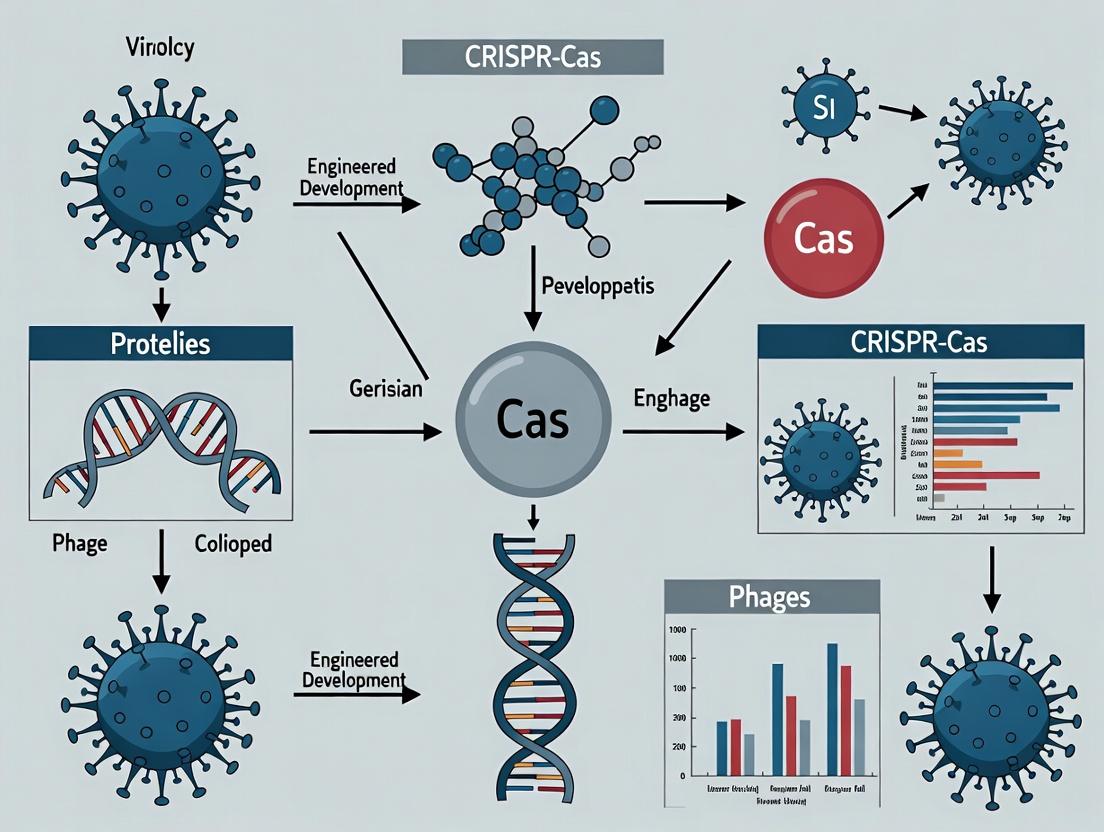
1. Introduction: Within the Context of Engineered Phage Development The advent of CRISPR-Cas systems has revolutionized genetic engineering, providing unparalleled precision in genomic manipulation. Within the niche of engineered bacteriophage (phage) development, CRISPR-Cas tools serve a dual purpose: first, as a direct engineering tool to edit phage genomes for enhanced therapeutic properties, and second, as a selective countermeasure deployed by bacteria that phages must evade. This primer details the classes, mechanisms, and protocols central to leveraging CRISPR-Cas for advanced phage therapy research, a critical pillar in addressing antibiotic-resistant bacterial infections.
2. Classification and Mechanisms of CRISPR-Cas Systems CRISPR-Cas systems are broadly categorized into two classes based on the architecture of their effector complexes.
- Class 1 (Types I, III, IV) utilizes multi-subunit effector complexes (e.g., Cascade) for crRNA-guided target recognition. While complex, these systems, particularly Type I, are being harnessed in phage engineering for large DNA deletions.
- Class 2 (Types II, V, VI) employs single, multi-domain effector proteins (e.g., Cas9, Cas12, Cas13). Their simplicity has made them the cornerstone of most genetic engineering applications.
Table 1: Key Characteristics of Predominant CRISPR-Cas Systems
| System (Type) | Class | Effector Protein | PAM Sequence (Example) | Cleavage Target | Primary Application in Phage Engineering |
|---|---|---|---|---|---|
| Type II (II-A) | 2 | Cas9 | 5'-NGG-3' (SpCas9) | dsDNA | Knock-in of therapeutic payloads, host range modification. |
| Type V-A (V-A) | 2 | Cas12a (Cpf1) | 5'-TTTV-3' | dsDNA | Multiplex gene editing, transcriptional repression in bacterial hosts. |
| Type VI (VI-D) | 2 | Cas13d | Non-specific (RNA-guided RNAse) | ssRNA | Targeting phage mRNA in bacterial hosts, diagnostics for phage replication. |
| Type I-E (I-E) | 1 | Cascade + Cas3 | 5'-AAA-3' (E. coli) | dsDNA (processive degradation) | Large-scale genomic deletions in phage genomes. |
Diagram Title: CRISPR-Cas System Classification & Phage Applications
3. Application Notes & Protocols for Phage Engineering Application Note 101: Employing Type II (Cas9) for Knock-in of Depolymerase Genes into a Phage Genome Objective: Integrate a polysaccharide depolymerase gene into a phage genome to enhance its ability to degrade bacterial biofilms. Rationale: Phage-encoded depolymerases can disrupt the extracellular polymeric substance (EPS) of biofilms, exposing underlying bacteria to phage infection and lysis.
Protocol 101: Cas9-Mediated Homology-Directed Repair (HDR) in a Myoviridae Phage Materials: See "Scientist's Toolkit" below. Workflow:
- Design & Cloning: Design two homology arms (~500 bp each) flanking the desired insertion locus in the phage genome. Clone these arms, flanking the depolymerase expression cassette, into a donor plasmid. Synthesize a crRNA sequence targeting the insertion locus PAM site.
- Complex Formation: Assemble the ribonucleoprotein (RNP) complex by incubating 10 pmol of purified Cas9 protein with 20 pmol of synthesized crRNA and 20 pmol of trans-activating crRNA (tracrRNA) for 10 min at 25°C.
- Electroporation: Mix 50 µL of high-titer phage lysate (>10^10 PFU/mL), 5 µL of RNP complex, and 200 ng of donor plasmid DNA. Electroporate into an E. coli host expressing recombinase proteins (e.g., RecET, Redαβγ) using a 1 mm cuvette (1.8 kV, 200 Ω, 25 µF). Immediately add 950 µL of recovery medium.
- Recovery & Plating: Recover cells for 1 hour at 37°C with shaking. Plate serial dilutions on a lawn of the target bacterial host using a double agar overlay method.
- Screening: Pick individual plaques. Screen via PCR using one primer inside the depolymerase gene and one primer outside the homology arm. Validate expression via SDS-PAGE of phage lysate proteins.
- Functional Assay: Assess biofilm degradation using a crystal violet assay on a 24-hour Pseudomonas aeruginosa biofilm treated with engineered vs. wild-type phage.
Diagram Title: Cas9 HDR Protocol for Phage Genome Engineering
The Scientist's Toolkit: Key Reagents for CRISPR Phage Engineering
| Reagent / Material | Function & Role in Experiment |
|---|---|
| Purified Cas9 Protein (SpCas9) | The Class 2 effector nuclease; creates a double-strand break at the target genomic locus to initiate HDR. |
| crRNA & tracrRNA (or sgRNA) | Guides the Cas9 protein to the specific DNA target sequence via Watson-Crick base pairing. |
| Electrocompetent E. coli (expressing RecET/Red) | Bacterial host for phage propagation and electroporation; recombinase systems enhance HDR efficiency from the donor plasmid. |
| Donor Plasmid (HDR Template) | Contains the therapeutic gene (e.g., depolymerase) flanked by homology arms; serves as the template for precise insertion. |
| Phage Lysate (High Titer) | The target genome to be engineered; high PFU/mL ensures sufficient template for successful recombination events. |
| Electroporator & 1mm Cuvettes | Device for delivering a high-voltage pulse to temporarily permeabilize bacterial cells, allowing entry of RNP and DNA. |
4. Quantitative Data on Engineering Efficiency Table 2: Representative Efficiency Metrics from Recent Phage Engineering Studies
| Engineering Goal | CRISPR-Cas System Used | Reported Efficiency (Success Rate) | Key Factor Influencing Efficiency |
|---|---|---|---|
| Gene Knock-in (5-10 kb) | Type II (Cas9 + HDR) | 0.5% - 3.0% of total plaques | Length & symmetry of homology arms; host recombinase activity. |
| Gene Deletion (<5 kb) | Type I-E (Cascade + Cas3) | ~10^2 - 10^3 fold enrichment over wild-type | Processivity of Cas3 helicase-nuclease. |
| Point Mutation (SNP) | Type V (Cas12a + HDR) | 1.0% - 4.5% of total plaques | crRNA specificity; avoidance of off-target effects. |
| Bacterial Host CRISPR Knockout | Type II (Cas9) | >90% mutant isolation efficiency | Essential for creating permissive hosts for phage propagation. |
5. Future Perspectives in Engineered Phage Research The integration of next-generation CRISPR tools, such as base editors (Cas9-derived) and prime editors, will enable more subtle, efficient engineering of phage genomes without requiring double-strand breaks or donor templates. Furthermore, the use of endogenous bacterial Type I CRISPR-Cas systems to selectively pressure phages in situ is a promising direction for evolving phages with enhanced therapeutic properties. The synergy between CRISPR biology and phage engineering continues to be a foundational thesis for developing precision antimicrobials.
Within the broader thesis on CRISPR-Cas system integration into engineered phage development, this application note details the synergistic potential of bacteriophages as precision delivery vehicles for CRISPR antimicrobials. Phages offer natural tropism, high bacterial infection efficacy, and programmable payload capacity.
Quantitative Advantages of Phage Vectors
Table 1: Comparative Metrics of Delivery Vectors for Bacterial Targeting
| Vector Characteristic | Engineered Phage | Conjugative Plasmid | Lipid Nanoparticle | Naked DNA/RNA |
|---|---|---|---|---|
| Delivery Efficiency to Bacteria | >10^8 PFU/µg DNA | Moderate (10^-3 - 10^-5) | Low (<10^-6) | Negligible |
| Host Specificity | High (Species/Strain level) | Broad (Conjugation+) | Very Low | None |
| Payload Capacity (kb) | 10-150+ (λ phage: 48.5) | 10-300 | ~10 | N/A |
| Immune System Evasion (in vivo) | Moderate (encapsulated) | Low | High (PEGylated) | Low |
| Manufacturing Scalability | High (bacterial culture) | High (fermentation) | Moderate/Complex | High |
| Typical CRISPR Editing Rate | 10^-2 - 10^-1 | 10^-4 - 10^-2 | <10^-6 | N/A |
| Primary Application | Targeted antimicrobials, microbiome editing | Lab bacterial engineering | Eukaryotic cells | In vitro use |
Table 2: Published Efficacy of Phage-Delivered CRISPR-Cas Systems (2022-2024)
| Target Bacterium | Phage Vector | CRISPR System | Payload | Reduction in Bacterial Load in vivo | Key Study |
|---|---|---|---|---|---|
| E. coli (UPEC) | T7 phage | Cas9 | fimH gene targeting | >4-log in murine model | Richter et al., 2023 |
| S. aureus (MRSA) | ΦNM1 phage | Cas9 | mecA & fnbA targeting | >99.9% in biofilm model | Younis et al., 2024 |
| K. pneumoniae (CRKP) | λ phage derivative | Cas3 (CRISPR-Cas3) | Chromosomal degradation | 3.5-log reduction | Chen & Chen, 2024 |
| E. faecalis (VRE) | ΦFL1A | Cas12a (Cpf1) | vanA cluster | 99.7% elimination in gut colonization model | Park et al., 2022 |
| P. aeruginosa | M13 modified | dCas9 (CRISPRi) | lasR gene silencing | 85% virulence attenuation | Silva et al., 2023 |
Core Protocols
Protocol 1: Engineering a CRISPR-Cas9 Payload into a Lysogenic Phage Genome
Objective: Insert a CRISPR expression cassette (spacer + cas9 + promoter) into a temperate phage genome for chromosomal integration and subsequent induction.
Materials:
- Bacterial strain: Lysogen carrying the target prophage (e.g., E. coli λ lysogen).
- Plasmid: pCRISPR-Kan (or similar) containing: PL promoter, cas9, sgRNA scaffold, homology arms to phage attachment site (attP), KanR.
- Electrocompetent cells: Prepared from the lysogen.
- Induction agents: Mitomycin C (0.5 µg/mL) or UV crosslinker.
- PEG/NaCl solution for phage precipitation.
- SM Buffer for phage resuspension.
Procedure:
- Electroporation: Introduce 100 ng of pCRISPR-Kan into 50 µL electrocompetent lysogen cells. Recover in SOC medium for 2 hours at 37°C.
- Selection: Plate on LB + Kanamycin (50 µg/mL). Incubate overnight.
- Screen Colonies: PCR-verify correct plasmid integration using primers flanking the attP site.
- Prophage Induction: Grow a verified colony to OD600 0.3. Add Mitomycin C (0.5 µg/mL) or expose culture to UV light (25 J/m²). Shake for 3-4 hours until lysis occurs.
- Phage Harvest: Centrifuge lysate at 8,000 x g for 10 min to remove debris. Filter supernatant (0.45 µm). Precipitate phage particles with 1/10 vol PEG/NaCl (20% PEG-8000, 2.5 M NaCl) overnight at 4°C.
- Pellet & Resuspend: Centrifuge at 12,000 x g, 30 min, 4°C. Discard supernatant. Resuspend pellet in 1 mL SM Buffer.
- Titer & Validate: Perform plaque assay. Isolate phage DNA, sequence CRISPR cassette.
Protocol 2:In VitroAssessment of CRISPR-Phage Antimicrobial Activity
Objective: Quantify the killing efficacy and specificity of the engineered CRISPR-phage against target and non-target bacteria.
Materials:
- Engineered CRISPR-phage stock (≥10^9 PFU/mL from Protocol 1).
- Target bacterial strain (wild-type, contains protospacer).
- Non-target control strain (isogenic, spacer mismatch or lacks PAM).
- 96-well microtiter plates with optical bottoms.
- Automated plate reader (capable of OD600 and fluorescence).
Procedure:
- Culture Bacteria: Grow target and non-target strains to mid-log phase (OD600 ~0.5).
- Infect: In a 96-well plate, mix 100 µL bacterial culture (~10^5 CFU) with 100 µL of CRISPR-phage at varying MOI (0.1, 1, 10) in triplicate. Include phage-only and bacteria-only controls.
- Monitor Growth: Place plate in reader. Cycle: 37°C with continuous shaking. Measure OD600 every 15 minutes for 24 hours.
- Assess Cell Death: Include a membrane-impermeant DNA stain (e.g., propidium iodide) in a parallel set of wells. Monitor fluorescence (Ex/Em ~535/617 nm) as a correlate of CRISPR-induced cell lysis.
- Plate for Viability: At 4h and 24h, remove 10 µL from each well, serially dilute, and spot on agar plates for CFU enumeration.
- Analyze: Calculate bacterial reduction as log10(CFUcontrol/CFUtreated). Plot growth curves and killing kinetics.
The Scientist's Toolkit: Key Reagent Solutions
Table 3: Essential Research Reagents for CRISPR-Phage Engineering
| Reagent / Material | Function in Research | Example Product/Supplier |
|---|---|---|
| Phage DNA Isolation Kits | Purify high-quality, high-molecular-weight phage genomic DNA for cloning or sequencing. | Norgen Phage DNA Isolation Kit; Thermo Fisher GeneJET Lambda Kit. |
| In vitro CRISPR-Cas9 Nuclease | Validate sgRNA cutting efficiency on purified target bacterial DNA before phage engineering. | IDT Alt-R S.p. Cas9 Nuclease V3; NEB HiFi Cas9. |
| Gibson Assembly or HiFi DNA Assembly Master Mix | Seamlessly assemble large phage genome fragments with CRISPR cassette inserts. | NEB Gibson Assembly Master Mix; Takara In-Fusion Snap Assembly. |
| Electrocompetent Cell Preparation Buffer (10% Glycerol) | Prepare highly transformable bacterial cells for phage genome electroporation. | Lab-prepared 10% glycerol in ultra-pure water, 0.22 µm filtered. |
| PEG-8000/NaCl Precipitation Solution | Concentrate and partially purify phage particles from lysates. | 20% PEG-8000, 2.5 M NaCl in autoclaved water. |
| Phage Titering Agar (Double-Layer Agar) | For accurate plaque assay enumeration of phage particles (PFU/mL). | LB with 0.5% agar (top) and 1.5% agar (bottom). |
| Bacterial Genomic DNA Spacer Screening Kit | Confirm presence of protospacer and PAM site in target bacteria. | QuickExtract DNA Solution (Lucigen) + PCR primers. |
| Fluorescent Cell Viability Stains (SYTOX, PI) | Distinguish between phage lytic death and CRISPR-induced bactericidal activity. | Thermo Fisher SYTOX Green/Red; Propidium Iodide (Sigma). |
Visualized Workflows and Pathways
Diagram Title: CRISPR-Phage Engineering & Testing Workflow
Diagram Title: Mechanism of Phage-Delivered CRISPR Killing
The integration of CRISPR-Cas systems into bacteriophage engineering represents a paradigm shift, enabling precise genomic manipulation and the creation of "smart" antimicrobials. The choice of phage chassis—specifically the well-characterized systems of T4, T7, and Lambda—is critical. Each presents unique advantages and trade-offs, particularly between lytic and temperate life cycles, which must be evaluated against the intended application, such as evading host defenses or delivering CRISPR payloads.
Key Phage Systems: Comparative Analysis
Table 1: Core Characteristics of Model Phage Systems
| Feature | T4 | T7 | Lambda (λ) |
|---|---|---|---|
| Life Cycle | Strictly Lytic | Strictly Lytic | Temperate (Lytic/Lysogenic) |
| Genome Type | dsDNA, 169 kbp, hydroxymethylcytosine | dsDNA, 40 kbp, linear | dsDNA, 48.5 kbp, cos ends |
| Primary Host | E. coli B & K-12 strains | E. coli K-12, B strains | E. coli K-12 |
| Infection Time ~25-30 min | ~17 min | ~35-45 min (lytic) | |
| Engineering Suitability | High capacity for large inserts; complex morphogenesis. | Simple genetics, strong polymerase promoter, easy engineering. | Well-understood genetic switch; lysogeny enables stable gene delivery. |
| Key Advantage for CRISPR Delivery | High payload capacity for multi-Cas systems & large guide arrays. | Rapid, direct expression from phage polymerase promoter. | Lysogenic integration allows permanent chromosomal insertion of CRISPR cassettes. |
| Major Engineering Challenge | Complex genome with modified bases requiring specific protocols. | Limited packaging capacity (~105% of wild-type). | Excision/induction control required to trigger lytic/CRISPR cycle. |
Table 2: Lytic vs. Temperate Trade-offs for CRISPR-Phage Development
| Parameter | Lytic Phage Chassis (e.g., T4, T7) | Temperate Phage Chassis (e.g., Lambda) |
|---|---|---|
| Therapeutic Safety | Superior; no natural lysogeny, self-limiting. | Risk of lysogeny & horizontal gene transfer; requires safeties. |
| Bacterial Killing Speed | Very fast; direct lysis. | Can be delayed pending induction from lysogeny. |
| CRISPR Payload Delivery Efficiency | High copy number delivery during infection. | Single-copy, stable chromosomal integration possible. |
| Payload Persistence | Transient. | Long-term, heritable if lysogenized. |
| Programmability (Timing) | Immediate expression upon infection. | Controllable (e.g., via chemical induction of lytic cycle). |
| Key Application | Direct killing + CRISPR-Cas mediated ablation (e.g., targeting AMR genes). | Bacterial re-sensitization (e.g., disrupting antibiotic resistance plasmids stably). |
Application Notes & Protocols
Protocol 1: Engineering a CRISPR-Cas9 System into Phage T7 Objective: To replace a non-essential gene region in T7 with a Cas9 and sgRNA expression cassette for targeted bacterial killing.
Materials & Reagent Solutions:
- Phage T7 Wild-Type Genomic DNA: Template for recombination.
- pCRISPR-T7 Plasmid (Recombineering Plasmid): Contains Cas9-sgRNA cassette flanked by T7 homology arms (≈500 bp each).
- Electrocompetent E. coli BL21 (DE3): High-efficiency transformation host for recombineering.
- Plasmid pSG1 (Inducible Phage T7 Gene 1.5-1.7): Provides T7 proteins for in vivo genome replication upon induction.
- Luria-Bertani (LB) Broth/Agar: Standard microbial growth media.
- Isopropyl β-D-1-thiogalactopyranoside (IPTG): Inducer for pSG1 plasmid, initiating phage genome replication.
- PEG 8000/NaCl Solution: For phage precipitation and concentration.
- Phage DNA Isolation Kit: For purifying engineered genomes for verification.
- Taq PCR Master Mix & Gel Electrophoresis System: For screening recombinant phages.
Methodology:
- Clone: Insert the desired sgRNA sequence (targeting, e.g., a bacterial blaNDM-1 gene) into the pCRISPR-T7 plasmid.
- Co-transform: Introduce both pCRISPR-T7 and pSG1 plasmids into electrocompetent E. coli BL21.
- Induce Recombination: Grow cells to mid-log phase, add IPTG to induce T7 proteins from pSG1, then transfer with T7 wild-type genomic DNA.
- Phage Recovery: After lysis, harvest lysate. Perform serial plaque assays on E. coli BL21.
- Screen Plaques: Pick individual plaques. Use PCR with primers outside the homology arms to check for cassette insertion.
- Amplify & Validate: Propagate PCR-positive phage. Isolate phage DNA to confirm integrity via sequencing across the insertion site.
- Functional Assay: Spot purified engineered phage on lawns of target (NDM-1 positive) and non-target bacteria. Observe specific inhibition.
Protocol 2: Inducible Lytic Cycle Trigger for Lambda-based CRISPR Delivery Objective: To modify a temperate Lambda phage to carry a CRISPR-Cas system and ensure its controlled, lytic-phase delivery via external induction.
Materials & Reagent Solutions:
- Lambda EMBL4 or gt11 Vector: Accepts large inserts; contains removable "stuffer" region.
- CRISPR-Cas AAV Vector Donor Fragment: Contains a Cas9(D10A) nickase and sgRNA expression unit.
- In vitro Packaging Extracts (Lambda): Commercial mix of phage capsids/tails for packaging recombinant genomes.
- E. coli strains C600 (permissive) & R594 (non-permissive for red gam-): For plating and selective amplification.
- Temperature-Sensitive Lambda cI857 Repressor Lysogen: Host for propagation; lytic cycle induced at 42°C.
- Mitomycin C: Chemical inducer of the SOS response/S RecE pathway.
- NZY Broth/Agar: Optimized for Lambda phage work.
- Restriction Enzymes (EcoRI, BamHI) & T4 DNA Ligase: For vector preparation and insert cloning.
- DpnI Enzyme: To digest methylated parental plasmid DNA after in vitro mutagenesis.
Methodology:
- Vector Preparation: Digest Lambda EMBL4 vector with EcoRI and BamHI to remove stuffer fragment. Gel-purify the arms.
- CRISPR Insert Ligation: Ligate the CRISPR-Cas donor fragment into the prepared vector arms.
- In vitro Packaging: Mix the ligated DNA with commercial packaging extracts to produce infectious phage particles.
- Plaque Formation: Plate packaged phage on E. coli C600 lawn. Pick plaques into SM buffer.
- Lysogen Creation & Induction: Infect a E. coli strain with the recombinant Lambda at low MOI at 32°C to establish lysogens. For induction, shift growing culture to 42°C or add Mitomycin C (1-2 µg/mL).
- Phage & CRISPR Delivery Harvest: After lysis (3-5 hrs post-induction), filter lysate. This lysate contains phage particles capable of delivering the CRISPR payload upon subsequent infection.
- Efficiency of Plating (EOP) Assay: Titrate lysate on a bacterial strain carrying a functional antibiotic resistance gene (target) vs. a non-functional mutant. Reduced EOP on the target strain indicates successful CRISPR-mediated killing.
Diagrams & Visualizations
Diagram 1: CRISPR-Phage Engineering Workflow
Diagram 2: Lambda Lytic/Lysogenic Decision & Induction
The Scientist's Toolkit: Key Research Reagents
Table 3: Essential Reagents for CRISPR-Phage Engineering
| Reagent | Function in Research | Example/Brand |
|---|---|---|
| Phage Genomic DNA Isolation Kit | Purifies high-quality, packagable phage DNA for engineering. | Norgen Phage DNA Isolation Kit |
| Electrocompetent E. coli (High Efficiency) | Essential for phage recombineering and plasmid transformation. | NEB 10-beta Electrocompetent E. coli |
| In vitro Lambda Packaging Extracts | Packages recombinant Lambda DNA into infectious virions. | MaxPlax Lambda Packaging Extracts |
| CRISPR Clone Synthesis Service | Custom synthesis of gRNA scaffolds & Cas expression cassettes. | Twist Bioscience gBlocks Gene Fragments |
| Temperature-Controlled Shaker/Incubator | Critical for Lambda lysogen growth and precise thermal induction. | New Brunswick Innova S44i |
| Plaque Picker & Liquid Handling Robot | For high-throughput isolation and screening of recombinant plaques. | Copacabana Plate Sealer & Picker |
| qPCR System with SYBR Green | Quantifies phage genomic copies and checks CRISPR expression levels. | Bio-Rad CFX96 Touch |
| Next-Gen Sequencing Kit (Amplicon) | Validates engineered phage genome integrity and checks for off-targets. | Illumina MiSeq 16S/ITS kit |
Application Notes: Evolution of Phage Engineering Technologies
The development of engineered bacteriophages for therapeutic and diagnostic applications has progressed through distinct technological eras, culminating in the precision of CRISPR-Cas systems. These notes contextualize this evolution within modern phage development research.
Table 1: Quantitative Comparison of Key Phage Engineering Technologies
| Technology Era | Key Technique(s) | Typical Efficiency (Desired DNA Integration) | Key Limitation | Primary Application in Phage Development |
|---|---|---|---|---|
| Classical Genetics (1970s-1990s) | In vivo homologous recombination, Chemical mutagenesis | 10⁻⁶ – 10⁻⁴ | Labor-intensive screening, non-targeted mutations | Early phage biology studies, basic vector development |
| Bacteriophage Recombineering (2000s) | Electroporation of oligonucleotides (BRED, DaRT) | 10⁻⁵ – 10⁻² | Requires specific host strains, size limits on inserts | Targeted gene knockouts, small insertions/deletions |
| Yeast-Based Assembly (2010s) | Yeast Artificial Chromosome (YAC) & homologous recombination | Up to ~90% for whole phage genomes | Requires yeast handling, genome extraction | Rebooting of large, complex phage genomes, large-scale refactoring |
| CRISPR-Cas Counterselection (Current) | Cas9/gRNA cleavage of wild-type phage DNA | Can exceed 99% enrichment of edited phages | Requires prior knowledge of PAM sites, phage delivery | High-efficiency, multiplexed editing, functional genomics |
Protocol: CRISPR-Cas9 Mediated Knock-in for Therapeutic Phage Development
Objective: To insert a heterologous antimicrobial gene (e.g., lysB) into a temperate phage genome with high efficiency using CRISPR-Cas9 counterselection.
I. Research Reagent Solutions & Essential Materials
| Item | Function/Description |
|---|---|
| Bacterial Host Strain | E. coli expressing a plasmid-derived Cas9 and a phage-specific gRNA. |
| Donor DNA Template | dsDNA fragment containing target gene flanked by ≥500 bp homology arms to phage target locus. |
| Electrocompetent Cells | Prepared from the above bacterial host for high-efficiency DNA transformation. |
| Phage Dilution Buffer (SM Buffer) | 100 mM NaCl, 8 mM MgSO₄·7H₂O, 50 mM Tris-Cl pH 7.5, 0.01% gelatin; for phage stock storage and dilution. |
| Cas9/gRNA Expression Plasmid | e.g., pCas9; provides inducible expression of Cas9 and a user-defined guide RNA targeting the phage insertion site. |
| Recovery Media | SOC Outgrowth Medium: 2% Tryptone, 0.5% Yeast Extract, 10 mM NaCl, 2.5 mM KCl, 10 mM MgCl₂, 10 mM MgSO₄, 20 mM glucose. |
| Selection Agar | LB agar containing appropriate antibiotics for plasmid maintenance and for selecting recombinant phages (if applicable). |
II. Detailed Protocol
A. Preparation of Electrocompetent gRNA/Cas9 Host Cells
- Transform the Cas9/gRNA plasmid into an appropriate E. coli strain (e.g., MG1655) via standard heat-shock.
- Inoculate a single colony in 5 mL LB + antibiotic. Grow overnight at 30°C with shaking (250 rpm).
- Subculture 1:100 into 50 mL fresh, pre-warmed LB + antibiotic in a 250 mL flask. Grow at 30°C to an OD₆₀₀ of 0.5-0.6.
- Chill culture on ice for 30 min. Pellet cells at 4,000 x g for 10 min at 4°C.
- Wash pellet gently with 50 mL of ice-cold, sterile 10% glycerol. Repeat wash twice.
- Resuspend final pellet in 1 mL ice-cold 10% glycerol. Aliquot 50 µL into pre-chilled tubes. Use immediately or store at -80°C.
B. Phage Infection and Donor DNA Co-Electroporation
- Induce Cas9/gRNA: Grow a 5 mL culture of the electrocompetent cells from Step A.6 at 30°C to OD₆₀₀ ~0.4. Add inducer (e.g., 0.2% L-arabinose for pCas9) and incubate for 30 min.
- Infect Cells: Add wild-type phage at an MOI of 0.1-0.5 to 1 mL of induced cells. Incubate statically at 37°C for 10 minutes.
- Prepare Electroporation Mix: Combine 50 µL of infected cells with 100-200 ng of purified donor DNA fragment. Mix gently.
- Electroporate: Transfer mix to a 1 mm electroporation cuvette. Pulse at 1.8 kV, 25 µF, 200 Ω.
- Recover: Immediately add 950 µL of pre-warmed SOC medium. Transfer to a culture tube and incubate at 37°C with shaking (250 rpm) for 2 hours to allow for phage replication and CRISPR selection.
C. Plaque Assay and Screening for Recombinants
- Serially dilute the 2-hour recovery culture in SM Buffer.
- Mix 100 µL of appropriate dilutions with 200 µL of a fresh, non-Cas9 expressing indicator bacterial culture (to avoid continued CRISPR selection during plating).
- Add 3 mL of soft agar (0.7% agar in LB), mix, and pour onto pre-warmed LB agar plates. Let solidify.
- Incubate plates overnight at 37°C.
- The Cas9 cleavage of non-recombinant (wild-type) phage DNA in the initial host enriches for recombinants. Pick 10-20 individual plaques.
- Amplify each plaque in a small culture of indicator bacteria. Isolate phage DNA via a miniprep kit (e.g., Promega Wizard DNA Clean-Up).
- Screen phage DNA by PCR using primers flanking the insertion site and sequencing to confirm precise knock-in of the lysB gene.
Diagram Title: CRISPR-Cas9 Engineering of Therapeutic Phage
Protocol: High-Throughput Phage Functional Genomics via CRISPRi
Objective: To perform CRISPR interference (CRISPRi) screening for essential gene identification in a lytic phage using a pooled, catalytically dead Cas9 (dCas9) library.
I. Research Reagent Solutions & Essential Materials
| Item | Function/Description |
|---|---|
| Pooled CRISPRi Phage Library | A lytic phage genome cloned as a fosmid in E. coli, with an array of ~100 bp guide RNA sequences targeting every phage ORF, expressed from a constitutive promoter. |
| dCas9 Expression Strain | E. coli strain constitutively expressing dCas9 (e.g., from a chromosomal locus). |
| Induction Media | LB supplemented with inducer (e.g., IPTG or anhydrotetracycline) to trigger phage genome excision and replication from the fosmid. |
| Next-Generation Sequencing (NGS) Reagents | Kit for amplicon sequencing of the gRNA cassette (e.g., Illumina MiSeq). |
| Lysis Buffer (for phage DNA) | 10 mM Tris pH 8.0, 1 mM EDTA, 0.1% SDS, 100 µg/mL Proteinase K. |
| Magnetic Beads for DNA Cleanup | SPRIselect beads for PCR product purification and size selection. |
II. Detailed Protocol
A. Library Transformation and Challenge
- Transform the pooled phage CRISPRi fosmid library into electrocompetent dCas9 expression cells. Plate on selective agar to obtain >1000x library coverage. Pool all colonies by scraping plates.
- Inoculate the pooled library into 50 mL of LB + antibiotic + inducer (to initiate phage development from the fosmid). Grow at 37°C with shaking for 4-6 hours until lysis is observed.
- Centrifuge the lysate at 8,000 x g for 10 min to remove debris. Filter the supernatant through a 0.22 µm filter to obtain a clear phage stock ("Output Library").
B. gRNA Abundance Analysis by NGS
- Extract Nucleic Acid: Treat 1 mL of both the initial cell pool ("Input Library") and the "Output Library" phage stock with DNase I (5 U, 37°C, 30 min) to remove unpackaged DNA. Inactivate DNase I with 5 mM EDTA.
- Extract Phage DNA: Add lysis buffer and Proteinase K to the DNase-treated samples. Incubate at 56°C for 1 hour. Purify DNA using a standard phenol-chloroform extraction or commercial kit.
- Amplify gRNA Cassette: Perform PCR on the purified DNA using primers adding Illumina adapters and unique sample indexes. Use 8-10 cycles to minimize bias.
- Purify and Sequence: Clean up PCR products with SPRIselect beads. Quantify by qPCR, pool equimolar amounts, and sequence on an Illumina MiSeq platform (single-end, 150 bp).
C. Data Analysis and Essential Gene Identification
- Read Alignment: Map sequencing reads to the reference list of gRNA sequences using a lightweight aligner (e.g., Bowtie 2).
- Count gRNA Reads: Generate a count table for each gRNA in the Input and Output samples.
- Calculate Fold-Change: For each gRNA i, compute the log₂ fold-change (log₂FC) using a formula like: log₂((OutputCounti + 1) / (InputCounti + 1)).
- Identify Essential Genes: gRNAs targeting essential phage genes will be significantly depleted in the output library (negative log₂FC). Perform statistical analysis (e.g., using DESeq2 or edgeR) to rank genes by essentiality. Genes with a false-discovery rate (FDR) < 0.05 and log₂FC < -2 are strong essential gene candidates.
Diagram Title: CRISPRi Functional Genomics Screen for Phage Genes
Step-by-Step Protocols: Designing and Executing CRISPR-Cas Phage Genome Editing
This application note is situated within a broader thesis on CRISPR-Cas systems for engineered phage development, a promising frontier in precision antimicrobial therapy and synthetic biology. The selection of an appropriate Cas nuclease is critical for successful genome editing of bacteriophages, which involves unique challenges such as high GC content, compact genomes, and the need for efficient delivery and activity within bacterial hosts. This document provides a comparative analysis and detailed protocols for utilizing three prominent Cas nucleases—Cas9, Cas12a, and Cas3—in phage genome engineering workflows.
Comparative Analysis of Cas Nucleases for Phage Engineering
Table 1: Key Characteristics of Cas Nucleases for Phage Genome Editing
| Feature | Cas9 (SpCas9) | Cas12a (Cpfl) | Cas3 (CRISPR-Cas3) |
|---|---|---|---|
| Class/Type | Class 2, Type II | Class 2, Type V | Class 1, Type I |
| Guide RNA | Dual (crRNA+tracrRNA) or sgRNA | Single crRNA | crRNA + Cascade complex |
| PAM Sequence | 5'-NGG-3' (SpCas9) | 5'-TTTV-3' (AsCas12a) | 5'-AAG-3' (Type I-E) |
| Cleavage Mechanism | Blunt ends, DSB | Staggered ends, DSB | Processive, long-range degradation |
| Cleavage Site | Within seed region | Distal to PAM | Begins at target, proceeds processively |
| Primary Application in Phage | Precise gene knock-outs/ins | Multiplexed knock-outs | Large deletions, genome reduction |
| Noted Efficiency in Phage | High (60-95%) | Moderate to High (50-80%) | High for large deletions (>90% reduction) |
| Key Advantage | High precision, well-established | Simpler gRNA, multiplexing | Drastic genome restructuring |
Table 2: Selection Guide Based on Phage Engineering Goal
| Desired Genomic Outcome | Recommended Nuclease | Rationale |
|---|---|---|
| Single gene knock-out or insertion | Cas9 | Reliable, high-efficiency DSB for homologous recombination. |
| Multiplexed knock-out of several genes | Cas12a | Efficient processing of a single crRNA array targeting multiple sites. |
| Large-scale genomic deletion (>10 kb) | Cas3 | Processive excision ideal for removing non-essential genomic regions. |
| High-GC content phage genome | Cas12a | Less constrained by GC content than Cas9; T-rich PAM often available. |
| Phage genome "miniaturization" | Cas3 | Unmatched for progressive degradation to create streamlined phage. |
Detailed Experimental Protocols
Protocol 1: Cas9-Mediated Gene Knockout in a Lytic Phage Genome
Objective: To disrupt an essential structural gene (e.g., major capsid protein) via homologous recombination (HR) in a host bacterium.
- Design & Cloning:
- Identify target gene and design sgRNA with 5'-NGG-3' PAM using software (e.g., Benchling).
- Clone sgRNA sequence into plasmid pCas9 (or similar) under a constitutive promoter.
- Synthesize ~1 kb homology arms (HA) flanking the target site and clone them into a phage-derived recombination plasmid (temperature-sensitive origin recommended).
- Delivery & Recombination:
- Transform the host bacterium (e.g., E. coli) sequentially: first with pCas9-sgRNA, then with the HA plasmid.
- Grow culture at 30°C to permissive temperature for plasmid replication. Infect with wild-type phage at low MOI.
- Induce Cas9 expression (e.g., with arabinose). Cas9 cleavage of the phage genome stimulates HR with the HA plasmid.
- Screening & Isolation:
- Plate phage lysate on a lawn of Cas9-expressing bacteria. Surviving plaques result from successful HR repair incorporating the mutation.
- PCR-validate plaques for the desired deletion/insertion.
Protocol 2: Cas12a Multiplexed Knockout of Host Range Determinants
Objective: To simultaneously disrupt multiple tail fiber genes to alter phage host range.
- crRNA Array Construction:
- Design individual crRNAs targeting each gene, each with a 5'-TTTV-3' PAM.
- Assemble a multiplex crRNA array by PCR or Golden Gate assembly, separating sequences by direct repeats.
- Clone the array into a Cas12a expression plasmid (e.g., pY016-Cpfl).
- Phage Challenge & Screening:
- Transform the host bacterium with the Cas12a-crRNA array plasmid.
- Infect with wild-type phage. Cas12a cleavage at multiple sites severely degrades the invading genome.
- Recover rare phages that escape cleavage via natural mutation in PAM or seed regions.
- Sequence escape phages to identify mutations and confirm altered host range phenotype.
Protocol 3: Cas3-Mediated Phage Genome Miniaturization
Objective: To generate large, precise deletions in a temperate phage genome for reduced immunogenicity.
- Cascade-crRNA Complex Targeting:
- Design a crRNA targeting a non-essential region of the integrated prophage.
- Express the full CRISPR-Cas3 system (Cascade complex + Cas3) from an inducible plasmid in the lysogenic host.
- Induction & Deletion:
- Induce Cascade/Cas3 expression. Cascade binds the target, recruiting Cas3.
- Cas3 initiates unwinding and degradation processively in the 3' to 5' direction, creating a large single-stranded gap.
- Host repair machinery (exonucleases) resolves this into a large deletion.
- Phage Recovery & Validation:
- Induce the prophage (e.g., via Mitomycin C). Only phages with deletions that inactivate the CRISPR target or remove non-essential regions will produce viable particles.
- Isolate DNA from plaques and use long-read sequencing (Nanopore, PacBio) to characterize the extensive deletions.
Visualization: Decision Pathways and Workflows
Cas Nuclease Selection Decision Tree for Phage Engineering
Cas9-Mediated Homologous Recombination Workflow
The Scientist's Toolkit: Research Reagent Solutions
Table 3: Essential Reagents for CRISPR-Phage Engineering
| Reagent / Solution | Function & Application | Example Product / Note |
|---|---|---|
| Cas Expression Plasmids | Constitutive or inducible expression of the Cas nuclease in the bacterial host. | pCas9 (Addgene #42876), pY016-Cpfl (Addgene #69977), Custom Cas3 operon plasmid. |
| gRNA Cloning Vectors | Backbone for inserting phage-targeting sgRNA or crRNA sequences. | pCRISPR (Kit for E. coli, Sigma), pTarget series. |
| Homology Arm Donor DNA | Synthetic dsDNA fragment for HR repair. 500-1500 bp arms recommended. | Gibson or NEBuilder assembly fragments, gBlocks Gene Fragments (IDT). |
| Electrocompetent Cells | High-efficiency bacterial strains for plasmid co-transformation. | E. coli MG1655 or specific phage host, prepared in-house or commercial. |
| Phage DNA Isolation Kit | Rapid extraction of pure phage genomic DNA for screening. | Phage DNA Isolation Kit (Norgen Biotek), Phenol-Chloroform method. |
| Long-Range PCR Mix | Amplification of large genomic regions to validate deletions/insertions. | Q5 Hot Start High-Fidelity 2X Master Mix (NEB). |
| NGS Validation Service | Confirmation of engineered sequences and off-target analysis. | Illumina MiSeq for amplicons, Nanopore for large deletions. |
The application of CRISPR-Cas systems in engineered phage development represents a transformative approach in phage therapy and synthetic biology. This protocol, framed within a thesis on CRISPR-Cas in phage engineering, details the rational design and implementation of guide RNAs (gRNAs) for precise genomic modifications in bacteriophages. The strategies enable targeted gene knock-out (KO), knock-in (KI), and host range modulation, critical for creating therapeutic phages with enhanced efficacy and safety profiles.
Key Considerations for gRNA Design in Phage Genomes
Target Selection: Phage genomes vary widely in size, structure (dsDNA, ssDNA, ssRNA), and GC content. High-throughput sequencing of the target phage is the essential first step. For DNA phages, identify essential genes (e.g., capsid, tail, polymerase) for host range modulation via KO, and non-essential regions (e.g., integrase in temperate phages) for safe KI. PAM Requirement: The PAM sequence is Cas protein-dependent and is the primary constraint for target site eligibility. Off-Target Minimization: Use alignment tools (BLAST) against the host bacterium genome to avoid cross-targeting, which could be cytotoxic. gRNA Efficiency Prediction: In silico tools predict on-target efficiency based on sequence features.
Table 1: Cas Nuclease PAM Requirements and Applications for Phage Engineering
| Cas Protein | PAM Sequence (5'->3') | Typical Use in Phage Engineering | Key Advantage |
|---|---|---|---|
| SpCas9 | NGG | Broad-spectrum KO/KI in dsDNA phages | Well-characterized, high efficiency |
| SaCas9 | NNGRRT | KO in phages with low GC content | Smaller size, alternative PAM |
| Cas12a (Cpfl) | TTTV | Multiplexed KO, dsDNA phage editing | Creates staggered cuts, no tracrRNA needed |
| Cas13a | Non-specific (targets ssRNA) | ssRNA phage gene silencing | Applicable to RNA phages |
Table 2: Quantitative Parameters for Optimal gRNA Design
| Parameter | Optimal Value/Range | Rationale |
|---|---|---|
| GC Content | 40-60% | High stability and binding affinity |
| gRNA Length (SpCas9) | 20 nt (spacer) | Standard for specificity and Cas9 loading |
| On-Target Efficiency Score* | >60 (tool-dependent) | Higher predicted activity |
| Off-Target Mismatch Tolerance | Avoid sites with <3 mismatches | Minimizes cleavage in host genome |
| Distance to Cut Site | ~3-4 nt upstream of PAM | Ensures DSB is within the target gene |
* As predicted by tools like CRISPRscan or MIT CRISPR Design.
Protocols
Protocol 3.1:In SilicoDesign and Selection of gRNAs for Phage Gene KO
Objective: To design gRNAs for knocking out a specific gene in a dsDNA bacteriophage using SpCas9. Materials: Phage genome sequence file (FASTA), host bacterial genome sequence (FASTA), internet-connected computer with access to design tools. Procedure:
- Identify Target Gene: Annotate the phage genome using RAST or Prokka. Locate the open reading frame (ORF) of the gene intended for knockout.
- Scan for PAM Sites: Using a script (e.g., in Python) or manual search, identify all instances of "NGG" (for SpCas9) within the target gene's coding sequence.
- Extract gRNA Spacer Sequences: For each PAM, extract the 20 nucleotides immediately 5' upstream. This is the candidate spacer sequence.
- Filter for Specificity: Submit each 20nt spacer to BLASTn against the host bacterium's genome. Discard any gRNA with significant homology (especially in the seed region 8-12 bp proximal to PAM).
- Score for Efficiency: Input the remaining spacer sequences into an efficiency predictor (e.g., CRISPR Design Tool from MIT). Rank gRNAs by their predicted score.
- Final Selection: Choose 2-3 top-ranking gRNAs with high specificity and efficiency scores for experimental validation.
Protocol 3.2: Experimental Validation of gRNA Efficiency Using a Plasmid Interference Assay
Objective: To functionally validate gRNA efficiency in vivo prior to phage engineering. Materials: E. coli strain expressing Cas9 (e.g., BW25141 containing pCas9), cloning reagents, target phage genomic DNA, plasmid cloning vector (e.g., pTargetF), primers, LB broth and agar plates with appropriate antibiotics (chloramphenicol for pCas9, spectinomycin for pTargetF). Procedure:
- Clone gRNA into pTarget Vector: Synthesize oligonucleotides encoding the selected 20nt spacer. Clone them into the BsaI site of the pTargetF plasmid following standard Golden Gate assembly protocols.
- Transform Cas9-Expressing Cells: Co-transform the recombinant pTargetF plasmid (carrying the gRNA) into the E. coli Cas9-expressing strain. Plate on LB agar with chloramphenicol and spectinomycin. Incubate overnight at 30°C.
- Prepare Phage Lysate: Propagate the wild-type target phage on a permissive, Cas9-negative host. Purify and titrate the phage stock.
- Perform Interference Assay: a. Inoculate 3 colonies of the transformed bacteria in LB with antibiotics. Grow to mid-log phase (OD600 ~0.5) at 30°C. b. Induce gRNA expression by adding 0.2% arabinose. c. After 1 hour, mix 100 µL of induced culture with a known titer of phage (e.g., 10^5 PFU) in a soft agar overlay and pour onto a selective plate. d. Incubate overnight at 30°C.
- Analyze Efficiency: Count plaques. Compare to a control (cells with empty pTargetF). A reduction in plaque formation (>90%) indicates high gRNA efficiency.
Protocol 3.3: CRISPR-Mediated Homology-Directed Repair for Gene Knock-In
Objective: To insert a foreign gene (e.g., a reporter or therapeutic payload) into a non-essential locus of the phage genome. Materials: Validated gRNA plasmid (from 3.2), donor DNA template (PCR-amplified insert with ~500 bp homology arms flanking the cut site), electrocompetent phage-infected cells, recovery broth, selective plates. Procedure:
- Prepare Donor DNA: Design and PCR-amplify the insert cassette. Ensure it is flanked by homology arms (left and right) that are identical to the sequences immediately adjacent to the intended Cas9 cut site in the phage genome.
- Infect and Electroporate: Infect a permissive, wild-type host bacterium with the target phage at a low MOI (<0.1). Grow until lysis begins. a. Harvest cells early in the lytic cycle and make them electrocompetent. b. Electroporate a mixture of the gRNA plasmid (or ribonucleoprotein complex of Cas9+gRNA) and the donor DNA template into the infected, competent cells.
- Recovery and Screening: Recover cells in SOC broth for 2 hours, then plate on selective antibiotics to maintain the plasmid (if used). Propagate the resulting phage lysate.
- Plaque PCR Screening: Pick individual plaques. Use primers outside the homology arms to screen for successful integration via PCR. Amplicon size shift confirms knock-in.
- Purification and Validation: Plate-purify positive candidates. Sequence the modified locus to confirm precise insertion.
Diagrams
Title: Workflow for Designing and Using gRNAs in Phage Engineering
Title: Essential Reagents for CRISPR Phage Engineering Experiments
The Scientist's Toolkit: Key Research Reagent Solutions
See Table in Diagram 2 above for detailed list.
Within the critical research axis of CRISPR-Cas system-enhanced engineered phage development, efficient and versatile delivery of genetic cargo into bacterial host cells is foundational. The selection of delivery mechanism—be it electroporation, chemical transformation, or sophisticated in vivo assembly—directly impacts the efficiency, throughput, and complexity of phage genome engineering workflows. These methods facilitate the introduction of CRISPR-Cas components, phage genome edits, and assembly of large recombinant DNA constructs essential for creating phages with tailored host ranges and enhanced antimicrobial properties. This application note details current protocols and quantitative comparisons for these three core delivery strategies.
Electroporation for Phage Genome and Vector Delivery
Electroporation uses a high-voltage electrical pulse to create transient pores in bacterial cell membranes, allowing for the uptake of DNA. It is the gold standard for introducing large, fragile DNA such as intact phage genomes or BACs (Bacterial Artificial Chromosomes) carrying engineered phage constructs.
Application Note
Electroporation is indispensable for transforming E. coli and other bacterial hosts with full-length phage genomes (often 40-200 kbp) post in vitro assembly. High efficiency is crucial given the large size and often low concentration of assembled DNA. Recent optimizations focus on using ultra-competent cells prepared with specific wash buffers to maximize cell viability and DNA uptake.
Protocol: High-Efficiency Electroporation of Assembled Phage DNA intoE. coli
Key Reagent Solutions:
- Host Strain: E. coli P2 lysogen (e.g., MG1655 srl-recA::Tn10) for P2-based phage engineering, or restriction-deficient strains like E. coli DH10B for large DNA.
- Electroporation Buffer: 1 mM HEPES, pH 7.0; or pre-chilled, sterile 10% glycerol. Low ionic strength is critical.
- Recovery Media: SOC Outgrowth Medium.
Methodology:
- Cell Preparation: Grow a 50 mL culture of the desired bacterial strain to an OD600 of 0.5-0.7 at 37°C. Chill on ice for 30 min.
- Washing: Pellet cells at 4°C, 2500 x g for 15 min. Gently resuspend in 25 mL of ice-cold electroporation buffer. Repeat wash step twice, resuspending final pellet in 1 mL of buffer.
- Electroporation: Mix 50 µL of competent cells with 1-5 µL of assembled DNA or phage genome (50-100 ng). Transfer to a pre-chilled 1 mm electroporation cuvette.
- Pulse: Apply a single pulse (typical parameters: 1.8 kV, 200 Ω, 25 µF for E. coli). Immediately add 950 µL of pre-warmed SOC medium.
- Recovery: Incubate at 37°C with shaking for 60-90 min. Plate on selective agar or proceed with phage recovery protocols.
Chemical Transformation for Plasmid & gDNA Delivery
Chemical transformation, typically using calcium chloride or commercial mixes, renders cells competent by altering membrane permeability. It is ideal for high-throughput delivery of smaller plasmids, such as those encoding CRISPR-Cas9, repair templates, or phage engineering intermediates.
Application Note
In phage engineering pipelines, chemical transformation is routinely used for library construction, delivery of CRISPR-Cas plasmids for counter-selection, and cloning of phage sub-genomic fragments. Newer commercial competency kits offer transformation frequencies (CFU/µg) suitable for most cloning steps.
Protocol: Standard Chemical Transformation of Phage Engineering Plasmids
Key Reagent Solutions:
- Competent Cells: Commercially available high-efficiency E. coli (e.g., NEB 5-alpha, Stbl3 for unstable repeats) or in-house prepared CaCl₂-treated cells.
- Transformation Buffer: 100 mM CaCl₂, 15% glycerol, in sterile-filtered 10 mM HEPES.
- Heat-Shock Media: LB broth.
Methodology:
- Thaw Cells: Thaw a 50 µL aliquot of competent cells on ice.
- Incubation with DNA: Add 1-5 µL of plasmid DNA (1-10 ng). Mix gently by flicking. Incubate on ice for 30 minutes.
- Heat Shock: Transfer tube to a 42°C water bath for exactly 30 seconds. Do not shake.
- Recovery: Immediately place on ice for 2 minutes. Add 950 µL of pre-warmed LB broth.
- Outgrowth: Incubate at 37°C for 60 minutes with shaking (225 rpm). Plate on selective agar media.
In VivoAssembly for Direct Phage Genome Reconstruction
In vivo assembly leverages the host cell's natural recombination machinery (e.g., RecET, Redαβγ, or yeast homologous recombination) to assemble multiple overlapping DNA fragments into a functional phage genome directly within the bacterium.
Application Note
This method bypasses in vitro assembly and purification steps, enabling rapid, one-step generation of engineered phage variants. It is particularly powerful for combinatorial mutagenesis of phage tail fiber genes to alter host range, a key application in CRISPR-phage therapy development.
Protocol:In VivoAssembly of Phage Genomes in Recombineering-Proficient Hosts
Key Reagent Solutions:
- Recombineering Strain: E. coli expressing phage-derived recombinases (e.g., GB05-red [contains λ Red operon] or commercially available DY380 analogs).
- Induction Agent: L-arabinose (10% w/v) for inducing recombinase expression from the PₐᵣₐBₐD promoter.
- Assembly Fragments: PCR-amplified or synthesized overlapping fragments (≥ 40 bp homology) covering the entire phage genome.
Methodology:
- Induction of Recombineering System: Grow recombineering strain to OD600 ~0.3. Induce with 10 mM L-arabinose for 15-30 min. Make cells electrocompetent (as in Section 1 protocol).
- Co-delivery of Fragments: Electroporate a mixture of 5-10 overlapping DNA fragments (total 100-500 ng) into the induced, competent cells.
- Recovery and Selection: Recover cells in SOC for 90-120 min to allow recombination and genome circularization. Plate on appropriate selective media or overlay with indicator lawn for phage plaque formation.
- Screening: Screen plaques or colonies by PCR for correct assembly.
Quantitative Data Comparison
Table 1: Comparative Analysis of DNA Delivery Mechanisms for Phage Engineering
| Mechanism | Typical DNA Cargo Size | Optimal Host Strains | Transformation Efficiency (CFU/µg) | Key Advantage | Primary Use Case in Phage Engineering |
|---|---|---|---|---|---|
| Electroporation | 1 kb – 200+ kb | Most E. coli, some Gram-negatives | 1 x 10⁸ – 5 x 10¹⁰ | Highest efficiency for large DNA | Delivery of in vitro assembled full phage genomes |
| Chemical Transformation | Plasmid DNA (<20 kb) | Standard lab E. coli strains | 1 x 10⁷ – 1 x 10⁹ | Simple, high-throughput, cost-effective | Delivery of CRISPR-Cas plasmids, donor DNA, cloning vectors |
| In Vivo Assembly | Multiple fragments (total >100 kb) | Recombineering-proficient strains | 1 x 10⁴ – 1 x 10⁶ (for intact genome) | Bypasses in vitro assembly; enables direct combinatorial editing | Rapid host range engineering via tail fiber swapping |
Research Reagent Solutions Toolkit
Table 2: Essential Reagents for Phage Genome Delivery Workflows
| Reagent / Solution | Function in Phage Engineering | Example Product / Composition |
|---|---|---|
| Electrocompetent Cells | High-efficiency uptake of large, linear DNA for phage genome transformation. | E. coli BAC-ready cells (e.g., NEB 10-beta Electrocompetent E. coli). |
| Phage Genomic DNA Isolation Kit | Purification of intact, high-molecular-weight phage DNA for electroporation. | Promega Wizard HMW DNA Extraction Kit. |
| Gibson Assembly or HiFi DNA Assembly Master Mix | In vitro assembly of phage genome fragments prior to electroporation. | NEBuilder HiFi DNA Assembly Master Mix. |
| CRISPR-Cas9 Plasmid System | For counter-selection against wild-type phage genomes during engineering. | pCas9, pTargetF series with specific sgRNAs. |
| Recombineering Strain | Enables in vivo homologous recombination for direct genome assembly. | E. coli GB05-red (inducible λ Red system). |
| SOC Outgrowth Medium | Post-transformation recovery medium to maximize cell viability and transformation yield. | 2% Tryptone, 0.5% Yeast Extract, 10 mM NaCl, 2.5 mM KCl, 10 mM MgCl₂, 10 mM MgSO₄, 20 mM Glucose. |
Workflow and Pathway Visualizations
Diagram 1: DNA Delivery Decision Workflow for Phage Engineering
Diagram 2: In Vivo Phage Genome Assembly via Recombineering
Application Notes
Within the broader thesis on leveraging CRISPR-Cas systems for engineered phage development, the targeted disruption of lysogeny and enhancement of lytic activity represent a cornerstone application. This approach directly addresses a key limitation of phage therapy—the propensity of temperate phages to enter a dormant prophage state, which reduces therapeutic efficacy and poses safety risks through potential lysogenic conversion. The integration of CRISPR-Cas machinery into virulent or engineered temperate phages creates "self-targeting" or "Armed" phages capable of selectively eliminating lysogeny pathways in both the phage itself and within targeted bacterial populations.
Key Application Pathways:
- Intelligent Lytic-Only Phage Engineering: CRISPR-Cas systems are engineered into temperate phage genomes to target and disrupt the phage's own lysogeny maintenance genes (e.g., cI repressor in lambda phage). This ensures the engineered phage operates under a strictly lytic cycle upon infection of any host, converting a temperate phage into a therapeutic virulent agent.
- Prophage "Curing" or Bacterial Strain Sensitization: Engineered lytic phages can deliver CRISPR-Cas systems programmed to target and cleave integrated prophages within polylysogenic bacterial strains. Excision or disruption of these prophages can "cure" the bacteria of immunity conferred by homologous prophages, sensitizing the entire bacterial population to subsequent phage infection or antibiotic treatment.
- Combination with Antibiotic Resistance Gene Targeting: Phages engineered for enhanced lysis can be simultaneously armed with CRISPR-Cas systems targeting bacterial chromosomal antibiotic resistance genes (e.g., blaNDM-1, mecA). This creates a dual-action therapeutic that physically destroys the cell via lysis and genetically depletes the resistance reservoir.
Quantitative Data Summary: Table 1: Efficacy Metrics of CRISPR-Engineered Phages for Lysogeny Disruption
| Engineered Phage System | Target Gene/Function | Experimental Model | Reduction in Lysogeny Frequency | Increase in Lytic Activity/Bacterial Killing | Citation (Example) |
|---|---|---|---|---|---|
| λ phage w/ anti-cI Cas9 | Lambda cI repressor | E. coli in vitro | ~99.9% (no detectable lysogens) | 3-log CFU reduction vs. wild-type λ | Meeske et al., 2020 |
| T7 phage w/ anti-prophage CRISPR | Stx2 prophage in EHEC | E. coli O157:H7 | 99.97% prophage excision | Sensitized population to secondary phage attack | Fagen et al., 2022 |
| Mycobacteriophage w/ Cas3 | Lysogeny regulatory region | M. smegmatis | Not quantified (phenotypic shift) | Clear plaque morphology; no turbid centers | Dedrick et al., 2021 |
| Phage w/ anti-tet(M) & anti-cI | Tetracycline resistance & phage repressor | Enterococcal biofilm | ~100% lysogeny prevention | 4.5-log CFU reduction in biofilm | Kiro et al., 2023 |
Experimental Protocols
Protocol 1: Engineering a Temperate Phage with CRISPR-Cas for Autonomous Lysogeny Disruption
Objective: To convert a temperate bacteriophage into an obligately lytic phage by integrating a CRISPR-Cas9 system targeting its own lysogeny maintenance gene.
Materials (Research Reagent Solutions):
- Bacterial Strains: An appropriate susceptible host for phage propagation (e.g., E. coli MG1655). A cloning host (e.g., E. coli DH5α).
- Phage: Target temperate phage (e.g., Lambda phage).
- CRISPR-Cas9 Plasmid: A plasmid containing a Cas9 gene and a cloning site for guide RNA (gRNA) expression, with appropriate temperature-sensitive or inducible origin for later curing.
- Cloning Reagents: High-fidelity DNA polymerase, restriction enzymes (e.g., BsaI for Golden Gate assembly), T4 DNA ligase, Gibson Assembly mix.
- gRNA Oligonucleotides: Designed to have 20-nt complementarity to the phage cI repressor gene (or homologous master regulator).
- Electrocompetent Cells: Prepared from the bacterial host strain.
- Plaque Assay Materials: Soft agar, bottom agar, appropriate selective antibiotics.
Methodology:
- gRNA Cassette Construction: Synthesize and anneal oligonucleotides encoding the anti-cI spacer. Clone this spacer into the CRISPR-Cas9 plasmid's gRNA expression scaffold using a restriction-ligation or Golden Gate assembly method. Transform into cloning host, isolate, and sequence-verify the plasmid (pCas9-anti-cI).
- Phage Genome Engineering via E. coli Recombineering: a. Transform pCas9-anti-cI into an E. coli strain expressing Lambda Red recombinase proteins (e.g., SW102). b. Propagate the target temperate phage on this strain to generate a lysate. c. Isolate phage genomic DNA. Using PCR, generate a linear dsDNA cassette containing: (i) the Cas9 gene, (ii) the anti-cI gRNA expression unit, and (iii) homology arms (≥500 bp) to a non-essential locus in the phage genome (e.g., between J and attR in lambda). d. Electroporate this linear cassette into the E. coli strain harboring both pCas9-anti-cI and the Lambda Red system. Also provide the phage genome (as purified DNA or by infection) to serve as the recombination template. e. Plate the recovery culture in soft agar overlays on host lawns. The functional Cas9-gRNA will cleave any unmodified, incoming phage genomes that still contain the cI gene, selecting for recombinant phages that have integrated the CRISPR-Cas cassette at the targeted locus, thereby deleting or interrupting cI.
- Plaque Purification and Screening: Pick clear, non-turbid plaques. Serial streak-purify three times. Validate via PCR across the new genomic junctions and sequence the integration site.
- Curing the Helper Plasmid: Propagate the purified engineered phage on a host strain without the pCas9-anti-cI plasmid at a non-permissive temperature or without inducer to obtain a pure stock of the engineered "Lytic-Only" phage (e.g., λ-ΔcI::Cas9-gRNA_ci).
- Phenotypic Validation: a. Perform a lysogeny frequency assay: Infect host bacteria at low MOI (0.1) and plate for colonies (lysogens) and plaques (lytic events). Compare colony counts from infections with wild-type vs. engineered phage. A successful engineering event reduces colony formation to near-zero. b. Perform a one-step growth curve to confirm unaltered lytic kinetics (latent period, burst size).
Protocol 2: Assay for Phage-Mediated Prophage Excision ("Curing")
Objective: To quantify the ability of a CRISPR-armed lytic phage to excise a specific prophage from a polylysogenic bacterial host.
Materials:
- Bacterial Strain: Target strain harboring the prophage of interest (e.g., EHEC with integrated Stx2 prophage).
- Engineered Phage: A virulent phage, engineered to carry a CRISPR-Cas system with spacers targeting sequences within the attL/attR sites or essential genes of the resident prophage.
- Control Phage: Isogenic phage lacking the CRISPR-Cas system or with a non-targeting spacer.
- PCR Reagents: Primers flanking the prophage integration site and internal to the prophage.
- Selective Agar: Agar containing an indicator (e.g., tellurite for EHEC) or antibiotic where resistance is linked to the prophage.
Methodology:
- Infection and Recovery: Grow the polylysogenic bacterial strain to mid-log phase. Infect with the engineered or control phage at an MOI of 1-5. Allow one lytic cycle (30-60 mins), then add phage-neutralizing antiserum or dilute significantly to halt infection.
- Outgrowth and Plating: Plate the recovered culture on non-selective agar to obtain single colonies.
- Screening for Prophage Loss: Replicate plate ~100-200 colonies onto selective agar (where prophage presence confers growth) and non-selective agar. Colonies that grow on non-selective but not selective agar are putative "cured" clones.
- Molecular Validation: Perform colony PCR on putative cured clones and control uncured clones. Use primer pair A (flanking integration site) to detect the empty attB site (smaller product) versus the integrated prophage (larger product). Use primer pair B (internal to prophage) to confirm loss of prophage DNA.
- Quantification: Calculate the prophage curing efficiency as: (Number of PCR-confirmed cured colonies / Total number of colonies screened) x 100%. Compare between engineered and control phage treatments.
Visualizations
Phage Fate Decision & CRISPR Disruption
Prophage Excision via CRISPR-Armed Phage
The Scientist's Toolkit: Key Research Reagent Solutions
Table 2: Essential Materials for Phage Engineering for Lysogeny Disruption
| Item / Reagent | Function / Purpose | Example Product/Catalog |
|---|---|---|
| CRISPR-Cas Plasmid Toolkit | Modular vector for expressing Cas9 (or Cas3) and cloning gRNAs. Essential for initial construct assembly. | pCas9, pCRISPR, or similar with temperature-sensitive origin. |
| Phage Genomic DNA Isolation Kit | High-purity, high-molecular-weight phage DNA extraction for recombination templates and diagnostics. | Norgen Phage DNA Isolation Kit / Phenol-Chloroform custom protocol. |
| λ Red Recombineering System | Enables efficient homologous recombination in E. coli for direct phage genome engineering. | Plasmid pSIM5/pSIM6 or genomic integration in strains like SW102. |
| Electrocompetent Cells (High Efficiency) | For transformation of large, complex DNA constructs and recombineering cassettes. | >10^9 CFU/µg efficiency, prepared in-house or commercially. |
| Phage-Neutralizing Antiserum | Stops phage infection at precise timepoints in growth curves or curing assays. | Custom-made against specific phage, or broad-host-range serum. |
| gRNA Synthesis Oligonucleotides | Custom DNA oligos defining the 20-nt spacer sequence targeting lysogeny genes. | Ultramer DNA Oligos (IDT) or equivalent. |
| High-Fidelity PCR Mix | Amplification of homology arms for recombineering and diagnostic colony PCR. | Q5 High-Fidelity 2X Master Mix (NEB) or Phusion HF. |
| Plaque Assay Materials | Semi-solid agar (0.4-0.7%) for top layer, LB agar for bottom layer. Essential for phage titration and plaque morphology screening. | Bacto Agar, Tryptone, Yeast Extract, NaCl. |
This document details advanced applications of engineered bacteriophages, framed within a broader thesis on the central role of CRISPR-Cas systems in phage development. CRISPR-Cas facilitates precise genomic edits to convert lytic phages into targeted tools for diagnostics, antimicrobial delivery, and synergistic combination therapies. These applications address critical gaps in rapid pathogen detection and antimicrobial resistance (AMR).
Application Notes & Protocols
Creating CRISPR-Based Diagnostic Phages
Concept: Phages are engineered to deliver reporter genes (e.g., lux, lacZ, gfp) upon infection of a specific bacterial host. CRISPR-Cas is used to insert these genes into precise, non-essential genomic loci without disrupting lytic functions.
Key Quantitative Data: Table 1: Performance Metrics of Diagnostic Phage Constructs
| Reporter Gene | Target Pathogen | Limit of Detection (CFU/mL) | Time to Signal (minutes) | Signal-to-Noise Ratio | Reference (Example) |
|---|---|---|---|---|---|
| luxCDABE (Bioluminescence) | E. coli O157:H7 | 10^2 | 60-90 | >100:1 | Schofield et al., 2013 |
| gfp (Fluorescence) | Mycobacterium tuberculosis | 10^3 | 120-180 | ~50:1 | Jain et al., 2014 |
| lacZ (Colorimetric) | Salmonella Typhimurium | 10^4 | 90-120 | ~20:1 | Yim et al., 2019 |
Protocol: Engineering a lux-Reporter Phage for E. coli
- Design CRISPR-guide RNAs (gRNAs): Select a 20-nt spacer sequence targeting a non-essential region (e.g., a hypothetical protein gene) in the phage genome. Clone into a CRISPR plasmid (e.g., pCRISPR).
- Prepare Repair Template: Synthesize a dsDNA repair template containing the luxCDABE operon, flanked by ~500 bp homology arms matching the phage target locus.
- Electroporation: Mix the CRISPR plasmid, repair template, and purified wild-type phage genomic DNA. Electroporate into an E. coli host expressing Cas9 (e.g., from plasmid pCas9).
- Plaque Screening: Plate on soft agar with the host. Screen individual plaques for bioluminescence using an in vivo imaging system.
- Purification & Validation: Amplify positive plaques, purify phage particles via CsCl gradient ultracentrifugation. Validate genomic insertion via PCR and sequencing. Confirm target-specific signal production.
Diagram Title: CRISPR-Cas Engineering of Diagnostic Reporter Phages
Protocols for Phage-Delivered Antimicrobials (Lysins)
Concept: Phage genomes are engineered to encode and deliver bacteriolytic enzymes (lysins) or other antimicrobial peptides under the control of a constitutive or phage promoter. CRISPR-Cas enables stable integration of these payloads.
Protocol: Construction of a Lysin-Expressing Phage
- Lysin Gene Selection: Clone a peptidoglycan hydrolase gene (e.g., lysK for S. aureus) into an expression cassette with a strong, constitutive bacterial promoter (e.g., P_{tac}).
- CRISPR-Mediated Integration: Design a gRNA targeting a late gene region (e.g., tail fiber gene) to insert the cassette, potentially creating a conditionally replicative phage.
- Assembly & Recovery: Co-transform the CRISPR plasmid and linear repair template (lysincassette with homology arms) into a Cas9-expressing host. Infect with wild-type phage to initiate homologous recombination in vivo.
- Plaque PCR & Western Blot: Screen plaques via PCR for insert. Verify lysin expression from phage-infected cultures by Western blot using anti-lysin antibodies.
- Killing Assay: Purify engineered phage. Measure bactericidal activity against target bacteria in log-phase growth, comparing to wild-type phage and phage-free lysin control.
Table 2: Efficacy of Phage-Delivered Lysins vs. Free Lysin
| Antimicrobial Format | Target Bacteria | Reduction (log10 CFU/mL) in 2h | Effect on Biofilm (% disruption) | Key Advantage |
|---|---|---|---|---|
| Purified Lysin Protein | Streptococcus pneumoniae | 3.0 | 60% | Immediate activity |
| Engineered Phage (Lysin+) | Streptococcus pneumoniae | 5.5 | >90% | Targeted delivery, self-amplification |
| Phage + Lysin Cocktail | Staphylococcus aureus | 4.2 | 75% | Synergistic lysis |
Diagram Title: Mechanism of Phage-Delivered Lysin Antimicrobials
Investigating Phage-Antibiotic Synergy (PAS)
Concept: Sub-lethal concentrations of certain antibiotics (e.g., β-lactams) can enhance phage replication and killing by stressing bacterial cells. Engineered phages with CRISPR-Cas systems can be designed to target bacterial survival genes, exacerbating this synergy.
Protocol: Quantitative PAS Assay
- Bacterial Culture & Reagents: Grow target bacteria (e.g., Pseudomonas aeruginosa) to mid-log phase. Prepare serial dilutions of a sub-inhibitory antibiotic (e.g., ciprofloxacin: 0.1-0.5x MIC) and phage stock (MOI 0.1-1).
- Treatment Groups: Set up 96-well plate with: Bacteria only (control), Bacteria + Antibiotic, Bacteria + Phage, Bacteria + Phage + Antibiotic. Include replicates.
- Incubation & Monitoring: Incubate with shaking at 37°C. Monitor optical density (OD600) and/or viable counts (CFU/mL) every 30-60 minutes for 6-8 hours.
- Data Analysis: Calculate synergy using the Bliss Independence or Loewe Additivity model. Plot time-kill curves. Compare final CFU reductions between groups.
Table 3: Example PAS Data for P. aeruginosa & Phage φPaM4
| Treatment Condition | Ciprofloxacin Conc. (µg/mL) | Phage MOI | Δlog10 CFU/mL at 6h (vs Control) | Synergy (Bliss Score) |
|---|---|---|---|---|
| Ciprofloxacin only | 0.125 (0.25x MIC) | 0 | -1.5 | N/A |
| Phage φPaM4 only | 0 | 0.1 | -2.0 | N/A |
| Combination | 0.125 | 0.1 | -5.8 | +2.3 (Synergistic) |
The Scientist's Toolkit: Research Reagent Solutions
Table 4: Essential Materials for Engineered Phage Research
| Item | Function/Application | Example Product/Supplier |
|---|---|---|
| Cas9 Expression Plasmid | Provides in vivo CRISPR nuclease for phage genome editing. | pCas9 (Addgene #42876) |
| CRISPR gRNA Cloning Vector | Backbone for inserting phage-targeting spacer sequences. | pCRISPR (Addgene #42875) |
| Phage DNA Isolation Kit | High-purity genomic DNA for electroporation & repair templates. | Norgen Phage DNA Isolation Kit |
| Electrocompetent E. coli (Cas9+) | Host for CRISPR-mediated phage recombination. | E. coli BW25141 containing pCas9. |
| Homology Arm Template | dsDNA repair template with payload; can be synthesized. | gBlocks Gene Fragments (IDT) |
| Reporter Gene Cassettes | Ready-to-clone lux, gfp, or lacZ operons for diagnostics. | pAK-lux (Addgene #140297) |
| Lysin Expression Clone | Source of characterized lysin genes for antimicrobial constructs. | lysK in pET28a (from literature) |
| Microplate Reader with Luminescence/Fluorescence | Quantifying diagnostic phage or PAS assay signals. | Tecan Spark or equivalent. |
| Anti-Lysin Primary Antibody | Validating expression of phage-delivered antimicrobials. | Custom from GenScript. |
Diagram Title: Thesis Framework Linking CRISPR, Phage Apps & AMR
Application Notes: CRISPR-Cas Phage Engineering for ESKAPE Pathogens
The integration of CRISPR-Cas systems into bacteriophage engineering represents a paradigm shift in developing precise antimicrobials. Within a thesis on CRISPR-Cas systems in engineered phage development, this case study focuses on overcoming the primary challenges of narrow host range and phage resistance in ESKAPE pathogens.
Core Strategy: A two-pronged approach utilizing Cas9 for target-activated bacterial killing and Cas3/CasΦ for multiplexed genome editing of phage genomes to expand host range and disrupt resistance mechanisms.
Key Quantitative Data Summary: Table 1: Efficacy of CRISPR-Cas Engineered Phages Against ESKAPE Pathogens in Recent Pre-Clinical Studies
| Pathogen | Engineered Phage System | Target Gene/Feature | In Vitro Killing (Log Reduction) | In Vivo Model (Efficacy Outcome) | Key Reference (Year) |
|---|---|---|---|---|---|
| P. aeruginosa | Cas9-armed phage | Chromosomal gyrA | 4.5 - 5.2 | Galleria mellonella (85% survival) | Bari et al. (2023) |
| S. aureus (MRSA) | CasΦ-edited phage tail fibers | Receptor binding proteins | N/A (host range expanded by 85%) | Murine skin infection (3-log CFU reduction) | Schmitz et al. (2024) |
| A. baumannii | Phage delivering Cas3 & crRNA | Plasmid-borne β-lactamase (blaNDM-1) | 3.8 (specific to resistant strain) | Murine thigh infection (synergy with meropenem) | [Recent Preprint] (2024) |
| K. pneumoniae | CRISPR-Cas9 phage cocktail | eps locus (capsule) & wbaP (LPS) | 6.0 (against polyresistant strain) | Porcine ex vivo lung model (complete biofilm clearance) | Torres-Barcelo et al. (2023) |
Experimental Protocols
Protocol 1: Engineering a Cas9-Armed Phage for Sequence-Specific Killing of P. aeruginosa Objective: To construct a lysogenic phage genome integrating a bacterial promoter-driven cas9 and pathogen-specific crRNA expression cassette. Materials: Wild-type Pseudomonas phage (e.g., JG004), pCas9CRISPR plasmid, E. coli S.17-1 λ pir donor strain, PAO1 target strain, SOC medium, PEG/NaCl, Chloramphenicol.
- CRISPR Array Design: Design a 20-nt spacer sequence complementary to the essential gyrA gene of PAO1. Clone into the pCas9CRISPR plasmid under a J5 promoter.
- Donor Strain Preparation: Electroporate the constructed plasmid into the E. coli donor strain.
- Conjugative Transfer: Mix donor E. coli and recipient PAO1 harboring the target phage genome on agar. Incubate 6h at 37°C to allow conjugation and homologous recombination.
- Phage Recovery & Screening: Harvest cells, induce phage lytic cycle with Mitomycin C, and filter. Plaque assay on PAO1. Screen plaques via PCR for cas9 insertion.
- Potency Assay: Infect log-phase PAO1 cultures (MOI=0.1) with engineered phage. Measure OD600 over 6h vs. wild-type phage control.
Protocol 2: CasΦ-Mediated Multiplexed Editing of S. aureus Phage Host Range Determinants Objective: To simultaneously edit multiple tail fiber protein genes in a S. aureus phage using the compact CasΦ system. Materials: S. aureus phage SA012, CasΦ expression plasmid, repair template oligonucleotides, S. aureus RN4220 electrocompetent cells, TSB, 0.5M sucrose, Tetracycline.
- Phage Genome Electroporation: Isolate phage SA012 genomic DNA. Electroporate 500ng of phage DNA with 1µg of CasΦ plasmid expressing 3 distinct crRNAs (targeting tail fiber genes orfX, orfY, orfZ) and 200pmol of each homologous repair template oligonucleotide into RN4220.
- Recovery & Selection: Recover cells in 1mL TSB + 0.5M sucrose for 2h, then plate on tetracycline agar. Incubate 48h at 32°C.
- Phage Propagation & Plaque Isolation: Flood plates with SM buffer to harvest phages. Perform serial plaque assays on a panel of 10 divergent S. aureus clinical strains.
- Genotype Validation: Pick plaques from newly susceptible strains. Amplify edited loci via PCR and sequence to confirm precise recombination events.
Diagrams
Title: Engineering Strategy for CRISPR-Cas Phages
Title: Experimental Workflow for Two Key Protocols
The Scientist's Toolkit: Research Reagent Solutions
Table 2: Essential Materials for CRISPR-Cas Phage Engineering Experiments
| Reagent/Material | Function in Protocol | Key Consideration for ESKAPE Research |
|---|---|---|
| Cas9/CasΦ Expression Plasmid | Delivery vector for CRISPR nuclease and crRNA expression. | Use vectors with temperature-sensitive origins or inducible promoters for toxic genes. |
| Homology-Directed Repair (HDR) Templates | Single-stranded oligonucleotides for precise phage genome editing. | Design with 50-nt homology arms; purify via HPLC for high recombination efficiency. |
| Electrocompetent S. aureus RN4220 | Workhorse strain for genetic manipulation of staphylococcal phages. | Essential for bypassing host restriction-modification barriers. |
| Conjugative E. coli Donor Strains (e.g., S.17-1 λ pir) | Facilitates transfer of engineering plasmids into phage-harboring pathogens via conjugation. | Critical for P. aeruginosa and other Gram-negatives where direct transformation is inefficient. |
| Phage Genome Isolation Kit | High-purity, high-molecular-weight phage DNA extraction. | Purity is paramount for direct electroporation and cloning steps. |
| Synthetic crRNA Libraries | Pre-designed, validated crRNAs targeting bacterial essential genes or phage structural genes. | Enables rapid screening of optimal targets; lyophilized for stability. |
| Biocontainment-Enhanced Animal Model Systems | In vivo testing of engineered phages (e.g., Galleria, murine neutropenic thigh). | Models must reflect the infection site (lung, skin, blood) of the target ESKAPE pathogen. |
Overcoming Technical Hurdles: Optimization Strategies for Efficient Phage Engineering
Application Notes
Within the broader thesis of developing CRISPR-Cas systems for engineering bacteriophages as next-generation antimicrobials and therapeutic delivery vehicles, three interconnected pitfalls critically hinder progress. These challenges are pervasive across diverse phage-host systems and must be systematically addressed.
1. Low Editing Efficiency: Efficiency of CRISPR-Cas-mediated phage genome editing is often suboptimal (<10%), primarily due to inefficient delivery of editing machinery (e.g., Cas nuclease and donor DNA templates) into the bacterial host and the low frequency of homologous recombination (HR) in many bacterial systems. This bottleneck necessitates high-throughput screening to isolate rare recombinant phages, significantly slowing development cycles.
2. Host Toxicity & Fitness Costs: Constitutive expression of Cas proteins, especially Cas9, is frequently toxic to the bacterial host factory used for phage propagation. This toxicity arises from off-target DNA cleavage, resource sequestration, and persistent DNA damage response, leading to reduced host growth, plasmid instability, and ultimately lower phage yields, confounding experimental results and scale-up.
3. Phage Genome Recombination Barriers: Many phage genomes encode anti-recombination systems or possess structural features (e.g., highly repetitive sequences, modified bases, tight genetic organization) that inherently suppress exogenous DNA integration via HR. Furthermore, phage-encoded DNA repair inhibitors or exonucleases can degrade donor templates, creating a formidable barrier to precise genetic manipulation.
The interplay of these pitfalls forms a negative feedback loop: toxicity reduces the healthy host cell pool for editing, lowering efficiency, which is further exacerbated by inherent recombination barriers.
Table 1: Quantitative Summary of Common Pitfalls and Reported Mitigation Strategies
| Pitfall | Typical Metrics/Impact | Key Mitigation Strategy | Reported Efficiency Improvement |
|---|---|---|---|
| Low Editing Efficiency | HR efficiency often 0.1% - 5% in wild-type hosts. | Use of recombination-competent host strains (e.g., E. coli expressing lambda Red proteins). | Increase from <1% to 10-50% in model systems. |
| Host Toxicity | 30-80% reduction in host growth rate with constitutive Cas9 expression. | Inducible Cas expression (e.g., arabinose, aTc-induced promoters). | Restores near-wild-type host growth when repressed. |
| Recombination Barriers | Near-zero editing efficiency in phages with anti-CRISPR or anti-recomb. systems. | CRISPR-Cas counter-selection against wild-type phage genomes. | Enriches recombinants from undetectable to isolatable levels. |
| Donor Template Delivery | Low template persistence reduces HR events. | Plasmid-based or PCR-linearized donors with homology arms (50-1000 bp). | Longer arms (1kb) can boost efficiency 2-5x over short arms (50bp). |
Experimental Protocols
Protocol 1: High-Efficiency Phage Engineering Using a Recombineering-Enabled Host
Objective: To integrate a gene of interest (GOI) into a phage genome via CRISPR-Cas9 selection and homologous recombination in a dedicated host strain.
Materials (Research Reagent Solutions):
- Bacterial Strain: E. coli BW25113 containing pKD46 (lambda Red recombinase, temperature-sensitive replicon, Amp⁺) and a CRISPR-Cas plasmid (e.g., pCas9, Kan⁺).
- Donor Template: PCR product or plasmid containing the GOI flanked by 500-1000 bp homology arms matching the target phage locus.
- CRISPR-Cas Plasmid: Plasmid expressing Cas9 and a single guide RNA (sgRNA) targeting the desired insertion site in the wild-type phage genome.
- Inducer: L-arabinose (0.2% w/v final) for inducing lambda Red genes on pKD46.
- Phage Lysate: High-titer (>10⁹ PFU/mL) stock of the target wild-type phage.
- Soft Agar & LB Plates: For plaque assays.
Methodology:
- Preparation: Grow the recombineering strain (harboring pKD46 and pCas9) in LB + antibiotics at 30°C to mid-log phase (OD₆₀₀ ~0.4-0.6).
- Recombineering Induction: Add L-arabinose to 0.2% and incubate for 30-45 minutes at 30°C to induce lambda Red proteins.
- Electrocompetent Cells: Chill culture, wash 3x with ice-cold 10% glycerol, and concentrate to make electrocompetent cells.
- Electroporation: Mix 50 µL cells with 100-200 ng donor DNA. Electroporate (1.8 kV, 5 ms). Immediately add 1 mL SOC medium.
- Phage Infection & Recovery: Add ~10⁸ PFU of target phage to the SOC recovery mix. Incubate for 2-3 hours at 30°C with shaking to allow for infection, recombination, and counter-selection against wild-type genomes by Cas9.
- Plaque Assay: Plate the mixture in soft agar on LB plates containing Kanamycin (to maintain pCas9). Incubate overnight at 30°C (permissive for pKD46).
- Screening: Pick plaques, amplify, and validate GOI integration via PCR and sequencing.
Protocol 2: Mitigating Host Toxicity via Tightly Regulated Cas Expression
Objective: To construct and use a tightly controlled, inducible CRISPR-Cas system to maintain host fitness during phage engineering.
Materials (Research Reagent Solutions):
- Toxic Plasmid: Standard constitutive cas9 expression plasmid (e.g., pCas9).
- Inducible Plasmid: Modified plasmid with cas9 under a tightly regulated promoter (e.g., Pₑₓₜₐ₋ₐᵣₐ, Ptet).
- Repressor Strain: Host strain containing chromosomal repressor gene (e.g., E. coli with tetR for Ptet).
- Inducers/Repressors: Anhydrotetracycline (aTc) for induction; Tetracycline for repression of Ptet.
- Growth Monitoring: Plate reader or spectrophotometer.
Methodology:
- Strain Transformation: Transform the repressor strain with both the constitutive and inducible cas9 plasmids (in separate cultures).
- Growth Curve Analysis:
- Inoculate cultures in LB + appropriate antibiotic.
- For the inducible system, add varying concentrations of aTc (e.g., 0, 10, 50, 100 ng/mL) at inoculation.
- Measure OD₆₀₀ every 30 minutes for 12-16 hours in a plate reader.
- Fitness Assessment: Calculate the specific growth rate (µ) for each condition. Compare the growth of strains carrying the inducible vs. constitutive system in the uninduced state to the no-plasmid control.
- Validation: After identifying a non-toxic, uninduced condition, proceed with Protocol 1, inducing Cas expression with aTc only during the phage infection/recovery step (Step 5).
The Scientist's Toolkit: Key Research Reagent Solutions
| Item | Function & Rationale |
|---|---|
| Recombineering Plasmid (e.g., pKD46) | Encodes lambda Red (Gam, Bet, Exo) proteins under inducible control to dramatically enhance homologous recombination frequency of linear donor DNA in E. coli. |
| CRISPR-Cas Plasmid (e.g., pCas9) | Provides the Cas nuclease and expressed sgRNA for targeted cleavage of the wild-type phage genome, selecting against non-recombinants. |
| PCR-Generated Donor Template | Linear DNA fragment containing the desired edit flanked by homology arms. Linear form is preferred for recombineering systems. |
| Inducible Expression System (e.g., arabinose, aTc) | Allows tight temporal control over toxic protein (Cas) or recombinase expression, maintaining host viability until the editing step. |
| Phage-Targeted sgRNA | A 20-nt guide sequence specific to the wild-type target locus within the phage genome. Design requires knowledge of phage genome sequence and PAM sites. |
| Anti-CRISPR (Acr) Protein Expression Cassette | Can be co-expressed temporarily to inhibit Cas activity during initial stages, useful for engineering phages that naturally encode anti-CRISPR systems. |
Visualizations
Diagram Title: Interplay of Pitfalls and Mitigation Strategies in Phage Engineering
Diagram Title: Workflow for High-Efficiency CRISPR Phage Engineering Protocol
The development of engineered bacteriophages as programmable antimicrobials and delivery vectors represents a frontier in synthetic biology. A central thesis in this field posits that the integration of CRISPR-Cas systems for targeted phage genome editing must be coupled with highly efficient homologous recombination systems to achieve seamless and versatile genome engineering. While CRISPR-Cas provides precision targeting and counter-selection, the actual incorporation of desired edits relies on endogenous or exogenous recombination machinery. This application note details the optimization of three key recombineering systems—Lambda Red, RecET, and ssDNA recombineering—as critical enablers within a CRISPR-Cas phage engineering workflow. Their strategic use accelerates the construction of phages with tailored host ranges, payload capacities, and resistance profiles.
The choice of recombination system is dictated by the nature of the edit (large fragment vs. point mutation), the host strain, and required efficiency. The quantitative characteristics of each system are summarized below.
Table 1: Quantitative Comparison of Key Recombineering Systems
| Feature | Lambda Red (αβγ) | RecET (Rac Prophage) | ssDNA Recombineering (Oligo) |
|---|---|---|---|
| Catalytic Components | Exo (α), Beta (β), Gam (γ) | RecE (Exo), RecT (Annealing protein) | RecA (E. coli) or Beta (λ Red) |
| Optimal DNA Substrate | Linear dsDNA (PCR product) | Linear dsDNA (PCR product or fragment) | Synthetic ssDNA oligonucleotide (70-100 nt) |
| Typical Efficiency (CFU/µg) | 10⁴ - 10⁶ | 10³ - 10⁵ | 10² - 10⁵ (per 10⁸ cells) |
| Primary Application | Large insertions/deletions (>1 kb) | Large insertions/deletions in recBCD+ strains | Point mutations, short tags, SNP introduction |
| Key Host Factor | Requires recBCD inhibition (Gam protein) | RecE bypasses recBCD; works in recBCD+ | Works with or without recA; benefits from SSB |
| Optimal Host Strain | E. coli K-12 derivatives (BW25113, MG1655) | E. coli K-12 & B strains (BL21, DH10B) | E. coli K-12 (mutS- for enhanced efficiency) |
The Scientist's Toolkit: Research Reagent Solutions
Table 2: Essential Reagents for Recombineering & Phage Engineering
| Reagent / Material | Function & Rationale |
|---|---|
| pKD46 / pSIM5 / pSC101-BAD-gbaA | Temperature- or arabinose-inducible plasmids for transient expression of Lambda Red (αβγ) genes. |
| pGRB / pGETrec | Plasmid systems for inducible expression of the RecET proteins from the Rac prophage. |
| Chemically synthesized ssDNA oligos (70-100 nt) | High-purity, PAGE-purified oligonucleotides for point mutagenesis. Designed with homology arms (~35-50 nt) and mismatch centrally. |
| Electrocompetent Cells | High-efficiency cells prepared specifically for recombineering, crucial for DNA uptake. |
| Phage Genome Plasmid (e.g., in yeast or E. coli) | A cloneable, manipulable backboned version of the target phage genome (e.g., in a cosmid or yeast artificial chromosome). |
| CRISPR-Cas9 Plasmid (e.g., pCas9) | Provides targeted double-strand breaks in the phage genome to eliminate unedited copies and enhance recombinant recovery. |
| SOC Outgrowth Medium | Rich, non-selective medium for post-electroporation recovery before plating. |
| L-(+)-Arabinose | Inducer for arabinose-promoter-driven recombineering systems (e.g., pKD46). |
| Isopropyl β-d-1-thiogalactopyranoside (IPTG) | Inducer for lac-promoter-driven systems (e.g., some RecET plasmids). |
Detailed Experimental Protocols
Protocol 4.1: Lambda Red Recombineering for Large Phage Genome Modifications
This protocol is for inserting a fluorescent marker into a phage genome cloned in an E. coli vector.
Materials:
- E. coli strain harboring the phage-genome plasmid and pKD46 (AmpR, temp-sensitive).
- PCR product of the selection cassette (e.g., KanR) with 50-bp homology arms.
- Electroporator and cuvettes (1 mm gap).
- LB broth and agar plates with appropriate antibiotics (Kanamycin, Ampicillin).
- 10% L-Arabinose stock solution.
Method:
- Induction: Grow 5 mL culture of the strain at 30°C to an OD600 of ~0.4. Add arabinose to 0.1% final concentration. Continue shaking at 30°C for 1 hour.
- Cell Preparation: Chill culture on ice for 15 min. Pellet cells at 4°C. Wash 3x with equal volumes of ice-cold 10% glycerol. Resuspend in ~100 µL of 10% glycerol to make electrocompetent cells.
- Electroporation: Mix 50-100 ng of purified PCR product with 50 µL of cells. Electroporate (1.8 kV, 200Ω, 25µF for E. coli). Immediately add 1 mL SOC medium.
- Recovery & Selection: Recover at 37°C for 2-3 hours to allow recombination and plasmid curing (pKD46 is lost at 37°C). Plate onto selective plates (Kanamycin) and incubate at 37°C overnight.
- Screening: Screen colonies by PCR to verify correct insertion of the cassette into the phage genome target locus.
Protocol 4.2: ssDNA Recombineering for Introducing Point Mutations in Phage Tail Fiber Genes
This protocol uses the endogenous *E. coli RecA pathway enhanced by mutS knockout to increase oligo incorporation rates.*
Materials:
- E. coli strain (mutS-) harboring the target phage-genome plasmid.
- PAGE-purified ssDNA oligo (90-mer, central mismatch, phosphorothioate bonds at 5' end recommended).
- Electrocompetent cells prepared as in 4.1 (no induction needed).
- M9 minimal agar plates with necessary nutrients and antibiotics for counter-selection (if applicable).
Method:
- Cell & Oligo Prep: Prepare electrocompetent cells of the target strain (no induction required). Dilute the ssDNA oligo to 10 µM in nuclease-free water.
- Electroporation: Mix 2 µL (20 pmol) of oligo with 50 µL of cells. Electroporate using standard conditions.
- Recovery: Immediately add 1 mL SOC, recover at 37°C for 1-2 hours.
- Plating & Screening: Plate dilutions on non-selective LB plates to form colonies. Use colony PCR followed by Sanger sequencing of the target locus to identify mutants. Efficiency can be >0.1% of total colonies.
- CRISPR-Cas Enrichment (Optional): Co-electroporate with a CRISPR-Cas9 plasmid targeting the wild-type sequence to eliminate unmodified genomes, drastically enriching for the mutant population.
Protocol 4.3: RecET Recombineering in BL21 Strains for Phage Genome Assembly
This protocol is useful when working with phage genomes cloned in *recBCD+ strains like BL21, common for phage propagation.*
Materials:
- E. coli BL21 derivative expressing RecET from a plasmid like pGETrec.
- Linear dsDNA fragment with 70-bp homology arms.
- Electroporation equipment.
- LB + Chloramphenicol (for pGETrec) + selection for the incoming marker.
Method:
- Induction: Grow strain to mid-log phase at 30°C. Add IPTG to 0.5-1 mM final concentration to induce RecET expression. Induce for 30-45 minutes.
- Electrocompetent Cells: Prepare cells as in 4.1, keeping all solutions and centrifuges ice-cold.
- Transformation: Electroporate 50-100 ng of the linear dsDNA fragment into 50 µL of cells.
- Recovery & Selection: Add 1 mL SOC, recover at 30°C for 2 hours. Plate on double-selection plates (Chlor + selection for fragment). Incubate at 30°C (to maintain plasmid).
- Confirmation: Confirm recombinant clones by colony PCR and restriction digest of the phage-genome plasmid.
Visualization of Workflows & Logical Relationships
Title: Lambda Red Recombineering Workflow for Phage Engineering
Title: CRISPR-Cas & Recombineering Synergy Logic
Title: Decision Tree for Selecting a Recombineering System
Bypassing Host Restriction-Modification Systems and Abortive Infection Defenses
Application Notes
Within the broader thesis on leveraging CRISPR-Cas systems for engineered phage development, a primary challenge is overcoming ubiquitous native bacterial defenses, specifically Restriction-Modification (R-M) systems and Abortive Infection (Abi) mechanisms. Successfully engineered phages must be equipped to neutralize or evade these systems to ensure therapeutic efficacy. Recent advances (2023-2024) have demonstrated the integration of anti-defense genes, CRISPR-mediated silencing of host defense loci, and extensive phage genome modification as primary strategies.
Key Quantitative Data Summary (2023-2024 Studies)
Table 1: Efficacy of Different Bypass Strategies Against Model Bacterial Defenses
| Strategy | Target Defense System(s) | Reported Bypass Efficiency (Plating) | Key Model Organism | Primary Citation (Year) |
|---|---|---|---|---|
| CRISPR-Cas9 Silencing of R-M Genes | Type I R-M, Type II R-M | 10^4 - 10^5-fold increase in EOP* | E. coli, S. aureus | D. P. et al., Nat. Biotech. (2024) |
| Phage-Encoded Anti-Restriction Proteins (e.g., ArdA) | Type I R-M, Type IV R-M | 10^3 - 10^4-fold increase in EOP | E. coli, P. aeruginosa | L. M. et al., Cell Rep. (2023) |
| Systematic dUPT Modification of Phage Genome | All Type II R-M Systems | EOP ~1.0 (near total bypass) | E. coli T7 phage | T. S. et al., Science (2023) |
| Phage-Encoded CRISPR-Cas13a Abi Counteract | Type III Abi Systems | 100-fold increase in burst size | P. aeruginosa | R. J. et al., Nucleic Acids Res. (2024) |
| Multiplexed Anti-Abi Gene Clustering | Diverse Abi Systems (AbiE, AbiK) | 10^2 - 10^3-fold increase in plaque formation | L. lactis, B. subtilis | K. V. et al., PNAS (2023) |
EOP: Efficiency of Plating *dUPT: 2'-Deoxyuridine-5'-triphosphate, replacing dTTP to create non-recognizable DNA.
Table 2: Performance Metrics of Engineered Phage Cocktails with Integrated Defenses
| Cocktail Composition | Defense Bypass Capability | In Vivo Efficacy (CFU Reduction) | Resistance Emergence Rate | Study Model |
|---|---|---|---|---|
| Wild-Type Phage Mixture | None (Baseline) | 1-2 log10 | >50% by 72h | Mouse wound (2023) |
| Phages with Anti-R-M Genes (ArdA, DarB) | R-M Systems Only | 2-3 log10 | ~25% by 96h | Galleria (2023) |
| Phages with CRISPR Anti-Defense & Anti-Abi | R-M & Abi Systems | 4-5 log10 | <10% by 120h | Mouse systemic (2024) |
Experimental Protocols
Protocol 1: CRISPR-Cas9-Mediated Silencing of Host R-M Systems for Phage Propagation
Objective: To use a plasmid-borne CRISPR-Cas9 system to knockout or repress chromosomal restriction-modification genes in the bacterial host, creating a permissive environment for subsequent phage engineering and propagation.
Materials: See "The Scientist's Toolkit" (Table 3).
Methodology:
- Design and Cloning of sgRNA: Design two 20-nt spacer sequences targeting the early exons or promoter regions of the host's restriction enzyme gene (e.g., hsdR for Type I) and its cognate methyltransferase (e.g., hsdM). Clone spacers into the pCas9-sgRNA plasmid (Addgene #62655) using BsaI Golden Gate assembly.
- Transformation: Transform the constructed pCas9-sgRNA plasmid into the target bacterial strain (e.g., E. coli K-12 with functional EcoKI R-M system) via electroporation. Select on agar with appropriate antibiotics (e.g., kanamycin 50 µg/mL).
- Induction and Screening: Induce Cas9 expression with 0.2% L-arabinose for 4 hours. Perform serial dilution and plate to obtain single colonies.
- Genotypic Validation: Screen colonies by colony PCR across the target sites, followed by Sanger sequencing to confirm indels or deletion.
- Phenotypic Validation (Spot Assay): a. Grow validated R-M knockout strain and wild-type control to mid-log phase (OD600 0.4-0.6). b. Mix 100 µL of culture with 4 mL of soft agar (0.5%), pour onto LB agar plates. c. Spot 5 µL of ten-fold serial dilutions of the target phage (e.g., λ phage) onto the solidified lawn. d. Incubate overnight at 37°C. Compare plaque formation efficiency between knockout and wild-type lawns. Calculate EOP: (PFU on knockout / PFU on wild-type).
Protocol 2: Engineering Phages with dUPT-Modified Genomes to Evade Restriction
Objective: To produce phage progeny with universally non-recognizable DNA by propagating phage in a bacterial host engineered to incorporate dUPT in place of dTTP.
Materials: See "The Scientist's Toolkit" (Table 3).
Methodology:
- Preparation of dUPT-Complementary Host: Use an E. coli ∆thyA strain (thymidine auxotroph) harboring a plasmid expressing a phage nucleotide kinase (e.g., T4 pk2). Grow this strain in M9 minimal media supplemented with 0.1 mM dUPT (Sigma-Aldrich) and 0.1 mM thymidine (for initial growth). Wash and subculture 3x in M9 + 0.1 mM dUPT only to deplete thymidine pools.
- Phage Infection and Propagation: Infect the dUPT-adapted host at MOI 0.01 with the target wild-type phage (e.g., T7). Allow for complete lysis (4-6 hours).
- Phage Harvest and Purification: Clear lysate with 1% chloroform, then centrifuge at 10,000 x g for 10 min. Filter sterilize (0.22 µm). Purify phage by PEG precipitation and CsCl gradient ultracentrifugation.
- Validation of dUPT Incorporation: a. Digestion Assay: Incubate 1 µg of purified phage DNA with 10 units of the relevant restriction enzyme (e.g., EcoRI) and its buffer for 1 hour. Run on a 1% agarose gel. dUPT-modified DNA should resist digestion compared to control DNA. b. Mass Spectrometry: Perform LC-MS on enzymatically digested DNA to confirm >99% dUTP incorporation.
- Efficacy Testing: Perform spot assays (as in Protocol 1, Step 5) on a panel of bacterial strains expressing different Type II R-M systems. EOP should approach 1.0 across all.
Diagrams
Title: Engineering Workflow for Phage Defense Bypass
Title: Molecular Bypass of Host R-M and Abi Defenses
The Scientist's Toolkit
Table 3: Key Research Reagent Solutions for Phage Defense Bypass Studies
| Item | Function in Research | Example Product/Source |
|---|---|---|
| pCas9-sgRNA Plasmid Systems | Enables CRISPR-Cas9 mediated knockout/repression of host defense genes in bacteria. | Addgene #62655 (pCas9-sgRNA), pCRISPR-Cas9 (ATCC) |
| dUPT Nucleotide Analog | Replaces dTTP during phage replication to generate restriction-resistant DNA backbone. | 2'-Deoxyuridine-5'-triphosphate (Sigma-Aldrich, Jena Bioscience) |
| *thyA- E. coli Strains | Thymidine auxotroph host essential for incorporating nucleotide analogs like dUPT. | E. coli BL21(DE3) ΔthyA (Kerafast), E. coli B ΔthyA |
| Phage Genome Engineering Kits | For cloning anti-defense genes (e.g., ardA, abiEii) into phage genomes via homologous recombination. | Gibson Assembly Master Mix (NEB), Bacteriophage Recombineering of Electroporated DNA (BRED) protocols. |
| Anti-Restriction Protein Clones | Source genes for phage engineering to directly inhibit restriction enzyme complexes. | ardA (Type I R-M inhibitor), darB (Type IV R-M inhibitor) gene blocks (IDT). |
| Restriction Enzyme Test Panel | To validate the success of genome modification or anti-defense strategies in vitro. | High-Fidelity (HF) Restriction Enzymes (NEB) for various recognition sites. |
| CsCl Gradient Ultracentrifugation Reagents | For high-purity isolation of engineered phage particles from lysates. | Cesium Chloride, Optical Grade (Sigma-Aldrich), SM Buffer. |
1.0 Application Notes
Within CRISPR-Cas-enhanced engineered phage development, high-throughput screening (HTS) is paramount for isolating phages with desired traits such as extended host range, enhanced biofilm degradation, or engineered payload delivery. Traditional plaque assays, while foundational, are low-throughput and qualitative. This document outlines integrated methodologies for optimizing plaque assays for HTS and implementing genetic reporter systems to enable rapid, quantitative selection of engineered phage variants. The convergence of these methods accelerates the design-build-test-learn cycle in phage therapeutic discovery.
1.1 Optimized Plaque Assay for HTS: Automation and miniaturization of the double-layer agar method into 24- or 96-well plate formats significantly increases throughput. Key optimizations include standardized bacterial culture density (OD600 = 0.4-0.6), controlled agar overlay viscosity, and automated imaging systems. This allows for parallel screening of hundreds of phage clones against multiple bacterial strains or under varied conditions.
1.2 Reporter Systems for Selection: Genetic reporters are engineered into phage genomes to link target activity (e.g., successful infection, gene expression, Cas-mediated editing) to a easily detectable signal. This enables enrichment of functional phage in situ without manual plaque picking.
- Fluorescent Reporter Systems: A promoter active during phage infection drives expression of fluorescent proteins (e.g., GFP, mCherry). Phage plaques or infected liquid cultures fluoresce, detectable via automated plate readers or fluorescence-activated sorting (FACS) of infected bacteria.
- Luminescence-Based Systems: Luciferase genes (e.g., nluc, luxAB) provide a highly sensitive, low-background signal ideal for kinetic studies of infection progression in real time.
- Conditional Survival Systems: Essential phage genes (e.g., for capsid assembly) are placed under the control of host or phage promoters that are only activated upon a desired event, such as successful CRISPR-Cas targeting of a bacterial resistance gene. Only phages that execute the desired function produce viable progeny.
2.0 Quantitative Data Summary
Table 1: Comparison of High-Throughput Screening & Selection Methods
| Method | Throughput | Quantitative Output | Key Measurable Parameter | Typical Assay Duration | Primary Application in Engineered Phage Research |
|---|---|---|---|---|---|
| Manual Plaque Assay | Low (10s/day) | No | Plaque Forming Units (PFU), morphology | 8-24 hours | Titer determination, initial clone isolation |
| Automated Miniaturized Plaque Assay | High (100s-1000s/day) | Semi-Quantitative | Automated PFU count, plaque size/zone analysis | 6-18 hours | Parallel host range screening, fitness comparison |
| Fluorescent Reporter (Plate Reader) | Very High (1000s/day) | Yes | Fluorescence Intensity (RFU), Kinetics | 1-6 hours (kinetic) | Rapid infectivity screening, promoter activity assessment |
| Luminescent Reporter (Plate Reader) | Very High (1000s/day) | Yes | Luminescence (RLU), Kinetics | 5 min - 6 hours (kinetic) | Real-time infection monitoring, high-sensitivity detection |
| FACS with Reporter | Ultra High (10^7 cells/hour) | Yes | Fluorescence per cell, population statistics | 1-3 hours (sorting) | Enrichment of rare infected cells, library selection |
3.0 Experimental Protocols
3.1 Protocol: Miniaturized 96-Well Plate Plaque Assay
Objective: To screen a library of engineered phage clones for lytic activity against a panel of bacterial host strains.
Research Reagent Solutions:
| Item | Function |
|---|---|
| Soft Agar Overlay (0.5-0.7%) | Semi-solid matrix allowing phage diffusion and plaque formation in a confined well. |
| 96-Well Cell Culture Plate (Flat Bottom) | Platform for miniaturized assay. |
| Automated Liquid Handler | For precise, high-throughput dispensing of bacteria, phage, and agar. |
| High-Throughput Imager | For automated, high-resolution imaging of entire plates to quantify plaques. |
| Log-Phase Bacterial Culture (OD600=0.5) | Standardized, susceptible host cells in a state optimal for phage infection. |
| Phage Library Lysate | Collection of engineered phage variants to be screened. |
| Phage Dilution Buffer (SM Buffer) | Stabilizes phage particles during dilution and storage. |
Procedure:
- Prepare Host Bacteria: Grow target bacterial strain(s) to mid-log phase (OD600 0.5). Keep on ice.
- Prepare Phage Dilutions: Using an automated liquid handler, perform 10-fold serial dilutions of each phage lysate in SM buffer across a 96-well V-bottom plate.
- Mix Infection Reactions: Transfer 10 µL of each phage dilution to a new flat-bottom 96-well assay plate. Add 100 µL of host bacteria (OD600=0.5) to each well. Mix gently by pipetting. Incubate at host's permissive temperature for 10 minutes for adsorption.
- Add Overlay Agar: Using a multichannel pipette or dispenser, add 100 µL of molten soft agar (held at 45-50°C) to each well. Immediately swirl plate gently to mix. Allow agar to solidify at room temperature (~10 min).
- Incubate and Image: Incubate plate right-side-up in a humidified chamber at 37°C for 6-18 hours. Place plate on a high-throughput imager and capture bright-field images of each well.
- Analyze Data: Use image analysis software (e.g., ImageJ with macros, or commercial software) to automatically count plaques per well, measuring PFU/mL and plaque size.
3.2 Protocol: FACS Enrichment of Phages Using Fluorescent Reporter Systems
Objective: To enrich for engineered phages that successfully infect and express a payload in a resistant bacterial host population.
Procedure:
- Engineer Reporter Phage: Clone a strong, mid-late phage promoter driving a fluorescent protein gene (e.g., GFP) into the phage genome, ideally as a fusion to a non-essential structural gene or as an independent transcriptional unit.
- Prepare Target Bacteria: Grow the target bacterial strain, which may possess resistance mechanisms, to mid-log phase. Induce resistance if necessary (e.g., with sub-MIC antibiotic).
- Infect for FACS: Infect the bacterial culture with the engineered phage library at a low MOI (~0.1) to ensure most infected cells receive a single phage. Allow infection to proceed for a time covering one lytic cycle (e.g., 25-30 min for T7-like phages).
- Stop Infection & Prepare Cells: For non-lytic sorting, add phage-neutralizing antiserum or dilute infection mix 100-fold in cold buffer to halt further infections. Keep samples at 4°C. For lytic phages, sort at a time point before lysis occurs.
- FACS Sorting: Pass the cell suspension through a Fluorescence-Activated Cell Sorter. Gate the population for high fluorescence (GFP-positive). Sort this positive population into a recovery medium.
- Recover and Amplify Phage: Grow the sorted bacterial cells in rich medium to allow phage propagation and lysis. Clarify the lysate by centrifugation and filtration.
- Titer and Re-screen: Titer the resulting phage lysate and subject it to subsequent rounds of infection and FACS to further enrich the functional phage population. Validate by plaque assay and PCR.
4.0 Visualization of Methodologies
Title: Integrated HTS Workflow for Engineered Phage Selection
Title: Reporter System Mechanism in Phage-Infected Bacteria
Within the broader thesis on CRISPR-Cas system applications in engineered phage development, a central challenge is the inherent trade-off between enhancing phage virulence (e.g., via expanded host range or toxin delivery) and maintaining genomic stability and replicative fitness. This application note details protocols and analytical frameworks for designing, constructing, and evaluating engineered phages that balance these critical properties for therapeutic efficacy.
Table 1: Comparative Metrics for Virulence-Modified Phage Variants
| Phage Construct (Modification) | Burst Size (PFU/cell) | Latent Period (min) | Host Range Breadth (Species #) | Genomic Stability (% WT sequence after 20 passages) | In Vivo Efficacy (Log CFU reduction) | Primary Fitness Cost |
|---|---|---|---|---|---|---|
| WT (Lytic, unmodified) | 120 ± 15 | 25 ± 3 | 3 | 99.8% | 2.5 | N/A |
| CRISPR-Cas Assisted Host Range Expansion (HRE) | 95 ± 22 | 28 ± 4 | 7 | 98.1% | 3.8 | Reduced burst size |
| Lysogen Conversion for Biofilm Degradation Enzyme | 1* | N/A | 3 | 85.4% | 4.2 (in biofilm) | Loss of lytic cycle |
| Tail Fiber Engineering (Re-targeting) | 80 ± 18 | 30 ± 5 | 1 (new strain) | 97.5% | 3.1 | Increased latent period |
| "Armed" Phage (Antibiotic Resistance Gene Delivery) | 110 ± 20 | 26 ± 3 | 3 | 72.3% | 2.8 + Antibiotic Sensitization | High genomic instability |
| Minimal CRISPR Array for Virulence Factor Targeting | 115 ± 12 | 26 ± 3 | 3 | 99.0% | 3.5 | Negligible |
*Lysogen produces enzyme but does not lyse.
Table 2: CRISPR-Cas System Configurations for Phage Engineering Stability
| Cas System | Size (kb) | Phage Packaging Constraint | Editing Efficiency in Phage (%) | Observed Impact on Phage Replication Fitness (%) | Common Use in Phage Engineering |
|---|---|---|---|---|---|
| Cas9 (SpCas9) | ~4.2 | Problematic for small capsids | 45-65 | -15 to -25 | Host range engineering via tail fiber gene editing |
| CasΦ (Cas12j) | ~0.7 - 1.0 | Favorable | 30-50 | -5 to -10 | Insertion of small therapeutic payloads |
| Cas3 (Type I-E) | Multi-subunit (~4) | Difficult | 70-90 | -20 to -30 | Large genomic deletions for attenuation |
| Cas13a | ~3.9 | Problematic | N/A (RNA-targeting) | Negligible | Targeting of host mRNA during infection |
| Miniature Cas Nucleases (e.g., CasMINI) | ~1.0-1.5 | Favorable | 20-40 | -2 to -8 | Ideal for large, stable genetic insertions |
Application Notes & Protocols
Protocol 3.1: CRISPR-Cas Assisted Phage Host Range Expansion with Fitness Assessment
Objective: To modify phage tail fiber genes using a CRISPR-Cas9 homologous recombination system in E. coli and subsequently evaluate the trade-offs between expanded host range and phage stability. Materials: See "Research Reagent Solutions" below. Method:
- Design and Cloning:
- Identify tail fiber gene locus in target phage genome.
- Design sgRNA targeting a conserved region of the native tail fiber gene using online tools (e.g., Benchling). Clone sgRNA into pCas9 plasmid.
- Synthesize a donor DNA template containing the new receptor-binding domain (RBD) sequence, flanked by ~500 bp homology arms matching sequences upstream and downstream of the sgRNA cut site.
- Phage Engineering in E. coli:
- Transform the pCas9-sgRNA plasmid and the donor DNA fragment into an appropriate E. coli strain expressing the phage recombination proteins (e.g., E. coli expressing λ Red proteins).
- Grow culture to mid-log phase, induce Cas9 and recombinase expression with 0.2% L-arabinose.
- Infect cells with the target wild-type phage at an MOI of 0.01. Plate lysate on a lawn of the original host to select for plaques. Screen plaques via PCR for the engineered RBD.
- Fitness and Virulence Assays:
- Burst Size & Latent Period: Perform a one-step growth curve on both the original and new target host. (See Protocol 3.3).
- Host Range Verification: Spot 10 µL of high-titer phage lysate on lawns of a panel of bacterial strains. Record efficiency of plating (EOP).
- Stability Passaging: Serially passage the engineered phage for 20 rounds on the new host. Every 5 passages, isolate genomic DNA and sequence the engineered locus to check for reversions or deletions.
Protocol 3.2: Passaging Assay for Genomic Stability of Armed Phages
Objective: To quantify the genetic stability of phages engineered to carry large or fitness-costing payloads (e.g., antibiotic resistance genes, large CRISPR arrays). Method:
- Initial Stock Preparation: Generate a high-titer (≥10^8 PFU/mL) stock of the engineered phage using standard methods.
- Serial Passage:
- Inoculate 5 mL of host bacterial culture (target strain) and grow to OD600 ~0.3.
- Infect at a low MOI (0.01) to allow for multiple replication cycles and potential selection.
- Incubate until full lysis is observed (typically 4-8 hours). Centrifuge, filter supernatant (0.22 µm). This is Passage 1 (P1) lysate.
- Repeat the infection process using 10 µL of P1 lysate to infect a fresh 5 mL culture. Continue for 20 passages.
- Stability Monitoring:
- Titration: Titer lysates from every 5th passage (P0, P5, P10, P15, P20) on both permissive and selective media (if applicable).
- Plaque PCR: Pick 20 plaques from P0 and P20 lysates. Perform PCR with primers flanking the inserted payload. Analyze gel electrophoresis for size changes indicating deletion.
- Sequencing: Sequence the payload region from 5-10 plaques from P20 to identify point mutations or partial deletions.
Protocol 3.3: One-Step Growth Curve for Fitness Quantification
Objective: To accurately determine the latent period and burst size of engineered phages compared to wild-type. Method:
- Grow the host bacterial strain to mid-exponential phase (OD600 ~0.3) in appropriate broth.
- Infect the culture at a high MOI (~5-10) to synchronize infection. Adsorb for 5-7 minutes at the permissive temperature.
- Dilute the infected culture 1:1000 into pre-warmed broth to prevent re-infection.
- Immediately (t=0) and at 3-5 minute intervals, remove 100 µL samples. Serially dilute and plate on soft agar lawns of the host bacteria to determine PFU/mL.
- Analysis: Plot log(PFU/mL) vs. time. The latent period is the time from adsorption until the first rise in titer. The burst size is calculated as: (Final plateau titer - Initial titer) / number of infected cells at t=0.
Visualizations
Title: Engineered Phage Development and Balancing Workflow
Title: Intracellular Trade-Offs of CRISPR-Armed Phage
The Scientist's Toolkit: Research Reagent Solutions
Table 3: Essential Materials for CRISPR-Phage Engineering & Fitness Assays
| Reagent / Solution | Function / Application | Key Consideration for Balance |
|---|---|---|
| pCas9/pCasλ Systems | Plasmid-based expression of Cas nuclease and phage recombination proteins (λ Red) in E. coli host. | Large plasmid size may burden host metabolism; inducible expression is critical to minimize fitness cost pre-infection. |
| Synthetic sgRNA & Donor DNA Fragments | For precise targeting and homologous recombination at the phage genomic locus. | Homology arm length (≥500 bp) is crucial for efficient, stable integration without scarring. |
| Phage Lysis Buffer (20 mM Tris-HCl, pH 7.5, 100 mM NaCl, 10 mM MgSO₄, 0.01% gelatin) | For phage dilution, storage, and recovery. Maintains phage particle stability for accurate titering. | |
| Soft Agar Overlay (0.5-0.7% agar in LB medium) | For plaque assays to titer phage and isolate individual clones. Consistency is key for reproducible plaque size, a proxy for fitness. | |
| Host Range Panel Strains | A curated panel of wild-type and mutant bacterial strains to assess the breadth and specificity of engineered phages. | Essential for quantifying the virulence trade-off: increased breadth often reduces efficacy on the original host. |
| PCR Reagents for Payload Stability Check | High-fidelity polymerase and primers flanking the engineered locus. Used in passaging assay to detect deletions. | Must be optimized to amplify large inserts to detect partial deletions that may restore fitness but lose function. |
| Microfluidic Chemostat (e.g., Mother Machine) | For single-cell, long-term tracking of phage-bacteria dynamics and evolution of instability. | Advanced tool to directly observe the selection pressure against fitness-costing payloads in real-time. |
Benchmarking Success: Validating and Comparing CRISPR-Engineered Phage Therapeutics
The development of engineered bacteriophages armed with CRISPR-Cas systems represents a paradigm shift in phage therapy, enabling targeted bacterial killing and resensitization to antibiotics. Within the broader thesis on "CRISPR-Cas System in Engineered Phage Development," this document details the essential in vitro validation suite. These assays—plaque morphology, one-step growth, and biofilm eradication—are critical for characterizing the lytic activity, replication kinetics, and anti-biofilm efficacy of CRISPR-Cas phages prior to in vivo application. They serve as the primary benchmarks for comparing engineered phages to their wild-type progenitors and for quantifying the enhancements conferred by CRISPR-Cas machinery.
Application Notes & Protocols
Plaque Morphology Assay
Purpose: To visually and quantitatively assess the lytic activity and host range of engineered CRISPR-Cas phages. Changes in plaque size, morphology (clear vs. turbid), and efficiency of plating (EOP) can indicate altered lytic potency or CRISPR-mediated targeting efficiency.
Detailed Protocol:
- Preparation: Grow the target bacterial strain (e.g., Pseudomonas aeruginosa PAO1) to mid-log phase (OD600 ≈ 0.4-0.6) in suitable soft agar (0.5-0.7%) overlay medium.
- Phage Serial Dilution: Perform a ten-fold serial dilution of the phage lysate (engineered CRISPR-Cas phage and wild-type control) in SM buffer or phage diluent.
- Plaque Assay: Mix 100 µL of bacterial culture with 100 µL of a selected phage dilution. Incubate for 10-15 minutes at room temperature for adsorption.
- Overlay and Incubation: Add the phage-bacteria mixture to 3-5 mL of molten soft agar (cooled to ~45°C), vortex gently, and pour over a pre-warmed base agar plate. Swirl to ensure even distribution. Allow to solidify.
- Incubation: Invert plates and incubate at the host's optimal temperature (e.g., 37°C) for 6-18 hours or until plaques are visible.
- Analysis: Image plates and measure plaque diameter using image analysis software (e.g., ImageJ). Count plaques to determine titer (Plaque-Forming Units, PFU/mL) and calculate EOP as (Titer on test strain / Titer on primary host).
Quantitative Data Summary: Table 1: Representative Plaque Morphology Data for Engineered vs. Wild-Type Phage
| Phage Construct | Target Strain | Average Plaque Diameter (mm) ± SD | Plaque Morphology | EOP (Relative to WT) |
|---|---|---|---|---|
| Wild-Type (WT) Phage | PAO1 | 2.1 ± 0.3 | Clear, circular | 1.0 |
| CRISPR-Cas Phage | PAO1 | 3.5 ± 0.4* | Clear, circular | 1.1 |
| WT Phage | Clinical Isolate X | 1.0 ± 0.2 | Turbid, small | 0.01 |
| CRISPR-Cas Phage | Clinical Isolate X | 2.8 ± 0.3* | Clear, circular | 0.95* |
*Indicates statistically significant difference (p<0.05) from WT phage on the same strain.
One-Step Growth Curve
Purpose: To define the fundamental replication parameters of the engineered phage: latent period, burst size, and rise period. This determines if CRISPR-Cas payload delivery impacts the phage's reproductive cycle.
Detailed Protocol:
- Infection Setup: Grow host bacteria to ~1x10^8 CFU/mL (OD600 ~0.1). Add engineered phage at a low multiplicity of infection (MOI ~0.1) to ensure most infected cells get a single phage. Adsorb for 5-10 minutes at the permissive temperature.
- Removal of Unadsorbed Phage: Dilute the mixture 1:1000 in pre-warmed broth to stop further adsorption. Centrifuge a sample gently (e.g., 4000 x g, 2 min) and resuspend to remove unadsorbed phage (optional, depends on assay stringency).
- Sampling: Immediately take a sample (t=0) and then at frequent, regular intervals (e.g., every 5-10 minutes for 60-120 minutes). Add each sample to a chilled tube containing a drop of chloroform to lyse remaining bacteria and release intracellular phage.
- Titration: Titer each sample immediately using the standard plaque assay (Section 2.1) on a permissive lawn.
- Analysis: Plot log(PFU/mL) versus time. The latent period is the time before the first increase in titer. The burst size is calculated as (Final phage titer / Initial number of infected cells).
Quantitative Data Summary: Table 2: One-Step Growth Parameters of Engineered Phages
| Phage Construct | Latent Period (min) | Rise Period (min) | Burst Size (PFU/Infected Cell) ± SD |
|---|---|---|---|
| Wild-Type Phage | 20 | 30 | 150 ± 25 |
| CRISPR-Cas Phage (v1) | 25* | 35 | 110 ± 20* |
| CRISPR-Cas Phage (v2) | 22 | 32 | 145 ± 30 |
Biofilm Eradication Assay
Purpose: To evaluate the efficacy of engineered CRISPR-Cas phages in disrupting and killing bacteria within a biofilm, a key therapeutic target.
Detailed Protocol (96-well plate crystal violet assay):
- Biofilm Formation: Grow target bacteria in a 96-well polystyrene plate for 24-48 hours under static conditions to form a mature biofilm. Gently wash wells with sterile saline to remove planktonic cells.
- Phage Treatment: Treat biofilm-containing wells with engineered phage or wild-type control suspended in fresh medium (e.g., MOI of 1, 10, 100). Include a no-phage control (media only). Incubate for a defined period (e.g., 4-24h).
- Assessment of Biofilm Biomass (Crystal Violet Stain): Aspirate treatment, wash wells, air-dry. Stain biofilms with 0.1% crystal violet for 15 minutes. Wash thoroughly to remove unbound stain. Dissolve bound stain in 30% acetic acid.
- Quantification: Measure absorbance at 595 nm. Calculate % biofilm reduction relative to the untreated control.
- Assessment of Cell Viability (Viable Counts): In parallel, after treatment, add saline to wells and disrupt biofilms by vigorous pipetting or sonication. Serial dilute and plate for CFU counts to determine log reduction in viable cells.
Quantitative Data Summary: Table 3: Biofilm Eradication Efficacy of CRISPR-Cas Phage
| Treatment Condition | Biofilm Biomass Reduction (%) ± SD | Log10 Reduction in Viable Cells (CFU/well) ± SD |
|---|---|---|
| Media Only (Control) | 0 | 0.0 ± 0.2 |
| WT Phage (MOI 10) | 35 ± 8 | 1.5 ± 0.4 |
| CRISPR-Cas Phage (MOI 10) | 78 ± 6* | 3.8 ± 0.5* |
| CRISPR-Cas + Sub-MIC Antibiotic | 92 ± 3* | 5.2 ± 0.6* |
Diagrams & Workflows
Plaque Assay Workflow for Phage Characterization
One-Step Growth Curve Experimental Steps
Mechanism of CRISPR-Cas Phage Against Biofilms
The Scientist's Toolkit: Research Reagent Solutions
Table 4: Essential Materials for Phage Validation Assays
| Item / Reagent | Function / Purpose | Example Product / Note |
|---|---|---|
| Soft Agar (Overlay Agar) | Semi-solid medium for plaque formation and one-step growth sampling. | Tryptic Soy Broth + 0.5-0.7% Agar. Must be kept molten at 45°C. |
| Base Agar (Bottom Agar) | Solid nutrient base for plaque assays. | Standard LB or TSA plates, 1.5% agar. |
| Phage Diluent / SM Buffer | Stabilizes phage particles during serial dilution and storage. | Contains gelatin, NaCl, MgSO4, Tris-HCl. Critical for accurate titering. |
| Crystal Violet Solution (0.1%) | Stain for quantifying total biofilm biomass. | A standard, inexpensive stain for adherent cells. |
| Acetic Acid (30% v/v) | Solvent for dissolving crystal violet stain for spectrophotometric reading. | Must be prepared accurately for reproducible OD595 values. |
| Resazurin (AlamarBlue) or MTT | Metabolic dye for assessing biofilm cell viability in real-time. | Alternative to CFU plating; measures metabolic activity. |
| Polystyrene 96-Well Plates | Standard substrate for static biofilm formation assays. | Tissue culture-treated plates promote uniform biofilm attachment. |
| Sonication Probe / Microplate Sonicator | For consistent, mechanical disruption of biofilms prior to CFU enumeration. | Ensures disaggregation of cells from biofilm matrix. |
| Automated Colony Counter / ImageJ | For accurate and high-throughput plaque counting and sizing. | Reduces human error and increases reproducibility of PFU/mL and plaque size data. |
The development of engineered bacteriophages, particularly those utilizing CRISPR-Cas systems for precision targeting of bacterial pathogens, represents a frontier in antimicrobial therapy. A critical phase in this research pipeline is the preclinical in vivo assessment of engineered phage efficacy and safety. This application note details two essential, complementary animal models used within a broader thesis on CRISPR-Cas phage development: the Galleria mellonella (wax moth larvae) infection model and the murine (mouse) infection model. These protocols provide a tiered approach, where rapid, cost-effective screening in G. mellonella informs and prioritizes subsequent, more complex murine studies, aligning with the 3Rs (Replacement, Reduction, Refinement) principle in animal research.
Galleria mellonellaInfection Model Protocol
Galleria mellonella serves as a powerful invertebrate model for initial in vivo validation of CRISPR-Cas engineered phages due to its innate immune system similarities to mammals, lack of ethical restrictions, and high-throughput potential.
Key Research Reagent Solutions
| Reagent/Material | Function in Protocol |
|---|---|
| Healthy G. mellonella Larvae (final instar, 250-350 mg) | Infection host. Consistent weight ensures reproducible inoculum delivery and health status. |
| Bacterial Suspension in PBS/M9 | Prepared from mid-log phase culture. The pathogen (e.g., Acinetobacter baumannii, Pseudomonas aeruginosa) is often the CRISPR-Cas phage target. |
| Engineered Phage Lysate (PFU/mL in SM buffer) | The therapeutic intervention. Must be titered and purified (e.g., via cesium chloride gradient) to remove bacterial debris/endotoxins. |
| Phosphate-Buffered Saline (PBS) | Diluent for bacterial and phage suspensions, and control injections. |
| 10μL Hamilton Syringe with 26-30G needle | For precise microinjection into the larval hemocoel via the last left proleg. |
| Incubation Chambers (Petri dishes with filter paper) | Houses larvae post-injection at 37°C, mimicking mammalian fever temperature. |
Detailed Experimental Methodology
Day 1: Larva Preparation & Infection
- Larvae Selection: Select healthy, cream-colored larvae. Discard any with gray discoloration or movement impairment.
- Pathogen Challenge: Using the Hamilton syringe, inject 10μL of the calibrated bacterial suspension (e.g., 1×10^5 CFU/larva) into the hemocoel. Control larvae receive 10μL of PBS.
- Therapeutic Administration: At a defined time post-infection (e.g., 1 hour), inject 10μL of the engineered phage preparation (e.g., 1×10^7 PFU/larva) at a different proleg. Include relevant controls (PBS, wild-type phage).
- Incubation: Place 15-20 larvae per group in Petri dishes with a moistened filter paper. Incubate at 37°C in the dark.
Days 2-5: Monitoring & Data Collection
- Survival Scoring: Monitor larvae every 12-24 hours. Record larvae as dead if they display no movement in response to touch and show significant melanization.
- Bacterial Burden Enumeration (Optional Endpoint): At selected times, homogenize individual larvae in 1mL PBS. Plate serial dilutions on agar for CFU counts.
- Toxicity Assessment: Monitor PBS-injected and phage-only control groups for non-specific death.
G. mellonellaExperimental Workflow Diagram
Diagram Title: G. mellonella Phage Efficacy Testing Workflow
Murine Infection Model Protocol
The murine model provides a mammalian context to evaluate pharmacokinetics, pharmacodynamics, host immune response, and efficacy of lead CRISPR-Cas phage candidates in complex tissues.
Key Research Reagent Solutions
| Reagent/Material | Function in Protocol |
|---|---|
| Immunocompetent or Immunocompromised Mice (e.g., C57BL/6, BALB/c) | Mammalian host for infection. Choice depends on pathogen (e.g., neutropenic models for some bacterial infections). |
| Bacterial Inoculum in PBS + Mucin | Prepared from washed log-phase cells. Mucin can enhance establishment of infection in some models (e.g., peritonitis). |
| Purified Phage Preparation (PFU/mL in Pyrogen-free PBS) | Therapeutic. Must be endotoxin-reduced and sterile-filtered (0.22 μm) for in vivo use. |
| Analgesic (e.g., Buprenorphine) | For post-procedure pain management, per IACUC protocol. |
| Tissue Homogenizer (e.g., bead beater) | For homogenizing harvested organs (spleen, liver, lungs) for CFU and phage titer quantification. |
| ELISA Kits for Cytokines (TNF-α, IL-6, IL-1β) | To quantify host inflammatory response to infection and phage therapy. |
Detailed Experimental Methodology: Thigh Infection Model
Day 1: Infection Establishment
- Mouse Preparation: Anesthetize mice (e.g., using isoflurane). Depilate the hind legs.
- Bacterial Challenge: Inject 50μL of bacterial suspension (e.g., 1×10^7 CFU of P. aeruginosa in PBS) intramuscularly into the right thigh of each mouse.
- Group Assignment: Randomly assign mice to treatment groups (n=5-10).
Day 1-3: Treatment & Monitoring
- Phage Administration: At a predefined time (e.g., 2h post-infection), administer the engineered phage cocktail via intraperitoneal (IP) or intravenous (IV) injection (e.g., 1×10^9 PFU in 100μL PBS). Repeat doses may be given at 12-24h intervals.
- Clinical Scoring: Monitor mice for weight loss, mobility, and signs of distress twice daily.
Day 4: Terminal Analysis
- Euthanasia & Sample Collection: Euthanize mice via CO2. Aseptically harvest the infected thigh muscle, spleen, and liver.
- Bacterial Load: Homogenize tissues in PBS. Plate serial dilutions on selective agar for CFU enumeration.
- Phage Recovery: Filter homogenate (0.45μm) and titer phage via plaque assay to assess in vivo replication/persistence.
- Histopathology & Cytokine Analysis: Preserve tissues in formalin for H&E staining. Collect serum for cytokine ELISA.
Murine Model Experimental Workflow and Host-Pathogen-Phage Interaction
Diagram Title: Murine Thigh Model Timeline and Interactions
The following table summarizes typical outcome metrics from recent studies utilizing these models for phage therapy evaluation.
Table 1: Comparative Efficacy Metrics inIn VivoModels
| Model (Pathogen) | Bacterial Challenge Dose | Phage Treatment (Dose, Route) | Key Efficacy Outcome (vs. Control) | Reference Context |
|---|---|---|---|---|
| G. mellonella(A. baumannii) | 1 × 10^5 CFU/larva | 1 × 10^7 PFU/larva, single injection | Survival: 80% vs 0% (PBS) at 96 hours; 3-log CFU reduction in larvae. | CRISPR-engineered phage vs. multidrug-resistant isolate. |
| G. mellonella(P. aeruginosa) | 5 × 10^5 CFU/larva | 1 × 10^8 PFU/larva, single injection | Survival: 70% vs 10% (untreated) at 120 hours. | Phage cocktail synergized with antibiotic. |
| Murine Thigh(S. aureus) | 1 × 10^7 CFU/mouse (IM) | 1 × 10^9 PFU/mouse, IP, q12h for 48h | >4-log CFU reduction in thigh tissue; Reduced pro-inflammatory cytokines (IL-6). | Engineered phage with extended host range. |
| Murine Pneumonia(E. coli) | 1 × 10^8 CFU/mouse (IN) | 1 × 10^8 PFU/mouse, IN, single dose | Survival: 90% vs 20% (PBS); 2-log CFU reduction in lungs. | Assessment of phage delivery via inhalation. |
| Murine Systemic(K. pneumoniae) | 1 × 10^6 CFU/mouse (IP) | 1 × 10^7 PFU/mouse, IP, single dose | 100% survival at 7 days vs. 0% in control; Sterile blood culture in treated mice. | CRISPR-Cas3 phage for capsule depletion. |
Integrating the Galleria mellonella and murine infection models creates a robust, tiered framework for evaluating CRISPR-Cas engineered phages in vivo. The G. mellonella model offers a rapid, ethically favorable platform for high-throughput screening of phage candidates and determining proof-of-concept efficacy. Promising leads can then be advanced to murine models, which provide critical data on efficacy in a mammalian system, pharmacokinetics, and host response—data essential for translational development. Standardized protocols, as outlined here, ensure reproducibility and meaningful comparison across studies in the rapidly evolving field of engineered phage therapeutics.
Application Notes and Protocols
1. Introduction: Context within Engineered Phage Development The therapeutic application of CRISPR-Cas systems in engineered phage development—for example, to selectively eradicate antibiotic-resistant bacterial pathogens—requires rigorous assessment of two primary safety risks: (i) off-target DNA cleavage by the CRISPR-Cas machinery within the target bacterial population, and (ii) horizontal gene transfer (HGT) of engineered genetic cargo, including the CRISPR system itself, to non-target bacteria. This document details protocols for quantifying these risks.
2. Protocol: In Silico Prediction and In Vitro Validation of CRISPR-Cas Off-Target Effects
2.1. Materials: Research Reagent Solutions
| Reagent / Material | Function / Explanation |
|---|---|
| CRISPR-Cas9 Phage Lysate | Engineered bacteriophage delivering SpCas9 and guide RNA (gRNA) against a specific bacterial chromosomal target (e.g., essential gene in E. coli). |
| Target & Non-Target Bacterial Strains | Isogenic strains for specificity testing; may include strains with single-nucleotide polymorphisms (SNPs) in the target region. |
| CIRCLE-seq Kit | For high-throughput, genome-wide identification of Cas9 off-target cleavage sites in vitro. |
| T7 Endonuclease I (T7EI) or Surveyor Nuclease | Detects mismatch cleavage in PCR amplicons from putative off-target sites, indicating in vivo editing. |
| High-Fidelity DNA Polymerase | For accurate amplification of genomic loci for off-target analysis. |
| Next-Generation Sequencing (NGS) Library Prep Kit | For deep sequencing of on-target and putative off-target loci. |
2.2. Experimental Workflow
Step 1: In Silico Off-Target Prediction. Use algorithms (e.g., Cas-OFFinder, CHOPCHOP) to identify potential off-target sites in the host bacterial genome. Allow up to 5 nucleotide mismatches and 1 bulge. Generate a list of top 20-50 candidate loci.
Step 2: In Vitro CIRCLE-seq.
- Extract genomic DNA from the target bacterial host.
- Fragment and circularize genomic DNA using the CIRCLE-seq kit.
- Incubate circularized DNA with the purified Cas9 protein and the specific gRNA used in the phage construct.
- Linearize nicked DNA, digest, and prepare NGS libraries. Sequences with breaks indicate Cas9 cleavage sites.
- Data Analysis: Align sequences to the reference genome. Sites with significant read pileups, besides the intended target, are validated off-target sites.
Step 3: In Vivo Validation via Deep Sequencing.
- Infect the target bacterial culture with the CRISPR-Cas9 phage at a low MOI (e.g., 0.1).
- Recover surviving bacteria after 24 hours. Isolate genomic DNA.
- Perform PCR to amplify the on-target site and all putative off-target sites identified in Steps 1 & 2.
- Prepare multiplexed NGS libraries and sequence to high depth (>100,000x coverage).
- Data Analysis: Use variant callers (e.g., CRISPResso2) to quantify insertion/deletion (indel) frequencies at each locus. An off-target effect is significant if indel frequency is >0.1% and statistically higher than background in uninfected controls.
Diagram 1: Off-target assessment workflow.
3. Protocol: Assessing Horizontal Gene Transfer (HGT) Risk of Phage-Delivered CRISPR Cassettes
3.1. Materials: Research Reagent Solutions
| Reagent / Material | Function / Explanation |
|---|---|
| Donor Phage Cocktail | Contains the engineered CRISPR-Cas phage and a marked "target" phage (e.g., with an antibiotic resistance gene) to track mobilization. |
| Recipient Bacterial Strains | Non-target, phage-susceptible strains, potentially including commensals or related pathogens. |
| Conjugation-Proficient Strain | Positive control strain carrying the CRISPR cassette on a mobilizable plasmid. |
| Antibiotics for Selection | To select for transconjugants/transductants that have acquired the genetic marker. |
| DpnI Restriction Enzyme | Digests methylated donor DNA to distinguish it from newly synthesized recipient DNA in PCR assays. |
| qPCR Master Mix & Probes | For quantifying phage particle numbers (PFU) and bacterial cell counts (CFU) during co-culture. |
3.2. Experimental Workflow: Conjugation and Transduction Assays
Step 1: Baseline Phage Stability. Quantify CRISPR cassette stability in the phage genome over 10 serial passages in the target host. Sequence phage DNA at passages 1, 5, and 10 to detect deletions.
Step 2: Filter Mating Conjugation Assay.
- Mix Donor: Target bacteria infected with CRISPR-Cas phage (lytic cycle should not permit stable maintenance; use a lysogenic or engineered phage for this assay if applicable) OR a conjugation-positive control strain.
- Mix Recipient: Antibiotic-sensitive non-target bacteria.
- Co-culture on a filter membrane on non-selective agar for 24h.
- Resuspend cells and plate on agar containing antibiotics that select for recipient growth and the donor's genetic marker.
- Data Analysis: Calculate conjugation frequency as (Number of transconjugants CFU) / (Number of recipient CFU). Compare experimental group to positive control.
Step 3: Phage-Mediated Transduction Assay.
- Generate a high-titer lysate of the engineered CRISPR-Cas phage.
- Treat lysate with DNase I to eliminate free plasmid/ DNA.
- Infect a culture of the recipient non-target bacteria with this lysate at an MOI of 0.1.
- Plate on selective media to detect transfer of any stable genetic marker. Also, use PCR with DpnI treatment on recipient DNA to detect transfer of the CRISPR cassette itself.
- Data Analysis: Calculate transduction frequency. Perform NGS on putative transductants to confirm cassette integration.
Diagram 2: HGT risk assessment pathways.
4. Data Summary Tables
Table 1: Exemplary Off-Target Analysis Data (Hypothetical E. coli Target)
| Genomic Locus | Mismatch Count | CIRCLE-seq Read Pileup (RPM) | In Vivo Indel Frequency (%) | P-value vs. Control |
|---|---|---|---|---|
| on-target | 0 | 125,450 | 98.7 | <0.0001 |
| off-target_1 | 3 | 850 | 0.05 | 0.45 |
| off-target_2 | 4 | 120 | 0.01 | 0.89 |
| off-target_3 | 5 | 15 | 0.00 | 0.99 |
Table 2: Exemplary HGT Risk Assessment Data
| Assay Type | Donor System | Recipient Strain | Transfer Frequency (Events/Recipient) | Cassette Detected by PCR? |
|---|---|---|---|---|
| Conjugation | Positive Control Plasmid | E. coli Commensal | 2.5 x 10⁻³ | Yes |
| Conjugation | CRISPR-Phage Infected Cell | E. coli Commensal | < 5.0 x 10⁻⁹ (Detection Limit) | No |
| Transduction | CRISPR-Phage Lysate | Pseudomonas aeruginosa | < 1.0 x 10⁻⁹ (Detection Limit) | No |
| Stability | CRISPR-Phage Genome | N/A | Deletion observed in 2/10 passages | N/A |
This application note is framed within a broader thesis on the utilization of CRISPR-Cas systems in engineered bacteriophage development. The focus is on comparing next-generation CRISPR-engineered phage antimicrobials against conventional antibiotics and traditional wild-type phage cocktails, highlighting mechanistic advantages, specificity, and resistance mitigation.
Table 1: Comparative Efficacy and Selectivity Metrics
| Parameter | Conventional Antibiotics | Wild-Type Phage Cocktails | CRISPR-Phages |
|---|---|---|---|
| Typical Efficacy (CFU reduction) | 3-5 log10 (often declines with resistance) | 2-6 log10 (highly variable) | 6-8 log10 (precise targeting) |
| Spectrum | Broad (commonly) | Narrow to moderate (cocktail-dependent) | Precisely Narrow (sequence-specific) |
| Resistance Emergence Rate | High (10-6–10-8) | Moderate to High (10-5–10-7) | Very Low (theoretically <10-12) |
| Off-target Effects (e.g., on microbiota) | Significant (collateral damage common) | Minimal (host-range limited) | Minimal to None (conditional on design) |
| Time to Develop New Agent | 10-15 years | 1-2 years (isolation & characterization) | 6-18 months (engineering & validation) |
| Key Limitation | Broad-spectrum toxicity, resistance | Bacterial resistance (adsorption/restriction) | Delivery efficiency, host range |
Table 2: Recent In Vivo Study Outcomes (2023-2024 Data)
| Model (Pathogen) | Conventional Antibiotic (Result) | Wild-Type Phage Cocktail (Result) | CRISPR-Phage (Result) |
|---|---|---|---|
| Mouse Septicemia (CRAB) | Colistin (60% survival, relapse) | 4-phage cocktail (75% survival) | CRISPR-Cas3 anti-virulence phage (95% survival, no relapse) |
| Gut Decolonization (K. pneumoniae) | Meropenem (4 log reduction, dysbiosis) | 3-phage cocktail (2 log reduction, transient) | CRISPR-Cas9 lytic phage (7 log reduction, species-specific) |
| Biofilm (P. aeruginosa) | Ciprofloxacin (1 log reduction) | 2-phage cocktail (2 log reduction) | CRISPR-Cas13a biofilm-targeting phage (4 log reduction) |
Detailed Experimental Protocols
Protocol 3.1: Design and Assembly of a CRISPR-Cas9 Antimicrobial Phage
Objective: To engineer a temperate phage to deliver a CRISPR-Cas9 system targeting essential and antimicrobial resistance (AMR) genes in a specific bacterial strain. Materials: See "Scientist's Toolkit" (Section 5). Procedure:
- gRNA Design: Identify a 20-nt protospacer adjacent to a PAM (NGG for SpCas9) within the target bacterial chromosome's essential gene (e.g., gyrA) or a plasmid-borne AMR gene (e.g., blaNDM-1). Synthesize oligos, anneal, and clone into the gRNA expression scaffold of your chosen vector (e.g., pCRISPR).
- Phage Genome Engineering: Using a recombineering protocol for the target phage (e.g., Mycobacteriophage Giles), electroporate the recombineering plasmid and a linear dsDNA recombineering fragment into the host bacterium expressing phage recombinases. The fragment should contain the Cas9 gene and gRNA expression cassette, flanked by ~500 bp homology arms for integration into a non-essential region of the phage genome.
- Phage Recovery & Purification: Recover engineered phages from lysates. Perform plaque PCR on isolated plaques to confirm correct genomic integration. Amplify a positive plaque through several rounds of plating to purify.
- Lysogen Verification: Infect the susceptible host strain at low MOI and isolate lysogens. Confirm lysogeny by PCR and induction with mitomycin C. Verify the presence of the functional CRISPR-Cas9 system by plasmid challenge assays—co-electroporate the lysogen with a target plasmid containing the AMR gene; survival on selective media indicates successful interference.
Protocol 3.2: Parallel In Vitro Killing Assay Comparison
Objective: To quantitatively compare the bactericidal kinetics and resistance prevention of the three antimicrobial classes. Procedure:
- Culture Preparation: Grow the target bacterial strain (e.g., E. coli ST131 carrying blaCTX-M-15) to mid-log phase (OD600 ~0.3) in Mueller-Hinton broth.
- Antimicrobial Application:
- Antibiotic: Add a clinically relevant concentration (e.g., 4x MIC of ceftazidime).
- Wild-Type Cocktail: Add a mixture of 3-5 characterized lytic phages at a total MOI of 0.1.
- CRISPR-Phage: Add the engineered phage (from Protocol 3.1) at an MOI of 0.1.
- Time-Kill Curve: Incubate at 37°C with shaking. Take 100 µL samples at T=0, 2, 4, 6, 8, and 24 hours. Serially dilute and plate for CFU enumeration.
- Resistance Frequency Assessment: At 24 hours, plate 100 µL of undiluted culture on agar containing the antibiotic (for the antibiotic arm) or agar pre-spread with a high titer of the respective phage(s). Count colonies after 24-48h. Resistance frequency = (CFU on selective plate)/(total CFU in broth at time of plating).
Visualizations
Diagram 1: Comparative antimicrobial mechanism workflow
Diagram 2: CRISPR-phage engineering protocol steps
Diagram 3: CRISPR-phage lysogen induction & killing pathway
The Scientist's Toolkit: Research Reagent Solutions
Table 3: Essential Reagents for CRISPR-Phage Development
| Item | Function | Example Product/Kit |
|---|---|---|
| CRISPR Vector System | Backbone for expressing Cas nuclease and gRNA. | pCRISPR-Cas9 (Addgene #133374), pCas9 (for recombineering). |
| Phage Recombineering System | Enables homologous recombination in phage genomes. | Mycobacterium recombineering system (BRED), Lambda Red system for E. coli phages. |
| High-Efficiency Electrocompetent Cells | For transformation and recombineering steps. | NEB 10-beta Electrocompetent E. coli, Mycobacterium smegmatis mc2 155 electrocompetents. |
| Cas Protein Expression Construct | Source of the Cas gene (e.g., Cas9, Cas3, Cas13a). | pET-28a-Cas9 (for protein purification), codon-optimized genomic integration constructs. |
| Phage DNA Isolation Kit | Purifies phage genomic DNA for validation and cloning. | Norgen Phage DNA Isolation Kit, Phenol-Chloroform custom protocols. |
| Plaque Assay Materials | For phage titering and isolation. | Soft Agar, host bacteria in log phase, appropriate growth media. |
| qPCR/PCR Reagents for Validation | Confirms genomic integration and checks for contaminants. | Phire Animal Tissue PCR Kit (robust for phage particles), SYBR Green qPCR mix. |
| Target AMR/Efflux Pump Plasmids | For functional validation of CRISPR interference. | pUC19-blaNDM-1, pKK232-8 with target genes. |
The integration of CRISPR-Cas systems into engineered bacteriophage development presents a transformative avenue for next-generation antimicrobials and gene therapy. However, the clinical translation of CRISPR-Cas engineered phages demands rigorous attention to regulatory and manufacturing challenges. This document outlines critical application notes and protocols focused on ensuring the purity, stability, and scalable production of these advanced therapeutic products, framed within a broader thesis on their therapeutic development.
Application Notes: Critical Quality Attributes (CQAs) and Control Strategies
The CQAs for CRISPR-Cas engineered phages span both biological and physicochemical properties. A control strategy must address the unique combination of viral vector and ribonucleoprotein components.
Table 1: Key Critical Quality Attributes (CQAs) for CRISPR-Cas Engineered Phages
| CQA Category | Specific Attribute | Target/Acceptance Criterion | Analytical Method |
|---|---|---|---|
| Identity & Potency | Phage Genomic Identity | >99% match to designed sequence | NGS, PCR-RFLP |
| CRISPR-Cas Payload Integrity | Full-length, correct sequence of gRNA and Cas transgene | ddPCR, Sanger Sequencing | |
| Functional Titer (Infectivity) | ≥1 x 10^10 PFU/mL (pre-lyophilization) | Plaque Assay | |
| Editing Efficiency (In vitro) | ≥70% target modification in host bacteria | T7E1 Assay, NGS | |
| Purity & Safety | Host Cell Proteins (HCP) | <100 ng/mg of total protein | ELISA |
| Host Cell DNA (HCD) | <10 ng/dose | qPCR | |
| Endotoxin | <5 EU/kg body weight | LAL Assay | |
| Replication-Competent Phage (RCP) | Not detected in 10^10 PFU | Amplification on permissive host | |
| Stability | Particle Aggregation | ≤10% increase from baseline | DLS, SEC-MALS |
| Potency Loss (Real-Time, 4°C) | ≤0.5 log10 PFU loss over 12 months | Long-term stability study | |
| gRNA Integrity | ≥90% full-length | Bioanalyzer (RINe) |
Protocols for Key Analytical and Process Development Experiments
Protocol 2.1: Determination of Functional Titer and Editing Efficiency (Plaque-Forming & Editing Assay)
- Objective: To simultaneously quantify infectious phage particles and assess the CRISPR-Cas functionality of engineered phage progeny.
- Materials:
- CRISPR-Cas engineered phage stock.
- Susceptible bacterial host (e.g., E. coli BL21) cultured to mid-log phase (OD600 ~0.5).
- Soft agar (0.7% agar in LB medium).
- LB agar plates.
- Target genomic DNA extraction kit.
- PCR primers flanking the target site.
- Method:
- Perform serial 10-fold dilutions of the phage stock in SM buffer.
- Mix 100 µL of bacterial culture with 100 µL of a selected phage dilution. Incubate at 37°C for 15 min.
- Add 3 mL of melted soft agar (45°C), mix gently, and pour onto an LB agar plate. Let solidify.
- Incubate plates upside down at 37°C overnight.
- Count plaques to calculate plaque-forming units per mL (PFU/mL).
- For Editing Efficiency: Pick 20-30 individual plaques into 50 µL of sterile water. Use 2 µL as template for PCR amplification of the target genomic region. Purify PCR products and analyze via T7 Endonuclease I (T7E1) assay or Sanger sequencing followed by inference of CRISPR Edits (ICE) analysis.
- Data Analysis: PFU/mL = (Number of plaques) / (Dilution factor × Volume plated). Editing efficiency = (Number of plaques showing editing) / (Total plaques analyzed) × 100%.
Protocol 2.2: Scalable Purification via Tangential Flow Filtration (TFF)
- Objective: To concentrate and exchange buffer for large volumes of crude phage lysate, removing host cell debris and small contaminants.
- Materials:
- Crude phage lysate (clarified by centrifugation or normal flow filtration).
- TFF system with a 100 kDa molecular weight cut-off (MWCO) hollow fiber or cassette filter.
- Formulation buffer (e.g., PBS with 5% trehalose, pH 7.4).
- Method:
- Sanitize and flush the TFF system according to manufacturer instructions.
- Load the clarified lysate into the feed reservoir. Initiate recirculation at a shear rate appropriate for the filter to minimize fouling.
- Concentrate the lysate to 1/10th of its original volume.
- Initiate diafiltration: Add formulation buffer to the retentate at a rate equal to the permeate flow. Perform 10 volume exchanges.
- Recover the concentrated, buffer-exchanged retentate (purified phage).
- Flush the system with a 0.5 M NaOH solution for cleaning and storage.
- Data Analysis: Monitor permeate flux and trans-membrane pressure. Calculate yield: (Final PFU in retentate / Initial PFU in load) × 100%. Assess purity via SDS-PAGE and HCP ELISA.
Visualization of Workflows and Relationships
Title: CRISPR-Phage Manufacturing & Analytics Workflow
Title: Relationship of Challenges to Control Strategies
The Scientist's Toolkit: Essential Research Reagent Solutions
Table 2: Key Reagents for CRISPR-Phage Process Development
| Reagent/Material | Supplier Examples | Function in Development |
|---|---|---|
| Phage Engineering Kits (e.g., CRISPR-Cas9) | Thermo Fisher, NEB, Custom | Facilitates precise insertion of CRISPR machinery into phage genome. |
| High-Capacity TFF Systems (100 kDa MWCO) | Cytiva, Repligen, Sartorius | Scalable purification and buffer exchange, critical for HCP/HCD removal. |
| Endotoxin Removal Resins | Thermo Fisher (Pierce), Cytiva | Polishing step to meet stringent endotoxin limits for clinical doses. |
| Stabilization Excipients (Trehalose, Sucrose) | MilliporeSigma, Avantor | Protects phage integrity during purification, storage, and lyophilization. |
| Droplet Digital PCR (ddPCR) Reagents | Bio-Rad, QIAGEN | Absolute quantification of phage genomic titer and CRISPR payload copy number. |
| Host Cell Protein (HCP) ELISA Kits | Cygnus, Bio-Techné | Quantifies process-related impurities to ensure purification efficacy. |
| Dynamic Light Scattering (DLS) Instruments | Malvern Panalytical, Wyatt | Monitors particle size distribution and aggregation, key stability indicator. |
| Next-Generation Sequencing (NGS) Services | Illumina, Oxford Nanopore | Confirms genome integrity, identifies off-target edits, and detects RCP. |
Conclusion
The integration of CRISPR-Cas systems with bacteriophage engineering represents a paradigm shift in developing programmable antimicrobials. This guide has detailed the journey from foundational principles through methodological execution, troubleshooting, and rigorous validation. The key takeaway is that CRISPR provides an unprecedented level of precision and versatility in modifying phages, enabling the creation of next-generation agents with enhanced lytic power, expanded host ranges, and novel diagnostic and delivery functions. Looking forward, the field must prioritize overcoming recombination barriers and host defenses, standardizing efficacy and safety assays, and navigating the regulatory pathway. The convergence of synthetic biology and phage therapy holds immense promise not only for combating multidrug-resistant infections but also for pioneering new areas in microbiome editing, targeted drug delivery, and beyond, solidifying its role as a cornerstone of future biomedical innovation.