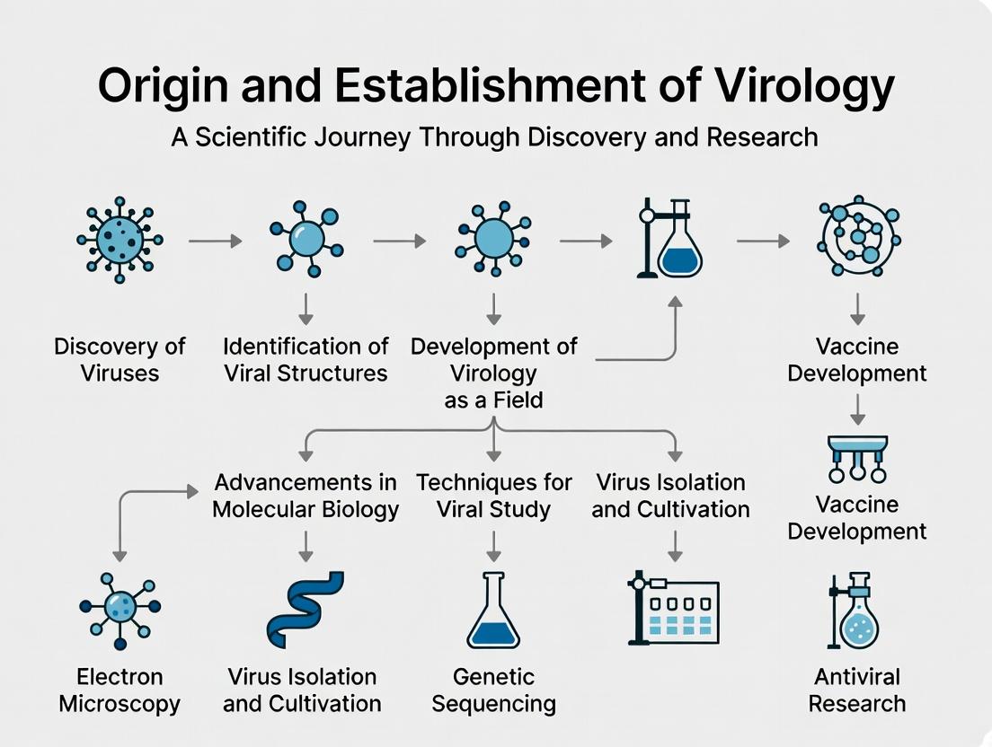
From Tobacco Mosaic to Modern Virology: The Definitive History of a Pioneering Science
This article traces the multidisciplinary journey of virology from its pre-scientific origins to its establishment as a foundational biomedical discipline.
From Tobacco Mosaic to Modern Virology: The Definitive History of a Pioneering Science
Abstract
This article traces the multidisciplinary journey of virology from its pre-scientific origins to its establishment as a foundational biomedical discipline. We explore the foundational discoveries of viral entities and the evolution of core methodologies like cell culture and molecular techniques. The discussion addresses historical and modern challenges in virus isolation, characterization, and overcoming research bottlenecks. A comparative analysis validates key technological shifts and theoretical models. Designed for researchers and drug development professionals, this synthesis highlights how historical insights inform contemporary antiviral strategies, vaccine development, and preparedness for emerging viral threats.
Unseen Invaders: The Pre-Molecular Era and Foundational Discoveries in Virology
The establishment of virology as a formal scientific field in the late 19th century, marked by the work of Ivanovsky and Beijerinck on the Tobacco Mosaic Virus, was preceded by millennia of human observation of viral-like diseases. This article posits that these ancient accounts represent a crucial pre-scientific phase in virology's origin. They provided the phenomenological foundation—detailed descriptions of contagion, immunity, and symptomology—that later guided germ theory and the specific quest for filterable pathogens. Analyzing these accounts through a modern technical lens allows for the retrospective extraction of epidemiological data and the formulation of hypotheses testable via contemporary molecular archaeology.
Historical Accounts & Quantitative Data Synthesis
Ancient texts from multiple civilizations describe diseases with high infectivity, specific symptom clusters, and epidemic spread, consistent with viral etiologies. Key accounts are summarized in Table 1.
Table 1: Ancient Accounts of Viral-Like Diseases: Key Features and Modern Correlates
| Civilization/Period | Document/Source | Disease Description | Recorded Symptoms (Modern Interpretation) | Epidemiological Notes | Presumed Modern Viral Disease |
|---|---|---|---|---|---|
| Ancient Egypt (c. 1157 BCE) | Papyrus of Ramses V (Mummy) | Not textual, but physical evidence. | Pustular rash on mummified skin. | Individual case, evidence of widespread outbreak inferred. | Smallpox (Variola virus) |
| Hittite Empire (c. 1320-1318 BCE) | Suppiluliuma I Prayers | Plague decimating population. | "Fatal pestilence," likely hemorrhagic fever. | Followed prisoner transfer from Egypt; suggests zoonotic introduction. | Possibly Hemorrhagic Fever (e.g., Ebola/Marburg filoviruses) |
| Ancient Greece (430 BCE) | Thucydides' History of the Peloponnesian War | "Plague of Athens." | High fever, inflammation, pharyngitis, rash, diarrhea, gangrene. | Highly contagious, no immunity, ~25% mortality. Cause remains debated (e.g., Ebola, Typhus). | |
| Roman Empire (165-180 CE) | Galen's Writings | "Antonine Plague." | Fever, sore throat, diarrhea, skin pustules (after 9 days). | Spread via army movements; killed millions. Likely Smallpox (or measles). | |
| Han Dynasty China (c. 250 CE) | Ge Hong's Zhou Hou Bei Ji Fang | "Heaven-Borne Pox." | Sores erupting on skin, high mortality. | Clear recognition of contagion and acquired immunity post-infection. | Smallpox (Variola virus) |
| Medieval Islam (10th c. CE) | Al-Razi (Rhazes) A Treatise on Smallpox and Measles | Distinct clinical differentiation. | Smallpox: severe, pustules. Measles: milder, rash. | Provided first definitive clinical distinction between two viral exanthems. | Smallpox & Measles (Morbillivirus) |
Experimental Protocols for Modern Retrospective Analysis
3.1. Protocol A: Paleogenomic Sequencing of Viral DNA/RNA from Ancient Remains
Objective: To extract and sequence fragmented viral nucleic acids from archaeological samples (teeth, bone) to confirm pathogen identity.
- Sample Preparation: Conduct in dedicated ancient DNA (aDNA) cleanroom. Powder 50-100mg of tooth root or petrous bone.
- Digestion & Extraction: Digest powder in buffer containing proteinase K and EDTA. Extract nucleic acids using silica-column methods optimized for fragments <100bp.
- Library Preparation: Build double-stranded DNA libraries with unique dual-index barcodes. Include uracil-specific excision reagent (USER) to mitigate damage-derived cytosine deamination errors.
- Target Enrichment: Perform in-solution hybrid capture using biotinylated RNA baits designed against conserved regions of suspected viral families (e.g., Poxviridae, Paramyxoviridae).
- Sequencing: Sequence on high-throughput platform (Illumina). Use high depth (>10M reads per sample).
- Bioinformatic Analysis: Map reads to human reference genome (hs37d5) to assess preservation. Unmapped reads are subsequently mapped to a microbial/viral pangenome. Authenticate ancient viral reads by checking for characteristic post-mortem damage patterns (elevated C→T at fragment ends).
3.2. Protocol B: Phylogenetic Molecular Clock Analysis
Objective: To integrate ancient viral sequences with modern genomes to estimate the time of most recent common ancestor (tMRCA) and trace viral evolution.
- Alignment: Multiple sequence alignment of ancient and modern full-genome or conserved gene sequences (e.g., hemagglutinin for smallpox) using MAFFT.
- Model Selection: Use jModelTest2 to determine best-fitting nucleotide substitution model (e.g., GTR+I+Γ).
- Tree Inference: Construct a maximum-likelihood phylogeny using RAxML or IQ-TREE.
- Calibration & Dating: Apply Bayesian dating software (BEAST2). Use radiocarbon dates of ancient samples as tip-date calibrations. Run Markov Chain Monte Carlo (MCMC) for sufficient generations (e.g., 100M) to achieve effective sample size (ESS) >200 for all parameters.
- Interpretation: The resulting time-scaled phylogeny estimates the evolutionary rate (subs/site/year) and tMRCA, providing a timeline for the establishment of the virus in human populations.
Visualization of Research Workflow
Title: Retrospective Viral Genomics Research Workflow
The Scientist's Toolkit: Key Research Reagent Solutions
Table 2: Essential Materials for Retrospective Viral Pathogen Research
| Item/Reagent | Supplier Examples | Function in Protocol |
|---|---|---|
| Silica-based aDNA Extraction Kit | Qiagen (MinElute), dedicated aDNA labs | Purifies short, damaged DNA/RNA fragments while removing PCR inhibitors common in ancient tissues. |
| USER Enzyme Mix | New England Biolabs (NEB) | Enzyme cocktail (Uracil-DNA Glycosylase + Endo VIII) that removes deaminated cytosines (uracils) at fragment ends, reducing sequencing errors. |
| Double-stranded DNA Library Prep Kit | NEB NEXT, Twist Bioscience | Prepares sequencing libraries from ultra-low input, fragmented DNA with dual indexing to prevent cross-sample contamination. |
| MyBaits Custom Viral Panel | Arbor Biosciences | Biotinylated RNA baits for in-solution capture of target viral sequences, enriching them from a background of host and environmental DNA. |
| BEAST2 Software Package | beast2.org | Bayesian evolutionary analysis software for molecular dating and phylogenetic reconstruction using tip-dated sequences. |
| Phusion U Hot Start DNA Polymerase | Thermo Fisher Scientific | High-fidelity polymerase resistant to uracil, used for PCR amplification during library enrichment steps post-capture. |
The establishment of virology as a discrete scientific field is inextricably linked to the investigation of Tobacco Mosaic Disease in the late 19th century. This period was defined by the nascent germ theory, which held that infectious diseases were caused by visible, cultivable bacteria. The breakthrough experiments on Tobacco Mosaic Virus (TMV) shattered this paradigm by demonstrating the existence of a novel class of pathogens: filterable, invisible, and non-cultivable on artificial media. This whitepaper deconstructs the key experiments, placing them within the thesis that TMV research provided the necessary methodological and conceptual toolkit—the "filterable agent" paradigm—that originated the field of virology.
Foundational Experiments & Quantitative Data
The critical path to discovery was paved by a series of meticulous experiments by Adolf Mayer, Dmitri Ivanovsky, and Martinus Beijerinck. Their collective work systematically eliminated known biological explanations.
Table 1: Foundational Experiments on Tobacco Mosaic Disease (1880-1899)
| Investigator (Year) | Key Experimental Question | Methodology | Observation & Result | Interpretation & Limitation |
|---|---|---|---|---|
| Adolf Mayer (1886) | Is the disease transmissible via fluid? | Sap from diseased plants injected into healthy plants. | Healthy plants developed mosaic symptoms. | Concluded an infectious agent, but presumed it was a bacterial disease. |
| Dmitri Ivanovsky (1892) | Can the infectious agent be removed by filtration? | Passed infectious sap through Chamberland porcelain filter (pores ~0.1 µm). | Filtrate remained infectious. | Correctly observed filterability. Incorrectly hypothesized a bacterial toxin or spore-small bacteria. |
| Martinus Beijerinck (1898) | Does the agent replicate in plant tissue, or is it a non-replicating poison? | 1. Diffusion Experiment: Placed filtrate on agar, let diffuse, then transferred agar block to plant.2. Serial Passage Experiment: Repeatedly transferred filtrate from newly infected plant to new healthy plants. | 1. Agent did not diffuse; only infected plant at point of contact.2. Agent remained potent indefinitely, demonstrating multiplication. | Concluded a contagium vivum fluidum ("contagious living fluid")—a replicating, non-particulate, filterable agent. This defined the new paradigm. |
Detailed Experimental Protocols
3.1. Chamberland Filtration Protocol (Ivanovsky, 1892)
- Materials: Diseased tobacco leaf tissue, mortar and pestle, sterile gauze, Chamberland porcelain filter candle (Type L1 or L2, pore size ~0.1 µm), sterile syringe or pressure apparatus, recipient flask, healthy Nicotiana tabacum seedlings.
- Procedure:
- Homogenize infected leaf tissue with distilled water (1:10 w/v) using a sterile mortar and pestle.
- Clarify the sap by coarse filtration through several layers of sterile cheesecloth.
- Load the clarified sap into the Chamberland filter apparatus. Apply positive pressure or gravity feed to pass the sap through the porcelain candle.
- Collect the sterile filtrate in a recipient flask. Confirm bacterial sterility by plating an aliquot onto nutrient agar.
- Inoculate healthy tobacco seedlings by gently rubbing the filtrate onto carborundum-dusted leaves (mechanical inoculation).
- Maintain plants in a controlled greenhouse and observe for 5-14 days for development of mosaic motting, chlorosis, and stunting.
- Control: Inoculate a separate group of plants with unfiltered sap.
3.2. Agar Diffusion & Serial Passage Protocol (Beijerinck, 1898)
- Materials: Infectious TMV filtrate, nutrient agar plates, sterile cork borers, scalpel, healthy tobacco plants.
- Diffusion Procedure:
- Pour sterile nutrient agar into a Petri dish and allow to solidify.
- Apply a droplet of infectious filtrate to the center of the agar surface.
- Incubate at room temperature for 24-48 hours to allow for potential diffusion of a "toxin."
- Using a sterile cork borer, excise agar blocks at increasing distances from the inoculation site.
- Macerate each agar block in a small volume of water and use it to inoculate separate healthy plants.
- Observe plants for symptoms. Result: Only the plant inoculated with the block from the direct application site became infected.
- Serial Passage Procedure:
- Inoculate Plant A1 with the original infectious filtrate.
- After disease symptoms develop, harvest tissue from A1, homogenize, filter, and use the filtrate to inoculate Plant A2.
- Repeat this process sequentially through multiple plant generations (e.g., A3, A4, A5...).
- Observe that the infectivity of the filtrate does not diminish but is maintained or increased, proving replication of the agent within the host tissue.
Visualizing the Logical Breakthrough
Diagram 1: Conceptual Evolution from Bacteriology to Virology
Diagram 2: Experimental Workflow for TMV Filterability & Infectivity
The Scientist's Toolkit: Key Research Reagent Solutions
Table 2: Essential Materials for Early Viral Pathogenesis Research
| Research Reagent / Material | Function in TMV Breakthrough Experiments |
|---|---|
| Chamberland Porcelain Filter | Sterilizing filter with ~0.1 µm pores; critical for separating bacterial cells from the smaller, filterable TMV agent. |
| Nicotiana tabacum (Tobacco) Cultivars | Model host organism; highly susceptible to TMV, providing clear and consistent symptomatic readout. |
| Carborundum (Silicon Carbide) Abrasive | Used in mechanical inoculation to create micro-wounds on leaves, allowing viral entry without vector organisms. |
| Nutrient Agar Plates | Used to confirm bacterial sterility of filtrates and in Beijerinck's diffusion experiment to disprove toxin hypothesis. |
| Sterile Mortar, Pestle, & Gauze | For homogenizing plant tissue and clarifying sap to create the initial infectious inoculum. |
The establishment of virology as a distinct scientific discipline in the late 19th and early 20th centuries was predicated on the fundamental need to define the pathogenic "entity." The seminal work of scientists like Dmitri Ivanovsky, Martinus Beijerinck, and Friedrich Loeffler did not merely identify new pathogens; it established a paradigm for differentiating between three core classes of agents: viruses, bacteria, and toxins. This differentiation, based on physical, biological, and biochemical criteria, laid the experimental and conceptual foundation for modern virology and remains critical for contemporary research and therapeutic development.
Core Defining Characteristics: A Comparative Analysis
The primary distinctions between these entities are summarized in the following tables.
Table 1: Structural and Replicative Characteristics
| Characteristic | Viruses | Bacteria | Toxins |
|---|---|---|---|
| Cellular Structure | Acellular; no organelles. | Prokaryotic cell (single-celled). | Non-living biochemical compound. |
| Genetic Material | DNA or RNA, single- or double-stranded. | DNA and RNA (both present). | None (protein, lipopolysaccharide, or other molecule). |
| Size Range | 20 - 300 nm (requires EM). | 0.5 - 5.0 μm (visible by light microscopy). | 5 - 150 kDa (molecular scale). |
| Replication Method | Obligate intracellular parasite; uses host machinery. | Binary fission (independent cell division). | Not applicable; synthesized in vivo or in vitro. |
| Metabolism | Absent. | Present; independent energy generation & biosynthesis. | Absent. |
| Response to Antibiotics | No. | Yes (target cell wall, ribosomes, etc.). | No (addressed by antitoxins/neutralizing antibodies). |
Table 2: Key Experimental Discriminators in Research
| Experimental Assay | Viral Signature | Bacterial Signature | Toxin Signature |
|---|---|---|---|
| Filtration (0.22 μm pore) | Filtrate remains infectious. | Not infectious (retained on filter). | Filtrate remains active. |
| Culture on Inert Media | Cannot grow. | Forms colonies. | Cannot grow (but may be present). |
| Antibiotic Challenge (e.g., Tetracycline) | No effect on direct agent. | Growth inhibition. | No effect. |
| Heat/Formalin Inactivation | Often inactivated (envelope sensitive). | Variable (spores resistant). | Protein toxins denatured; endotoxins stable. |
| Electron Microscopy | Reveals capsid/nucleocapsid structure. | Reveals cell wall, shape, organelles. | Reveals no particulate structure (amorphous). |
Foundational & Modern Experimental Protocols
Protocol 3.1: The Chamberland-Pasteur Filter Experiment (Historical & Conceptual)
Objective: To determine if an infectious agent is filterable and thus smaller than common bacteria. Materials: Infected tissue homogenate, Chamberland-type porcelain filter (0.22 μm pore size), sterile collection flask, susceptible host model (plant, animal). Procedure: 1. Pass the clarified homogenate through the sterilized filter under positive pressure or gravity. 2. Aseptically collect the filtrate. 3. Inoculate a naive, susceptible host with an aliquot of the filtrate. 4. Observe for disease signs. Concurrently, culture the filtrate on rich agar/media to test for bacterial growth. Interpretation: Disease in the host combined with no bacterial growth on media provides strong evidence for a viral etiology.
Protocol 3.2: Quantitative PCR (qPCR) for Discriminatory Detection
Objective: To specifically identify and quantify viral vs. bacterial genetic material in a sample. Materials: Nucleic acid extract, sequence-specific primers/probes for viral target (e.g., influenza M gene) and bacterial target (e.g., 16S rRNA gene), reverse transcriptase (for RNA viruses), qPCR master mix, qPCR instrument. Procedure: 1. Extract total nucleic acid from the clinical or research sample (e.g., using a silica-column method). 2. For RNA targets, perform reverse transcription to generate cDNA. 3. Set up parallel qPCR reactions: one with virus-specific primers/probe, one with bacteria-specific primers/probe, and necessary controls (no-template, positive control). 4. Run the thermocycling protocol (e.g., 95°C denaturation, 60°C annealing/extension for 40 cycles). 5. Analyze cycle threshold (Ct) values. A positive signal only in the viral assay confirms viral presence. Melt-curve analysis can check specificity. Interpretation: Distinct, sequence-specific amplification differentiates entities based on unique genetic signatures. Multiplexing allows for co-detection.
Protocol 3.3: Mass Spectrometry for Toxin Identification
Objective: To definitively identify a protein toxin (e.g., Staphylococcal enterotoxin B) and distinguish it from a viral or bacterial particle. Materials: Purified sample or cultured supernatant, trypsin for digestion, LC-MS/MS system, protein sequence database. Procedure: 1. If necessary, concentrate the sample and perform a proteolytic digest (e.g., with trypsin) overnight. 2. Separate the resulting peptides via liquid chromatography (LC). 3. Analyze eluting peptides by tandem mass spectrometry (MS/MS), fragmenting ions to generate spectra. 4. Search the acquired spectra against a protein database using bioinformatics software (e.g., Mascot, Sequest). Interpretation: High-confidence identification of toxin peptides confirms the presence of a toxic protein, excluding viral or living bacterial causation. Cannot detect viable organisms.
Visualizing Distinguishing Pathways & Workflows
Title: Diagnostic Workflow to Distinguish Pathogenic Entities
Title: Core Replication and Action Mechanisms Compared
The Scientist's Toolkit: Key Research Reagent Solutions
| Research Reagent / Material | Primary Function in Distinction Studies |
|---|---|
| Ultrafiltration Membranes (e.g., 100kDa MWCO) | Separates virions (retained) from soluble toxins and small proteins (pass through) based on size, not just porosity. |
| Penicillin-Streptomycin (Pen-Strep) Solution | Broad-spectrum antibiotic mix added to cell cultures to suppress bacterial growth, allowing isolation of viruses. |
| DNase I & RNase A Enzymes | Treats samples to degrade free nucleic acids; viral genomes within capsids are protected, helping confirm intact virions. |
| Lipopolysaccharide (LPS) ELISA Kit | Specifically detects endotoxin from Gram-negative bacteria, distinguishing bacterial presence from viral infection. |
| Plaque Assay Agar Overlay | Semi-solid medium to restrict virus spread, allowing quantification of infectious viral particles distinct from bacterial colonies. |
| Proteinase K | Digests proteins; can inactivate protein-based toxins and some viral capsids, used in inactivation controls. |
| Specific Neutralizing Antibodies | Binds to and neutralizes a specific virus or toxin, used to confirm causal agent in pathogenicity assays. |
| SYBR Green qPCR Master Mix | Intercalating dye for generic detection of amplified DNA, useful for initial broad screening of unknown genetic material. |
| Cell Line Permissive to Target Virus | Essential for propagating and studying obligate intracellular viruses, confirming living host requirement. |
| Mass Spectrometry Grade Trypsin | High-purity enzyme for reproducible digestion of protein samples into peptides for definitive toxin fingerprinting. |
This whitepaper details the foundational studies in Foot-and-Mouth Disease (FMD) and Yellow Fever that established the core principles and methodologies of animal virology, framed within the broader thesis on the origin of virology as a distinct scientific discipline.
Foundational Discoveries and Quantitative Data
The elucidation of FMD and Yellow Fever viruses as filterable agents provided the first quantitative evidence for a new class of pathogens.
Table 1: Foundational Experiments in Early Animal Virology
| Experiment | Key Scientist(s) & Year | Agent | Filter Pore Size | Key Finding (Quantitative) | Model System |
|---|---|---|---|---|---|
| First Demonstration of Filterability | Friedrich Loeffler & Paul Frosch (1898) | FMD | Chamberland-type filter (approx. 100-200 nm) | Filtrate from diluted lymph (1:100) induced disease in 4/4 inoculated cattle. | Cattle |
| Confirmation & Human Disease Link | Walter Reed Commission (1900-1901) | Yellow Fever | Berkefeld filter (unknown precise size) | Filtrate of blood from acute patients induced disease in 7/10 inoculated volunteers. Filterability confirmed. | Human volunteers |
| First In Vitro Cultivation | Karl Landsteiner & Erwin Popper (1908) | Poliovirus (parallel milestone) | Not applied in this study | Emulsion of infected spinal cord induced disease in 2/2 monkeys. Demonstrated non-bacterial, replicating agent. | Macacus monkeys |
Detailed Experimental Protocols
Protocol: Loeffler and Frosch's FMD Filterability Experiment (1898)
Objective: To determine if the causative agent of Foot-and-Mouth Disease was a filterable, non-cellular entity.
- Sample Preparation: Vesicular lymph from infected cattle was collected and serially diluted 1:100 in sterile fluid.
- Filtration: The diluted lymph was passed through a Chamberland-type porcelain filter, known to retain all known bacteria.
- Inoculation: Four healthy cattle were inoculated subcutaneously with the cell-free filtrate.
- Controls: Parallel inoculations with unfiltered lymph and bacteria-laden material retained by the filter were performed.
- Observation: Animals were monitored for the development of classic FMD symptoms (fever, vesicles on feet and mouth). Conclusion: The filtrate, devoid of bacteria, consistently induced FMD, proving the agent was filterable and replicating.
Protocol: Reed Commission's Yellow Fever Transmission (1900-1901)
Objective: To establish the etiology and mode of transmission of Yellow Fever.
- Blood Inoculation: Blood drawn from acute Yellow Fever patients within the first 3 days of illness was filtered through a Berkefeld filter.
- Human Challenge: Filtered serum was injected subcutaneously into consenting, non-immune human volunteers.
- Mosquito Transmission: Aedes aegypti mosquitoes were fed on Yellow Fever patients and, after a 12-day interval, allowed to feed on non-immune volunteers.
- Environmental Control: Volunteers inhabited thoroughly disinfected, mosquito-proof quarters with soiled bedding from patients. Conclusion: The disease was transmitted by filtered blood and by mosquitoes, but not by fomites, identifying a filterable virus and its vector.
Visualization of Foundational Concepts
The Scientist's Toolkit: Foundational Research Reagents & Materials
Table 2: Key Research Reagent Solutions in Early Virology
| Reagent / Material | Function in Foundational Experiments | Specific Example / Note |
|---|---|---|
| Porcelain (Chamberland) Filters | To physically separate bacteria from infectious filtrates based on size. Pore size estimated at 100-200 nm. | Loeffler & Frosch (1898) used these to prove FMD agent was filterable. |
| Diatomaceous Earth (Berkefeld) Filters | Alternative bacterial filter with slightly larger, more variable pores. Used for clarifying fluids. | Used by the Reed Commission (1900) for filtering Yellow Fever patient serum. |
| Infectious Clinical Material | Source of the putative viral agent for filtration and inoculation. | FMD vesicular lymph; Acute-phase Yellow Fever patient blood. |
| Susceptible Animal Model | A living system to demonstrate replication and pathogenicity of the filtered agent. | Cattle for FMD; Human volunteers for Yellow Fever (ethical standards differ today). |
| Sterile Dilution Fluids | To dilute infectious material, challenging the "concentration of poison" hypothesis and proving replication. | Saline or similar buffers used by Loeffler & Frosch for serial dilution. |
| Vector Colonies | To demonstrate biological transmission distinct from mechanical transfer. | Laboratory-bred Aedes aegypti mosquitoes fed on Yellow Fever patients. |
The establishment of virology as a discrete scientific field hinged upon the resolution of a fundamental paradox: the nature of the infectious agent. The late 19th and early 20th centuries saw the formulation of the "virus" concept to describe filterable, sub-bacterial pathogens. The pivotal thesis, developed through the work of Beijerinck, Stanley, and others, was that these entities were not merely small cells but were fundamentally replicating genetic entities—obligate intracellular parasites whose "life" is an expression of directed genetic replication and assembly. This whitepaper details the modern experimental foundations validating this core thesis, providing a technical guide for researchers engaged in antiviral drug and therapeutic development.
Core Quantitative Data: Viral Replication Metrics
Table 1: Quantitative Benchmarks for Model Viral Replication Cycles
| Viral Parameter | Influenza A Virus | HIV-1 | Bacteriophage λ | SARS-CoV-2 (Omicron) |
|---|---|---|---|---|
| Genome Size | 13.5 kb (ssRNA, segmented) | 9.7 kb (ssRNA, diploid) | 48.5 kb (dsDNA) | 29.9 kb (ssRNA) |
| Replication Time (One Cycle) | 6-8 hours | 24-48 hours | 30-40 minutes | 6-12 hours |
| Burst Size (Virions/Cell) | 10^3 - 10^4 | 10^3 - 10^5 | 10^2 (lysis) | 10^2 - 10^3 |
| Mutation Rate (per base per cycle) | ~1 x 10^-5 | ~3 x 10^-5 | ~2 x 10^-8 | ~1 x 10^-6 |
| Packaging Efficiency | ~90% (segment co-packaging) | >95% (dimerization) | ~99% (cos site) | >90% (packaging signal) |
Table 2: Key Experimental Techniques for Studying Viral Replication
| Technique | Primary Application | Quantitative Output |
|---|---|---|
| Plaque Assay | Infectious titer determination | Plaque Forming Units (PFU/mL) |
| Quantitative PCR (qPCR) | Genome copy number | Copies/µL (distinguishes infectious vs. total) |
| Focus Forming Assay (FFA) | Titration of non-lytic viruses | Focus Forming Units (FFU/mL) |
| Single-Cycle Growth Curve | Kinetic analysis of replication | Virion production over time post-synchronized infection |
| Next-Gen Sequencing (NGS) | Quasispecies diversity, recombination | Mutation frequency, haplotype networks |
Foundational Experimental Protocols
Protocol: Single-Cycle Growth Curve Analysis
Objective: To precisely quantify the kinetics of viral replication under non-spreading conditions. Methodology:
- Cell Seeding: Seed permissive cells (e.g., Vero E6, MDCK) in a 12-well plate to achieve 90% confluence at time of infection.
- Virus Inoculation & Synchronization: Incubate cells with virus at a high Multiplicity of Infection (MOI ≥ 5) for 1 hour at 4°C (allows attachment but not entry). Wash cells 3x with cold PBS to remove unbound virions.
- Temperature Shift & Entry: Add pre-warmed medium and shift cells to 37°C. This synchronizes entry. After 1 hour, treat cells with a acidic citrate buffer (pH 3.0) for 30 seconds to inactivate any remaining non-internalized virus. Wash immediately with neutral PBS.
- Sample Harvesting: At defined time points post-entry (e.g., 0, 2, 4, 6, 8, 10, 12, 24h), harvest both cell supernatant (released virions) and the cell monolayer (cell-associated virions) separately using freeze-thaw lysis.
- Titration: Titrate all samples using a plaque assay or FFA. Plot total infectious titer (log10 PFU/mL) vs. time to visualize eclipse phase, exponential rise, and plateau.
Protocol: Definitive Proof of Genetic Material Role (Modified Hershey-Chase)
Objective: To demonstrate that viral nucleic acid, not protein, is the replicating genetic element. Modern Application with Bacteriophage T4:
- Isotopic Labeling: Grow two separate batches of E. coli:
- In medium containing ^35S-labeled methionine/cysteine to label phage proteins.
- In medium containing ^32P-labeled phosphate to label phage DNA.
- Phage Propagation: Infect each culture with T4, harvest, and purify the labeled phage progeny.
- Infection & Shearing: Infect fresh, unlabeled E. coli with each phage preparation separately. Allow brief time for attachment and injection.
- Blending (Shearing): Subject the infected culture to high-speed vortexing or a Waring blendor to shear off phage capsids attached to the outside of the bacterial cells.
- Centrifugation: Pellet the bacterial cells. The pellet contains injected material and cell bodies; the supernatant contains sheared-off capsids and unattached phage.
- Quantification: Measure radioactivity (^35S or ^32P) in the pellet and supernatant fractions using a scintillation counter.
- Expected Result: ^32P (DNA label) is primarily in the bacterial pellet, while ^35S (protein label) is in the supernatant. This proves the genetic material transferred for replication is DNA.
Visualization of Core Concepts
Diagram 1: Universal Viral Replication Cycle
Diagram 2: Viral Central Dogma & Self-Assembly
The Scientist's Toolkit: Essential Research Reagents
Table 3: Key Reagent Solutions for Viral Replication Studies
| Reagent / Material | Function & Application | Example / Note |
|---|---|---|
| Polyethyleneimine (PEI) | Chemical transfection reagent for delivering plasmid DNA encoding viral genomes (infectious clones) into cells to initiate de novo virus production. | Essential for reverse genetics systems. |
| Protease Inhibitors (e.g., MG-132, BafA1) | Inhibit cellular proteasomes or lysosomal acidification. Used to study viral protein stability, processing, and entry pathways (e.g., for Coronaviruses, Influenza). | |
| RdRp Inhibitors (e.g., Remdesivir-TP, Favipiravir-RTP) | Nucleotide analogues that specifically target and inhibit viral RNA-dependent RNA polymerase (RdRp) activity. Key for mechanistic studies and control experiments. | Distinguish viral from host polymerase functions. |
| Neutralizing Antibodies | Bind specific viral surface proteins (e.g., Spike, HA) to block receptor attachment. Used for immunodepletion, entry pathway validation, and pseudovirus neutralization assays. | Convalescent serum or monoclonal antibodies (mAbs). |
| siRNA / CRISPR-Cas9 Knockdown Library | Targeted depletion of host gene expression to identify essential host factors (dependency factors) for viral entry, replication, or egress. | Genome-wide screens for host-virus interactions. |
| Plaque Agar Overlay (Methylcellulose/Avicel) | Semi-solid medium applied after infection to restrict viral spread to neighboring cells, enabling visualization and counting of discrete plaques formed by lytic viruses. | Critical for PFU-based titrations. |
| Pseudotyped Virus Particles | Reporter virions bearing the envelope protein of a target virus (e.g., VSV-G, HIV-1 Env) on a core from a different virus (e.g., VSV, MLV). Safe for studying entry of high-containment pathogens (BSL-2). | Core encodes luciferase or GFP. |
| Dual-Luciferase Reporter System | Plasmid constructs where viral IRES or promoter elements drive a reporter gene (Firefly luc). Co-transfected with a constitutively expressed control (Renilla luc) for normalization in replication/translation assays. | Quantifies viral regulatory element activity. |
Cultivating Discovery: Key Methodological Advances That Built Modern Virology
The establishment of virology as a distinct scientific field in the late 19th and early 20th centuries demanded the development of reliable, sterile, and living systems for the cultivation and study of obligate intracellular pathogens. The embryonated hen's egg emerged as the first practical and scalable platform, bridging the gap between animal models and the later advent of cell culture. This platform was foundational for the isolation, characterization, and vaccine development for numerous human and animal viruses, directly enabling the pioneering work of Goodpasture, Burnet, and others that defined modern virology.
Anatomy of the Embryonated Egg as a Viral Host System
The developing chick embryo, with its variety of differentiated tissues and membranes, provides unique sites for the propagation of different virus families. Key inoculation routes are defined in Table 1.
Table 1: Primary Inoculation Routes and Their Viral Applications
| Route | Target Membrane/Tissue | Key Virus Examples | Visible Reaction (Pock/Lesion) |
|---|---|---|---|
| Chorioallantoic Membrane (CAM) | Ectodermal & mesodermal layers | Vaccinia, Herpes Simplex, Smallpox | Distinct pocks (opaque, proliferative foci) |
| Allantoic Cavity | Endodermal layer & allantoic fluid | Influenza, Newcastle Disease, Mumps | Hemagglutination of harvested fluid |
| Amniotic Cavity | Amniotic membrane & fluid | Primary isolation of Influenza, Mumps | Direct infection of respiratory epithelium |
| Yolk Sac | Endodermal & mesodermal layers | Arboviruses, Chlamydia | Enrichment for high-titer stock |
Core Experimental Protocols
Protocol 1: Chorioallantoic Membrane (CAM) Inoculation for Virus Titration (Pock Assay)
- Materials: Specific pathogen-free (SPF) embryonated eggs (9-11 days old), candling lamp, drill or sandpaper, sterile syringe with 25-27G needle, 70% ethanol, wax or glue, virus sample in diluent.
- Method:
- Candle eggs to mark the air sac and a prominent chorioallantoic vessel. Avoid major blood vessels at the inoculation site.
- Swab the eggshell with 70% ethanol. Using a drill or sandpaper, create a small window (~1cm²) over the CAM, careful not to puncture the shell membrane.
- Create a small hole at the blunt end (air sac) using a needle.
- Apply gentle suction (e.g., with a rubber bulb) to the air sac hole to drop the CAM away from the shell membrane at the window site.
- Inoculate 0.1-0.2 mL of virus dilution directly onto the dropped CAM via the window.
- Seal the window with transparent tape or sterile glue. Seal the air sac hole with wax.
- Incubate the eggs horizontally at 35-37°C with the window upright for 48-72 hours.
- Chill eggs at 4°C for 4 hours or overnight to constrict blood vessels.
- Aseptically open the window, excise the CAM, and place in phosphate-buffered saline (PBS) in a Petri dish.
- Count discrete pocks (viral foci) under brightfield illumination. Calculate titer in pock-forming units (PFU) per mL.
Protocol 2: Allantoic Cavity Inoculation for Influenza Virus Propagation
- Materials: SPF embryonated eggs (9-11 days old), candling lamp, drill, sterile syringe with 23-25G needle, 70% ethanol, wax.
- Method:
- Candle the egg. Mark the boundary of the air sac and a point just above the allantoic cavity (typically on the side, avoiding major vessels).
- Swab the inoculation site with ethanol. Drill a small hole through the shell.
- Insert the needle (approx. 1-1.5 cm deep) at a 45-90 degree angle into the allantoic cavity and inoculate 0.1-0.2 mL of virus seed stock.
- Seal the hole with wax or glue. Incubate eggs vertically (blunt end up) at 33-37°C (virus-dependent) for 48-72 hours.
- Chill eggs at 4°C for a minimum of 4 hours.
- Aseptically open the blunt end, puncture the shell and inner membranes with sterile forceps.
- Using a sterile pipette, harvest the allantoic fluid (typically 5-10 mL per egg). Clarify by centrifugation at 1000-2000 x g for 10 min. Aliquot and store at ≤ -70°C.
- Virus yield is quantified by hemagglutination (HA) assay or plaque assay.
Quantitative Data & Modern Relevance
Table 2: Historical & Contemporary Yield Data from Embryonated Eggs
| Virus | Inoculation Route | Typical Yield (per egg) | Modern Application |
|---|---|---|---|
| Influenza A (Wild-type) | Allantoic | 10³-10⁴ HA Units / 10⁷-10⁹ PFU | Seed stock for inactivated split/subunit vaccines |
| Influenza B | Amniotic/Allantoic | 10²-10³ HA Units / 10⁶-10⁸ PFU | Vaccine seed stock development |
| Vaccinia Virus | CAM | 10⁸-10⁹ PFU (lysed CAM) | Research reagent, vector development |
| Yellow Fever 17D | Embryo (whole) | 10⁴-10⁵ LD₅₀ (Mouse) | Live-attenuated vaccine production (historical) |
| Mumps | Amniotic/Allantoic | 10⁴-10⁵ TCID₅₀ | Component of MMR vaccine (historical) |
Signaling & Replication Pathways in Ovo
The Scientist's Toolkit: Research Reagent Solutions
Table 3: Essential Materials for Egg-Based Virology
| Reagent/Material | Function & Purpose | Technical Note |
|---|---|---|
| Specific Pathogen Free (SPF) Eggs | Ensures absence of confounding agents (e.g., avian leukosis virus, mycoplasma). Critical for vaccine safety and consistent research. | Typically from defined flocks maintained in isolation. Age (in days) is virus-specific. |
| Antibiotic-Antimycotic Solution (100X) | Added to viral inoculum or diluent to suppress bacterial/fungal contamination from the shell. | Standard penicillin-streptomycin-amphotericin B mix. Use at 1X final concentration. |
| Sterile Diluent (e.g., PBS with SPA) | Phosphate-Buffered Saline (PBS) with antibiotics, often supplemented with protein (e.g., 0.1% BSA or gelatin) to stabilize virus. | Prevents non-specific virus adhesion to tubes and needles. |
| Candling Lamp/Light Source | Visualizes embryo viability, air sac boundaries, and major blood vessels for accurate inoculation. | LED-based systems provide cool, bright illumination. |
| Egg Drilling/Punching Device | Creates precise, clean openings in the eggshell for inoculation and harvesting. | Can be manual (abrasive tip) or electric. Must be sterilizable. |
| Sealing Material (Paraffin Wax or Sterile Glue) | Seals inoculation and harvest holes to maintain sterility and prevent desiccation of the embryo. | Semi-solid paraffin wax is traditional; sterile acrylic glue is an alternative. |
| Hemagglutination (HA) Assay Reagents | For quantification of hemagglutinating viruses (e.g., Influenza). Includes red blood cells (RBCs, e.g., turkey or guinea pig) and buffer. | Standardized viral titer method (HA Units/mL). A primary readout for vaccine seed stocks. |
While mammalian cell culture has largely superseded the embryonated egg for large-scale industrial vaccine manufacturing due to scalability and avoidance of egg-adaptive mutations, the platform remains irreplaceable for specific applications. It is the WHO-recommended system for generating seasonal influenza vaccine candidate viruses via reassortment. Furthermore, its cost-effectiveness, sterility, and physiological complexity ensure its continued use in basic virology research, diagnostics, and the production of some veterinary vaccines. As the first platform that enabled the rigorous experimental virology required to establish the field, its historical and practical significance remains profound.
The isolation and propagation of viruses in vitro represents the foundational turning point for virology as a rigorous scientific discipline. Prior to the work of John F. Enders, Thomas H. Weller, and Frederick C. Robbins at the Boston Children’s Hospital, virology was a largely descriptive field, constrained by the requirement for live animal hosts or embryonated eggs. Their demonstration that the Lansing strain of poliovirus could be grown in cultures of non-neural human tissue dismantled the dogmatic belief in the obligatory neurotropism of poliovirus and provided the essential, reproducible in vitro tool. This technical breakthrough directly catalyzed the development of both the Salk (inactivated) and Sabin (live-attenuated) polio vaccines, transforming virology from an observational science into an experimental one capable of precise quantification, manipulation, and large-scale vaccine production.
The Foundational Experiment: Methodology and Workflow
The critical 1949 experiment is detailed below. The core innovation was the use of human embryonic skin, muscle, and intestinal tissue, minced and suspended in a nutrient medium, to support viral replication.
Experimental Protocol: Propagation of Poliovirus in Non-Neural Tissue Culture
A. Tissue Preparation:
- Obtain human embryonic tissue (skin, muscle, intestine) under aseptic conditions.
- Mince tissue finely with surgical scissors to explants of approximately 1-2 mm³.
- Wash tissue fragments repeatedly in a balanced salt solution (e.g., Hanks' BSS) containing antibiotics (penicillin & streptomycin) to reduce microbial contamination.
B. Culture Establishment (Roller-Tube Method):
- Place several tissue explants into a flat-sided test tube or prescription bottle.
- Add a small volume of nutrient plasma clot (chicken plasma mixed with embryonic extract) to anchor explants to the glass surface.
- Allow plasma to coagulate, forming a solid clot.
- Add 1-2 mL of maintenance medium: a mixture of balanced salt solution, bovine amniotic fluid, and human cord serum.
- Seal the tube and incubate in a roller drum at 36°C, completing 8-12 rotations per hour to alternately bathe tissue in medium and air.
C. Viral Inoculation and Incubation:
- After 5-7 days, when a monolayer of fibroblastic outgrowth from explants is established, remove the existing maintenance medium.
- Inoculate the culture with 0.1-0.2 mL of a clarified suspension of ground poliovirus-infected mouse brain (Lansing strain).
- Allow the virus to adsorb for 30-60 minutes at room temperature.
- Add fresh maintenance medium and return to the roller drum incubator.
D. Viral Detection and Titration (Endpoint):
- At regular intervals (e.g., 3, 5, 7, 10 days post-inoculation), harvest the culture fluid.
- Clarify the fluid by low-speed centrifugation.
- Perform serial log₁₀ dilutions of the harvested fluid in fresh maintenance medium.
- Inoculate each dilution into fresh, susceptible tissue culture tubes (primary human or monkey kidney cells became standard later).
- Observe cultures for cytopathic effect (CPE) – cellular rounding, shrinkage, and detachment – over 7-14 days.
- Calculate the 50% tissue culture infectious dose (TCID₅₀) per mL using the Reed-Muench or Karber statistical method.
Experimental Workflow for Poliovirus Propagation
Quantitative Impact: Data from the Foundational Studies
The success of the technique was quantifiable, moving virology into a realm of precise measurement.
Table 1: Key Quantitative Findings from Enders, Weller, and Robbins (1949)
| Parameter | Experimental Result | Significance |
|---|---|---|
| Viral Replication Proof | Infectious virus titer increased 100 to 1000-fold over 7-10 days in culture. | Demonstrated active viral replication, not mere survival, in non-neural cells. |
| CPE Correlation | Distinct cytopathic effect (cell degeneration) correlated 100% with presence of infectious virus. | Established CPE as a reliable, visible marker for viral presence and titration (TCID₅₀). |
| Host Range Expansion | Virus propagated in human tissue culture remained pathogenic for cotton rats and mice. | Proved viral properties (pathogenicity) were maintained after in vitro passage. |
| Serum Neutralization | Specific antisera completely inhibited CPE in culture. | Provided a precise, quantitative in vitro method for immunologic typing and antibody assay. |
Table 2: Impact on Polio Vaccine Development Timeline
| Milestone | Pre-Cell Culture (Pre-1949) | Post-Cell Culture Revolution (Post-1949) |
|---|---|---|
| Virus Production | In living monkeys (spinal cord), limited scale, high cost. | In bottle cultures of monkey kidney cells, large-scale, reproducible. |
| Virus Assay | Intracerebral inoculation of monkeys; slow, expensive, variable. | TCID₅₀ assay in cell culture; rapid, cheap, quantitative, high-throughput. |
| Vaccine Inactivation | Formalin inactivation kinetics poorly monitored. | Formalin inactivation could be precisely monitored by frequent culture samples. |
| Result | Large-scale vaccine production impossible. | Salk vaccine field trials (1954) involved 1.8 million children. |
The Scientist's Toolkit: Essential Research Reagents & Materials
The revolution was enabled by a specific set of reagents and materials.
Table 3: Key Research Reagent Solutions for Early Viral Cell Culture
| Reagent/Material | Function | Specific Use/Example |
|---|---|---|
| Balanced Salt Solution (BSS) | Provides inorganic ions, maintains osmotic pressure and pH. | Hanks' BSS for washing tissue and as base for nutrient media. |
| Embryonic Extract | Source of undefined growth factors and nutrients to stimulate cell proliferation. | Chick embryo extract mixed with plasma to form the initial clot. |
| Plasma | Provides fibrinogen to form a solid clot, anchoring tissue explants. | Chicken plasma used to form the "plasma clot" culture substrate. |
| Bovine Amniotic Fluid | Complex nutrient source containing amino acids, vitamins, and growth factors. | Component of the maintenance medium used by Enders' group. |
| Human Cord Serum | Provides specific hormones, growth factors, and proteins, low in antibodies. | Preferred over adult serum in maintenance medium for its supportive properties. |
| Penicillin & Streptomycin | Antibiotic combination to suppress bacterial contamination in long-term cultures. | Critical for enabling reproducible, non-aseptic tissue work. |
| Trypsin Solution | Proteolytic enzyme for disaggregating tissue into single cells for monolayer culture. | Later became standard for preparing primary monkey kidney cell cultures. |
| pH Indicator (Phenol Red) | Visual indicator of metabolic activity and medium pH in closed culture systems. | Included in media to monitor for acid production from cell metabolism or contamination. |
The Virological Signaling Pathway: From Infection to CPE
The molecular understanding of poliovirus pathogenesis, built upon the cell culture tool, can be summarized as follows.
Poliovirus Replication Cycle & Pathogenesis
The work of Enders, Weller, and Robbins did not merely develop a technique; it established the core operational paradigm of modern virology. The ability to grow a virus in a monolayer of cells, quantify it via TCID₅₀, visualize its effects through CPE, and neutralize it with specific antisera in vitro, created a closed, controlled experimental system. This system became the engine for viral isolation, diagnostics, immunology, pathogenesis studies, and crucially, rational vaccine development. The polio vaccines were its first and most monumental products, but the true legacy was the transformation of virology into a rigorous, quantitative, and independent field of biomedical research.
The establishment of virology as a discrete scientific field was fundamentally contingent upon direct visualization. For decades, pathogenic agents like viruses remained theoretical constructs—“filterable agents”—invisible to light microscopy and thus uncharacterizable. The breakthrough to sub-cellular resolution, provided first by ultraviolet microscopy and conclusively by electron microscopy, transformed these unseen entities into tangible, classifiable objects of study, enabling the rigorous structural and mechanistic research that defines modern virology.
The Resolution Barrier: Light Microscopy and Its Limits
The theoretical limit of resolution for a light microscope is defined by Ernst Abbe’s formula: d = λ/(2NA), where d is the smallest resolvable distance, λ is the wavelength of light, and NA is the numerical aperture of the lens. For visible light (λ ≈ 400-700 nm) and high-quality oil immersion lenses (NA ≈ 1.4-1.6), the practical resolution limit is approximately 200 nm.
Table 1: Comparative Limits of Visualizing Technologies Pre-EM
| Technology | Approximate Resolution Limit | Key Limiting Factor | Virus Visibility |
|---|---|---|---|
| Brightfield Light Microscopy | 200 nm | Wavelength of visible light | No (Most viruses 20-300 nm) |
| Ultraviolet (UV) Microscopy | 100 nm | Shorter UV wavelength | Marginally (Largest viruses only) |
| Darkfield Microscopy | ~200 nm | Light scatter, not resolution | Indirect detection only |
| Theory (Abbe Limit) | ~λ/2 | Fundamental physics of light | Theoretical barrier |
This resolution wall was breached by two sequential advancements: the use of shorter-wavelength illumination and, decisively, the shift from photon to electron beams.
Key Experimental Protocol: First Direct Visualization of a Virus by Electron Microscopy
Experiment: Imaging of the Tobacco Mosaic Virus (TMV) by Helmut Ruska, Ernst Ruska, and colleagues (1939).
Objective: To provide definitive, direct visual evidence of the particulate nature of a filterable agent.
Detailed Methodology:
- Sample Preparation: A purified suspension of TMV was obtained from infected plant sap via differential centrifugation and filtration.
- Negative Staining (Primitive): The sample was mixed with a solution of phosphotungstic acid, a heavy metal salt. Upon drying on a collodion film stretched over a metal grid, the salt formed an amorphous background, while virus particles excluded the stain.
- EM Imaging: The grid was placed in the Siemens & Halske Übermikroskop, an early transmission electron microscope (TEM). The electron beam (accelerating voltage ~75-100 kV) was directed through the sample. Areas with less electron-dense material (the virus rods) allowed more electrons to pass, creating a darker contrast on the final photographic plate.
- Control: Preparations from healthy plant sap were processed identically to confirm the specificity of the rod-shaped structures.
Outcome: The micrographs revealed uniform, rod-shaped particles approximately 300 nm in length and 18 nm in diameter, conclusively demonstrating viruses as discrete physical entities.
The Core Technical Shift: From Photons to Electrons
The electron microscope exploits the wave nature of electrons. The de Broglie wavelength of an electron is given by λ = h/p, where h is Planck's constant and p is the electron's momentum. With sufficient acceleration (typically 60-300 kV), the effective wavelength drops to picometer scales (0.0037 nm at 100 kV), far below atomic dimensions.
Table 2: Quantitative Leap in Resolution: Light vs. Electron Microscopy
| Parameter | Light Microscope (UV) | Early TEM (1940s) | Modern TEM | Modern Cryo-EM |
|---|---|---|---|---|
| Illumination Source | UV Photons (λ ~200 nm) | Electrons (λ ~0.005 nm) | Electrons (λ ~0.0025 nm) | Electrons (λ ~0.0025 nm) |
| Theoretical Resolution | ~100 nm | ~2-10 nm | ~0.05-0.2 nm | ~0.15-0.3 nm |
| Practical Resolution (Biological) | ~200 nm | ~10-20 nm | ~1-2 nm (Negative Stain) | ~0.2-0.3 nm (Atomic) |
| Magnification Range | Up to ~2,000x | Up to ~50,000x | Up to ~10,000,000x | Typically 30,000-130,000x |
| Key Sample Prep | Chemical fixation, staining | Chemical fixation, heavy metal stain | Chemical fixation, plastic embedding, thin-sectioning | Vitrification (rapid freezing) |
The practical resolution in EM is limited not by wavelength but by lens aberrations, signal-to-noise ratio, and sample preparation. The recent "Resolution Revolution" in Cryo-Electron Microscopy (Cryo-EM) stems from direct electron detectors and advanced computational processing, allowing atomic-scale visualization of viral proteins and complexes in near-native states.
Signaling Pathway: The Role of Visualization in Establishing Virology
Title: How Visualization Tech Built Virology
The Scientist's Toolkit: Key Research Reagent Solutions for Viral Electron Microscopy
Table 3: Essential Materials for Structural Virology via EM
| Reagent / Material | Function & Rationale | Example in Protocol |
|---|---|---|
| Phosphotungstic Acid (PTA) | Negative Stain: Heavy metal salt that dries to form an amorphous, electron-dense background. Virus particles exclude stain, creating negative contrast. | First visualization of TMV (1939); rapid screening of viral samples. |
| Uranyl Acetate | Negative Stain / Positive Stain: Provides higher contrast and finer grain than PTA. Can also bind to nucleic acids for positive staining. | Staining of enveloped viruses (e.g., influenza) and viral capsids. |
| Glutaraldehyde / Formaldehyde | Chemical Fixation: Crosslinks proteins and lipids, preserving ultrastructure against the vacuum and electron beam. | Standard primary fixative for all TEM sample prep. |
| Epoxy Resins (e.g., Epon, Araldite) | Embedding Medium: Infiltrates and hardens to form a solid block, enabling ultrathin sectioning (50-100 nm) with a diamond knife. | For imaging viral assembly sites within infected cells. |
| Liquid Ethane / Propane | Cryogen for Vitrification: Ultrafast cooling (>10^4 K/sec) prevents ice crystal formation, trapping biological samples in a near-native "glass-like" state. | Essential step for single-particle Cryo-EM analysis. |
| Holey Carbon Grids | Specimen Support: Grids with a micro-perforated carbon film that spans the holes, allowing vitrified samples to be suspended across and imaged with minimal background. | Standard support for high-resolution Cryo-EM. |
Experimental Protocol: Single-Particle Cryo-EM for Atomic Resolution
Objective: Determine the atomic structure of a viral capsid or surface glycoprotein.
Detailed Methodology:
- Purification: The virus or viral protein complex is purified to homogeneity via density gradient ultracentrifugation or chromatography.
- Vitrification: 3-4 µL of sample is applied to a glow-discharged holey carbon grid. Excess liquid is blotted away with filter paper for a controlled time (1-6 seconds), leaving a thin film. The grid is plunged into liquid ethane cooled by liquid nitrogen, instantly vitrifying the sample.
- Data Acquisition: The grid is transferred to a Cryo-TEM (e.g., Titan Krios) under liquid nitrogen. Using a direct electron detector, thousands of low-dose micrograph movies are automatically collected, each targeting individual virus particles suspended over holes.
- Image Processing (Computational): a. Particle Picking: Algorithms (e.g., cryoSPARC, RELION) identify and extract millions of individual particle images from the micrographs. b. 2D Classification: Extracted particles are grouped by similarity into 2D class averages, removing junk and damaged particles. c. 3D Reconstruction: Selected particles are used to generate an initial 3D model, which is refined iteratively against the particle images to produce a high-resolution 3D density map. d. Atomic Model Building: Amino acid chains are fitted into the resolved electron density map using software like Coot and Phenix.
Outcome: A atomic-resolution (often <3 Å) model of the viral component, revealing epitopes for antibody binding, conformational states, and drug targets. This protocol was pivotal for characterizing the SARS-CoV-2 spike protein.
Workflow: From Sample to Atomic Model in Cryo-EM
Title: Cryo-EM Atomic Structure Pipeline
The trajectory from light microscopy to the electron microscope represents more than an incremental improvement in magnification. It was a conceptual rupture that allowed virology to progress from inferential science to one grounded in direct structural observation. Today, Cryo-EM continues this legacy, providing the atomic blueprints that drive targeted antiviral drug and vaccine development, firmly anchoring virology's methodologies in the era of structural biology.
The establishment of virology as a rigorous scientific field was contingent upon the development of precise molecular tools for manipulating and analyzing nucleic acids. The advent of Recombinant DNA Technology, the Polymerase Chain Reaction (PCR), and high-throughput sequencing has transformed virology from a descriptive discipline into a predictive and mechanistic science. This technical guide details the core methodologies that underpin modern virological research, enabling the dissection of viral origins, evolution, pathogenesis, and the development of antiviral therapeutics.
Recombinant DNA Technology in Virology
Recombinant DNA technology is foundational for constructing molecular clones of viral genomes, expressing viral proteins, and creating recombinant vectors for vaccines and gene therapy.
Key Experimental Protocol: Molecular Cloning of a Viral Gene
Objective: To insert a gene encoding a viral surface protein (e.g., SARS-CoV-2 Spike gene) into a plasmid vector for protein expression.
- DNA Isolation: Purify viral genomic RNA from cultured supernatant using a guanidinium thiocyanate-phenol-chloroform extraction.
- Reverse Transcription (RT): Synthesize complementary DNA (cDNA) using reverse transcriptase and a gene-specific primer or random hexamers.
- PCR Amplification: Amplify the target gene using high-fidelity DNA polymerase with primers containing added restriction enzyme sites (e.g., XhoI and BamHI).
- Digestion: Digest both the purified PCR product and the plasmid vector (e.g., pCMV or pET-28a) with the corresponding restriction enzymes.
- Ligation: Mix the digested insert and vector with T4 DNA ligase at a molar ratio of 3:1 (insert:vector). Incubate at 16°C for 16 hours.
- Transformation: Introduce the ligation product into competent E. coli (DH5α) via heat shock or electroporation.
- Screening & Verification: Select colonies on antibiotic plates. Screen by colony PCR and verify the insert sequence by Sanger sequencing.
Research Reagent Solutions Table:
| Item | Function in Viral Gene Cloning |
|---|---|
| High-Fidelity DNA Polymerase (e.g., Pfu) | Amplifies viral gene from cDNA with minimal errors, critical for maintaining authentic sequence. |
| Restriction Endonucleases (e.g., XhoI) | Creates specific, sticky ends in DNA for precise insertion of viral gene into plasmid. |
| T4 DNA Ligase | Catalyzes phosphodiester bond formation, sealing the viral gene into the plasmid backbone. |
| Chemically Competent E. coli | Allows for uptake and propagation of the recombinant plasmid containing the viral gene. |
| Plasmid Miniprep Kit | Rapidly purifies recombinant plasmid DNA from bacterial culture for verification and downstream use. |
The Polymerase Chain Reaction (PCR) and Its Derivatives
PCR is indispensable for viral detection, quantification, and genetic analysis.
Key Experimental Protocol: Quantitative Reverse Transcription PCR (RT-qPCR) for Viral Load
Objective: To quantify the titer of Hepatitis C Virus (HCV) RNA in a patient serum sample.
- RNA Extraction: Extract total RNA from 140 µL serum using a silica-membrane column kit, eluting in 60 µL RNase-free water.
- Reverse Transcription: Convert RNA to cDNA using a virus-specific primer and reverse transcriptase.
- qPCR Setup: Prepare a master mix containing:
- TaqMan Universal PCR Master Mix (2X): 12.5 µL
- Forward Primer (10 µM): 0.75 µL
- Reverse Primer (10 µM): 0.75 µL
- TaqMan Probe (10 µM): 0.25 µL
- cDNA template: 5 µL
- Nuclease-free H2O: to 25 µL total.
- Amplification & Detection: Run in a real-time PCR cycler:
- Hold: 50°C for 2 min, 95°C for 10 min.
- 40 cycles: 95°C for 15 sec (denature), 60°C for 1 min (anneal/extend; collect fluorescence).
- Quantification: Determine the cycle threshold (Ct) for each sample. Compare to a standard curve generated from known concentrations of HCV RNA transcript.
Table: Common PCR Variants in Virology
| Method | Primary Use in Virology | Key Feature |
|---|---|---|
| RT-PCR | Detects RNA viruses (e.g., Influenza, HIV). | Incorporates a reverse transcription step before PCR. |
| qPCR/ddPCR | Quantifies viral load (e.g., CMV, HBV). | Enables real-time (qPCR) or absolute (ddPCR) quantification. |
| Multiplex PCR | Simultaneous detection of multiple pathogens. | Uses multiple primer sets in one reaction. |
| Nested PCR | Increases sensitivity/specificity for low-titer viruses. | Uses two sets of amplification primers. |
Sequencing Technologies for Viral Genomics
Sequencing enables tracing viral outbreaks, studying evolution, and identifying drug resistance mutations.
Key Experimental Protocol: Next-Generation Sequencing (NGS) of a Viral Population
Objective: To perform whole-genome sequencing of influenza virus from a clinical sample.
- Sample Prep: Extract viral RNA from nasopharyngeal swab.
- Library Preparation:
- Reverse Transcribe & Amplify: Generate cDNA and amplify the entire viral genome using multi-segment RT-PCR (M-PCR) with primers covering all eight segments.
- Fragment & Tag: Fragment amplicons via ultrasonication to ~300 bp. Repair ends, add ‘A’ tails, and ligate platform-specific adapters.
- Index & Enrich: Add dual-index barcodes via PCR to allow sample multiplexing.
- Sequencing: Load library onto an Illumina MiSeq or NextSeq flow cell for paired-end (2x150 bp) sequencing.
- Bioinformatic Analysis: Demultiplex reads, trim adapters, map to a reference genome (e.g., H1N1), call variants, and perform phylogenetic analysis.
Table: Comparison of Key Sequencing Platforms in Virology
| Platform (Example) | Read Length | Throughput per Run | Key Virology Application |
|---|---|---|---|
| Illumina (NextSeq 2000) | Short (up to 2x300 bp) | High (up to 120 Gb) | Outbreak surveillance, whole-genome sequencing. |
| Oxford Nanopore (MinION) | Long (up to >1 Mb) | Low to Medium (up to 50 Gb) | Real-time outbreak sequencing, detecting structural variants. |
| PacBio (Revio) | Long (HiFi reads ~15-20 kb) | High (up to 360 Gb) | High-accuracy sequencing of complex viral quasispecies. |
Integrated Workflow in Virological Research
Viral Analysis Molecular Toolkit Workflow
The synergistic application of recombinant DNA technology, PCR, and advanced sequencing forms the core molecular toolkit of modern virology. These tools have been instrumental in transitioning the field from merely observing viral phenomena to deconstructing the molecular mechanisms of viral origin, emergence, and host interaction. They continue to be pivotal in accelerating the development of targeted antiviral drugs, novel vaccines, and sophisticated diagnostic platforms, solidifying virology's central role in global public health.
The establishment of virology as a discrete scientific field, born from the confluence of bacteriology, pathology, and biochemistry, provided the essential foundation for confronting viral diseases. This whitepaper details the core methodological pillars that emerged from foundational research and directly enabled the development of modern vaccines and antivirals, tracing the trajectory from viral discovery to clinical intervention.
Core Methodological Pillars in Virology
Cell Culture Systems: The Foundational Platform
The development of in vitro cell culture transformed virology from an in vivo observational science to a quantitative, experimental one.
Detailed Protocol for Primary Cell Culture for Virus Isolation:
- Tissue Acquisition: Obtain tissue sample (e.g., kidney, embryo) under aseptic conditions.
- Mechanical & Enzymatic Dissociation: Mince tissue into 1-2 mm³ fragments. Wash with Dulbecco's Phosphate-Buffered Saline (DPBS). Incubate with 0.25% trypsin-EDTA solution at 37°C for 15-30 minutes with gentle agitation.
- Neutralization and Filtration: Neutralize trypsin with complete growth medium containing fetal bovine serum (FBS). Pass cell suspension through a 70-100 µm cell strainer.
- Centrifugation & Seeding: Centrifuge filtrate at 200 x g for 5 minutes. Resuspend cell pellet in growth medium (e.g., DMEM + 10% FBS + 1% penicillin/streptomycin). Seed into tissue culture-treated flasks at a density of 1-2 x 10⁴ cells/cm².
- Incubation and Maintenance: Incubate at 37°C in a humidified 5% CO₂ incubator. Monitor daily for confluence and replace medium every 2-3 days.
- Virus Inoculation: Once monolayer is 80-90% confluent, inoculate with clarified clinical sample. Adsorb for 1 hour, then add maintenance medium (2% FBS).
Key Research Reagent Solutions:
| Reagent/Material | Function in Viral Research |
|---|---|
| Fetal Bovine Serum (FBS) | Provides essential growth factors, hormones, and nutrients for cell proliferation and maintenance. |
| Trypsin-EDTA | Proteolytic enzyme (trypsin) cleaves cell adhesion proteins; EDTA chelates calcium to enhance trypsin activity for cell dissociation. |
| Dulbecco's Modified Eagle Medium (DMEM) | A standard basal medium providing amino acids, vitamins, salts, and glucose to support cell metabolism. |
| Penicillin/Streptomycin | Antibiotic combination used to prevent bacterial contamination in cell cultures. |
| Cell Strainers (70-100 µm) | Remove tissue aggregates and debris to produce a single-cell suspension for uniform plating. |
Structural Biology and Imaging
Visualizing viruses informed vaccine design by identifying antigenic sites and fusion machinery.
Detailed Protocol for Negative Stain Electron Microscopy:
- Sample Preparation: Purify virus via ultracentrifugation (e.g., 100,000 x g, 2 hours). Resuspend in buffer (e.g., 20 mM Tris-HCl, pH 7.4).
- Grid Preparation: Glow-discharge a carbon-coated EM grid (400 mesh) to render it hydrophilic.
- Staining: Apply 5 µL of sample to grid for 1 minute. Wick away with filter paper. Immediately apply 5 µL of 2% uranyl acetate stain for 30 seconds. Wick away and air dry.
- Imaging: Insert grid into transmission electron microscope. Image at an accelerating voltage of 80-120 kV under low-dose conditions. Capture images at magnifications from 30,000x to 100,000x.
Key Research Reagent Solutions:
| Reagent/Material | Function in Viral Research |
|---|---|
| Uranyl Acetate (2% aqueous) | Heavy metal salt that surrounds and negatively stains biological specimens, enhancing contrast by scattering electrons. |
| Carbon-coated EM Grids | Provide a thin, conductive support film for stabilizing the viral sample in the electron beam. |
| Tris-HCl Buffer | Maintains a stable pH during virus purification to preserve native particle structure. |
Genomic Sequencing and Reverse Genetics
Determining and manipulating viral genomes enabled rational vaccine and antiviral design.
Detailed Protocol for Sanger Sequencing of PCR-Amplified Viral Genome Segments:
- RNA Extraction: Use guanidinium thiocyanate-phenol-chloroform extraction (e.g., TRIzol) to isolate viral RNA.
- Reverse Transcription: Combine RNA with random hexamers, dNTPs, and reverse transcriptase (e.g., M-MLV) in provided buffer. Incubate at 42°C for 50 minutes.
- PCR Amplification: Design primers flanking region of interest. Set up reaction with cDNA template, primers, dNTPs, and a thermostable DNA polymerase (e.g., Taq). Cycle: 95°C for 3 min; 35 cycles of (95°C for 30s, 55°C for 30s, 72°C for 1 min/kb); 72°C for 5 min.
- Purification: Clean up PCR product using a spin column-based purification kit.
- Sequencing Reaction: Use "Cycle Sequencing": Mix purified PCR product with primer, BigDye terminators, and sequencing buffer. Thermal cycle with rapid ramping.
- Clean-up & Electrophoresis: Remove unincorporated dyes via ethanol/sodium acetate precipitation. Analyze on a capillary sequencer.
Quantitative Impact of Core Methods
Table 1: Timeline and Impact of Core Virology Methods on Product Development
| Core Method | First Major Virology Application | Key Enabled Technology/Product | Approx. Development Time Reduction* |
|---|---|---|---|
| Cell Culture | Poliovirus propagation (Enders, 1949) | Inactivated (Salk) & Live-attenuated (Sabin) Polio Vaccines | ~40 years vs. no platform |
| Electron Microscopy | Visualization of Bacteriophage (1939) | Structure-guided vaccine design (e.g., HPV VLP vaccine) | ~20 years for epitope mapping |
| Plaque Assay | Quantification of animal viruses (Dulbecco, 1952) | Antiviral drug screening (e.g., plaque reduction assays) | ~50% reduction in screening time |
| Next-Gen Sequencing (NGS) | Full-length HIV genome (2009) | mRNA vaccine sequence design (SARS-CoV-2) | Days vs. months/years for pathogen ID |
| Reverse Genetics | Rescue of Poliovirus from cDNA (1981) | Live-attenuated influenza vaccines (LAIV) | ~60% reduction in attenuation time |
*Estimated compared to previous empirical methods.
Table 2: Efficacy Data of Technologies Enabled by Core Methods
| Product Class | Specific Example (Virus) | Core Enabling Method(s) | Efficacy/ Potency Metric |
|---|---|---|---|
| Live-Attenuated Vaccine | Measles Vaccine | Cell culture serial passage | >97% protection with two doses |
| Subunit Vaccine | Hepatitis B Vaccine | Recombinant DNA tech in yeast | >95% seroprotection in adults |
| Viral Vector Vaccine | Ebola Vaccine (rVSV-ZEBOV) | Reverse genetics, Cell culture | 97.5% efficacy in clinical trial |
| mRNA Vaccine | SARS-CoV-2 Vaccine (BNT162b2) | Genomic sequencing, In vitro transcription | 95% efficacy against original strain |
| Direct-Acting Antiviral | Sofosbuvir (HCV) | Replicon system for screening | >90% sustained virologic response |
Pathway Visualizations
Title: Reverse Genetics Workflow for VLP Vaccine Development
Title: Host Antiviral Signaling and Viral Evasion
Overcoming Invisibility: Solving Historical and Modern Challenges in Virus Research
The establishment of virology as a distinct scientific discipline pivoted on solving two core technical problems: the in vitro cultivation of obligate intracellular pathogens and the subsequent purification of these agents to homogeneity. For decades, the inability to propagate viruses outside a living host—a state of "viral fastidiousness"—served as the primary bottleneck. This guide examines the historical evolution and modern instantiation of the methodologies that overcame these hurdles, framed within the thesis that virology's origin is inextricably linked to the development of these enabling techniques.
The Evolution of Viral Cultivation Systems
The transition from animal inoculation to controlled in vitro systems was the pivotal leap.
Key Experimental Protocol: The Enders, Weller, Robbins Method for Poliovirus in Non-Nervous Tissue (1949)
This Nobel Prize-winning protocol broke the dogma that viruses could only grow in their in vivo target organs.
- Tissue Preparation: Obtain human embryonic skin, muscle, or intestinal tissue. Mince finely into ~1 mm³ fragments using sterile scalpels.
- Trypsinization: Suspend fragments in 0.25% trypsin solution at 4°C for 18 hours to dissociate cells. Filter through sterile gauze to remove clumps.
- Cell Culture Establishment: Centrifuge cell suspension at 150 x g for 5 min. Resuspend pellet in nutrient medium (balanced salt solution, 2% human serum, chick embryo extract). Dispense into flat-sided tubes. Incubate at 36°C until a confluent monolayer forms (~7 days).
- Viral Inoculation: Decant growth medium from established monolayer cultures. Inoculate with 0.1 mL of clarified poliovirus suspension (e.g., from patient stool).
- Maintenance & Observation: Add maintenance medium (serum-free). Observe daily for cytopathic effects (CPE): cell rounding, shrinkage, and detachment.
- Harvest: Once CPE is advanced (~75% of cells affected), freeze-thaw culture to lyse cells. Clarify by centrifugation at 2000 x g for 10 min. Store supernatant as viral stock at -70°C.
Quantitative Comparison of Viral Cultivation Systems
Table 1: Evolution of Primary Viral Cultivation Platforms
| System | Era | Exemplar Viruses Enabled | Key Limitation | Titer Achievable (PFU/mL) |
|---|---|---|---|---|
| Live Animal Inoculation | Pre-1930 | Rabies, Yellow Fever, Herpes Simplex | Host variability, ethical concerns, high cost | Variable, often ~10³ - 10⁵ |
| Embryonated Chicken Egg | 1930s-1950s | Influenza, Vaccinia, Fowlpox | Limited to specific viruses, sterile technique critical | Influenza: ~10⁸ - 10⁹ |
| Primary Cell Culture | 1950s+ | Polio, Measles, Adenovirus | Finite lifespan, batch variability | Polio in HeLa: ~10⁸ - 10⁹ |
| Continuous Cell Lines | 1960s+ | HSV, VZV, HIV | Potential for phenotypic drift from tissue origin | HSV-1 in Vero: ~10⁷ - 10⁸ |
| Organoid/Air-Liquid Interface | 2010s+ | Norovirus, Respiratory Syncytial Virus, SARS-CoV-2 | Technically complex, higher cost | SARS-CoV-2: ~10⁶ - 10⁷ |
Title: Historical Progression of Viral Cultivation Platforms
Purification of Infectious Virions
Purification was essential to isolate the infectious agent from host components for biochemical and structural study.
Key Experimental Protocol: Differential and Density Gradient Centrifugation
This remains the cornerstone of physical virus purification.
- Clarification: Centrifuge crude viral lysate (e.g., from cell culture) at 5,000 x g for 30 min at 4°C to remove cell debris.
- Pelleting (Differential): Transfer supernatant to ultracentrifuge tubes. Pellet virus particles by ultracentrifugation at 100,000 x g for 1-2 hours at 4°C.
- Resuspension: Carefully decant supernatant. Gently resuspend the often invisible pellet in a small volume (e.g., 0.5-1 mL) of suitable buffer (e.g., TNE: 10 mM Tris, 100 mM NaCl, 1 mM EDTA, pH 7.4) overnight at 4°C.
- Gradient Formation: Prepare a discontinuous or continuous gradient. For a discontinuous sucrose gradient, layer solutions (e.g., 20%, 30%, 40%, 60% w/v sucrose in TNE) from densest (bottom) to lightest (top) in an ultracentrifuge tube.
- Band Separation: Layer the resuspended virus pellet on top of the gradient. Centrifuge at 150,000 x g for 2-3 hours at 4°C (swinging bucket rotor).
- Harvesting: Using a syringe or fractionator, carefully extract the opaque virus band visible at the interface of densities corresponding to the virion's buoyant density (e.g., ~1.18 g/cm³ for many enveloped viruses).
- Desalting/Dialyzing: Remove gradient medium by dialysis against an appropriate buffer or using desalting columns.
Quantitative Parameters for Major Virus Purification Techniques
Table 2: Core Physical Purification Techniques and Outcomes
| Technique | Principle | Resolution Basis | Typical Yield of Infectious Virus | Key Contaminant Removed |
|---|---|---|---|---|
| Differential Centrifugation | Sequential pelleting at increasing g-forces | Size/Mass | Moderate (60-80%) | Cell debris, large organelles |
| Rate-Zonal Centrifugation | Migration through a shallow density gradient | Size/Shape | High (>90%) | Aggregates, subviral particles |
| Isopycnic (Equilibrium) Centrifugation | Banding at buoyant density | Density | Very High (>95%) | Host membranes, proteins |
| Size-Exclusion Chromatography | Elution through porous matrix | Hydrodynamic radius | Good (70-85%) | Soluble proteins, aggregates |
| Ultrafiltration | Pressure-driven membrane passage | Molecular Weight Cut-off | Variable | Small proteins, media components |
Title: Core Workflow for Ultracentrifugation-Based Virus Purification
The Scientist's Toolkit: Research Reagent Solutions
Table 3: Essential Materials for Viral Cultivation and Purification
| Reagent/Material | Function/Principle | Key Application |
|---|---|---|
| Cell Culture Media | Provides nutrients, growth factors, and pH buffer. | Sustaining host cells for viral replication. |
| Fetal Bovine Serum (FBS) | Supplements media with essential hormones and proteins. | Supporting cell growth and attachment. |
| Trypsin-EDTA Solution | Proteolytic enzyme (trypsin) cleaves cell adhesion proteins; EDTA chelates calcium. | Detaching adherent cells for subculturing. |
| Penicillin-Streptomycin | Antibiotic combination targeting Gram-positive and -negative bacteria. | Preventing bacterial contamination in cultures. |
| Opti-MEM / Serum-Free Media | Low-protein, defined formulation media. | Viral inoculation & maintenance to avoid serum interference. |
| Ultracentrifuge & Rotors | Generates ultra-high g-forces for pelleting nanometer-scale particles. | Concentrating and purifying virions from solution. |
| Sucrose or Cesium Chloride | Inert compounds to form density gradients. | Isopycnic separation of virions by buoyant density. |
| Transmission Electron Microscopy (TEM) Grids | Provide a conductive support for staining and imaging. | Visualizing purified virion morphology and structure. |
| Plaque Assay Agar Overlay | Semi-solid medium to restrict virus spread. | Quantifying infectious viral titer (PFU/mL). |
The establishment of virology as a scientific field is rooted in the challenge of identifying invisible pathogens. From the filtration experiments of Ivanovsky and Beijerinck on the Tobacco Mosaic Virus to modern metagenomic sequencing, the core dilemma remains: how to detect and characterize a novel infectious agent in the absence of prior knowledge, specific assays, or immunological tools. This whitepaper outlines contemporary, rigorous technical approaches to this fundamental problem, framed within the evolution of virological research principles.
Foundational Methodologies & Modern Adaptations
Cultivation-Independent Sequence-Based Discovery
This paradigm shift, central to 21st-century virology, bypasses the need for cell culture propagation.
Protocol: Viral Metagenomics (Virome) Analysis
- Sample Processing & Nucleic Acid Extraction: Treat sample (e.g., tissue, serum, environmental concentrate) with nucleases to degrade unprotected host and free nucleic acids. Extract total nucleic acid.
- Library Preparation: Use sequence-independent single-primer amplification (SISPA) or multiple displacement amplification (MDA) to amplify genetic material. For RNA viruses, include a reverse transcription step.
- High-Throughput Sequencing: Perform sequencing on platforms (e.g., Illumina NovaSeq, Oxford Nanopore).
- Bioinformatic Analysis:
- Quality Filtering & Host Subtraction: Remove low-quality reads and align to host reference genomes.
- De Novo Assembly: Assemble remaining reads into contigs.
- Homology-Dependent Analysis: BLAST against viral protein databases (e.g., NCBI Viral RefSeq, UniProt).
- Homology-Independent Analysis: Use machine learning classifiers (e.g., DeepVirFinder, VirSorter2) to identify viral sequences based on k-mer composition and genomic features.
- Phylogenetic Placement: Align conserved regions (e.g., RNA-dependent RNA polymerase) to known viral families for tentative classification.
Table 1: Quantitative Output from a Hypothetical Metagenomic Study of a Novel Outbreak
| Metric | Value | Interpretation |
|---|---|---|
| Total Sequencing Reads | 120 million | Sufficient depth for rare agent detection |
| Host-Subtracted Reads | 850,000 | ~0.71% of total; indicates high host load |
| De Novo Contigs (>1kb) | 15,200 | Complexity of remaining genetic material |
| Contigs Identified as Viral | 412 | ~2.7% of assembled contigs |
| Novel Viral Contig (Candidate) | 1 | Single, large (~18 kb) contig of interest |
| Read Coverage (Candidate) | 450x | High confidence in assembly |
| Nearest Relative (BLASTp, RdRp) | Paramyxoviridae (45% aa identity) | Suggests a new genus within a known family |
Agent Propagation & Phenotypic Characterization
Once a genetic signature is identified, fulfilling Koch's postulates (modernized) requires propagation and linkage to disease.
Protocol: Cell Culture Isolation Attempts on Diverse Lines
- Inoculum Preparation: Filter clinical sample through a 0.22-μm filter to remove bacteria and large fungi.
- Cell Line Panel Inoculation: Inoculate onto a broad panel of cell lines (see Table 2) in parallel. Include a negative mock-inoculated control.
- Monitoring: Observe daily for cytopathic effect (CPE) (e.g., rounding, syncytia, lysis). Maintain for multiple blind passages (e.g., 3 passages).
- Confirmation: Use PCR with sequence-specific primers from the metagenomic discovery to confirm viral presence in culture supernatant showing CPE.
Table 2: Essential Research Reagent Solutions for Virus Isolation & Characterization
| Reagent/Material | Function in Novel Virus Discovery |
|---|---|
| Broad-Spectrum Cell Lines (Vero E6, A549, Caco-2, primary human airway epithelial cells) | Permissive systems for unknown viruses with unknown receptor usage. |
| DNase/RNase Treatment Reagents | To selectively digest unprotected nucleic acid, enriching for virion-protected viral genomes. |
| Pan-Viral Degenerate PCR Primers (e.g., for Paramyxoviridae L-polymerase, Coronaviridae RdRp) | For initial amplification and confirmation from culture or tissue prior to full sequencing. |
| Negative Staining EM Grids (Uranyl Acetate) | For direct visualization of viral morphology in purified culture supernatant or tissue homogenate. |
| Pathogen-Inactivated Animal Serum (FBS) | Essential cell culture supplement; must be screened/tested to avoid introduction of confounding agents. |
| Next-Generation Sequencing Library Prep Kits (e.g., with ultra-low input protocols) | For building sequencing libraries from minute amounts of nucleic acid from filtered samples or culture. |
| Cryo-Electron Microscopy Grids (Quantifoil) | For high-resolution structural determination of the novel virion and its proteins. |
| Blocking Agents (e.g., tRNA, Glycogen) | To improve precipitation recovery of low-concentration nucleic acids during extraction. |
Integrating Evidence: The Modern Causal Framework
Proof of causation for a novel agent moves beyond classical postulates to a weight-of-evidence approach.
Protocol: In Vivo Pathogenesis & Serological Response
- Animal Model Challenge: Inoculate susceptible animal model (e.g., ferrets, humanized mice) with purified isolate from cell culture.
- Clinical & Pathological Monitoring: Record clinical signs, viremia (via qPCR), and histopathology of target organs.
- Seroconversion Analysis: Use the purified novel virus or a recombinant expressed major antigen (e.g., spike protein) in an ELISA or immunofluorescence assay to screen paired acute and convalescent serum from the original human cases. A significant rise in antibody titer links the agent to the disease.
The virus hunter's dilemma is navigated today by a synergistic toolkit: cultivation-agnostic sequencing to generate a hypothesis (the genetic target), followed by targeted classical virology to test it (isolation, pathogenesis, immune response). This iterative cycle, born from the foundational principles of the field, enables the rigorous identification and characterization of novel viral agents, driving forward both virological knowledge and preparedness for emerging threats.
The establishment of virology as a distinct scientific discipline was fundamentally predicated on the availability of suitable in vitro systems for viral propagation, isolation, and study. From the early 20th century, this relied heavily on animal models and primary cell cultures—cells taken directly from a host organism. While invaluable for foundational discoveries, these systems are limited by donor variability, finite lifespan, and ethical constraints. Modern virology and therapeutic development now hinge on optimizing these biological systems through the adoption of continuous cell lines and, more recently, three-dimensional organoids. This shift represents a strategic optimization for scalability, reproducibility, and physiological relevance, directly accelerating pathogenesis research, antiviral screening, and vaccine development.
Quantitative Comparison of Cultivation Systems
The evolution of virological systems is characterized by distinct trade-offs between physiological fidelity and experimental utility, as summarized below.
Table 1: Core Characteristics of Virological Cultivation Systems
| Parameter | Primary Cell Cultures | Continuous Cell Lines | Organoids |
|---|---|---|---|
| Origin | Directly isolated from tissue (e.g., human bronchial epithelial cells) | Genetically immortalized (e.g., HEK293, Vero, Caco-2) | Stem cell-derived 3D structures (e.g., intestinal, lung, brain organoids) |
| Lifespan | Finite (5-20 passages) | Essentially infinite | Long-term culture possible (months+) |
| Genetic & Phenotypic Stability | High, reflects donor | Drifts from original tissue, clonally selected | High, maintains tissue-specific genotype/phenotype |
| Physiological Complexity | High, but 2D monolayer simplifies | Low, often highly adapted to plastic | Very high, includes multiple cell types and microarchitecture |
| Throughput & Scalability | Low (donor-dependent, costly) | Very High | Moderate to Low (complex culture) |
| Typical Applications | Initial virus isolation, studies of innate immunity | Large-scale virus production (vaccines), high-throughput screening, basic mechanistic studies | Host-pathogen interactions, tropism studies, modeling complex disease, personalized medicine |
Table 2: Viral Yield & Experimental Readiness: A Representative Comparison
| System | Exemplar Virus | Typical Titer Achieved (PFU/mL) | Time to Result (e.g., CPE) | Key Advantage for Virology |
|---|---|---|---|---|
| Primary Human Airway Cells | Influenza A Virus | 1 x 10^6 - 1 x 10^7 | 3-5 days | Relevant receptor expression, authentic cytokine response |
| Vero Cells (Continuous) | Zika Virus, Poliovirus | 1 x 10^8 - 1 x 10^9 | 2-4 days | High-titer production, amenable to plaque assays |
| Intestinal Organoids | Human Norovirus | 1 x 10^4 - 1 x 10^6 | 1-6 days* | Only in vitro system supporting replication of some strains |
| HEK293T (Continuous) | Recombinant Lentivirus | 1 x 10^7 - 1 x 10^8 (transducing units) | 48-72 hrs (harvest) | Efficient transfection and vector production |
*Highly variable and strain-dependent.
Detailed Experimental Protocols
Protocol 1: Establishing a Primary Mouse Embryonic Fibroblast (MEF) Culture for Virology
- Purpose: To generate a finite, non-immortalized cell substrate for studying viruses sensitive to murine interferon responses or for creating virus stocks without artifacts from adapted cell lines.
- Materials: Pregnant mouse (day 13-15 post-coitum), sterile dissection tools, 70% ethanol, PBS without Ca2+/Mg2+, 0.25% Trypsin-EDTA, MEF culture medium (DMEM, 10% FBS, 1% Pen/Strep).
- Method:
- Euthanize the pregnant mouse according to approved ethical guidelines. Dissect out the uterine horn and place in a sterile petri dish with PBS.
- Isolate individual embryos, transfer to a new dish, and remove head and visceral tissues.
- Mince the remaining embryonic tissue finely with sterile scalpels.
- Digest the tissue in 5 mL of 0.25% Trypsin-EDTA for 15-20 minutes at 37°C with gentle agitation.
- Neutralize trypsin with 10 mL of complete MEF medium. Pipette vigorously to dissociate cells.
- Filter the suspension through a 70µm cell strainer. Centrifuge at 300 x g for 5 minutes.
- Resuspend the pellet in fresh medium and seed into tissue culture flasks. Culture at 37°C, 5% CO2.
- Passage cells at 80-90% confluence; MEFs are typically usable for 5-10 passages before senescence.
Protocol 2: Differentiating Human Intestinal Organoids for Human Norovirus Infection
- Purpose: To generate a physiologically relevant 3D model supporting the replication of fastidious enteric viruses.
- Materials: Human intestinal stem cell-derived organoid line, Matrigel or equivalent basement membrane matrix, Intestinal Organoid Growth Medium (e.g., IntestiCult), Differentiation Medium (Growth Medium without Wnt3A/Noggin), 24-well plate, Cell Recovery Solution.
- Method:
- Maintenance: Maintain organoids in Growth Medium embedded in Matrigel domes. Passage every 5-7 days by mechanically breaking and re-embedding fragments in fresh Matrigel.
- Differentiation for Infection: For infection studies, seed fragmented organoids into a thin Matrigel layer in a 24-well plate.
- Culture in Growth Medium for 2 days to allow reformation.
- Switch to Differentiation Medium for 5-7 days to induce maturation of enterocyte lineages.
- Infection: Prior to infection, mechanically break differentiated organoids and wash fragments.
- Incubate fragments with human norovirus inoculum (e.g., stool filtrate) for 1-2 hours with gentle rotation.
- Wash fragments 3x with PBS to remove unbound virus. Re-embed in fresh Matrigel in a 96-well plate.
- Culture in Differentiation Medium. Monitor replication via RT-qPCR for viral RNA in supernatant/cells or immunofluorescence for viral antigen.
Visualizations
Title: Organoid Culture and Differentiation Workflow
Title: Generic Viral Replication Pathway in a Cell
The Scientist's Toolkit: Key Research Reagent Solutions
Table 3: Essential Materials for Advanced Virology Systems
| Reagent/Material | Function/Purpose | Example Product/Catalog |
|---|---|---|
| Basement Membrane Matrix | Provides a 3D scaffold for organoid growth, mimicking the extracellular environment. | Corning Matrigel Growth Factor Reduced (GFR) |
| Rho-associated Kinase (ROCK) Inhibitor Y-27632 | Inhibits anoikis (cell death upon detachment), critical for survival of single stem cells during organoid passaging. | Tocris, Y-27632 dihydrochloride |
| Recombinant Human EGF/FGF/Noggin/Wnt3A | Key growth factors in defined media to maintain stemness or direct differentiation in organoid cultures. | PeproTech or R&D Systems recombinant proteins |
| Cell Recovery Solution | Used to dissolve Matrigel at 4°C for organoid harvesting without damaging cells. | Corning Cell Recovery Solution |
| Polybrene / Hexadimethrine Bromide | Enhances viral transduction efficiency in continuous cell lines by neutralizing charge repulsion. | MilliporeSigma, TR-1003-G |
| Viral Entry Inhibitors (e.g., Camostat Mesylate) | Tool compound to block TMPRSS2-mediated viral entry (e.g., SARS-CoV-2), used as a control in pathogenesis studies. | Tocris, 5942 |
| Transfection Reagent (Lipid-based) | For efficient delivery of viral genomes or plasmids into continuous cell lines for virus rescue or recombinant protein production. | Lipofectamine 3000 (Thermo Fisher) or Polyethylenimine (PEI) |
| Cryopreservation Medium (DMSO-based) | For long-term storage of primary cells, continuous lines, and organoid fragments. | Biolife Solutions, CryoStor CS10 |
The establishment of virology as a scientific field pivoted on the conceptual leap from observing disease symptoms to isolating and characterizing the causative agent. Early virology, defined by the "filterable agent" paradigm of Beijerinck and Ivanovsky, relied entirely on symptom manifestation in host organisms (plant wilt, animal disease) for identification and classification. This symptom-based framework, while foundational, created a fundamental bottleneck: it conflated host response with viral identity, obscuring the true diversity, origin, and evolutionary relationships of viruses. The advent of molecular biology and, subsequently, high-throughput sequencing (HTS), represents the field's second pivotal transition—moving from syndromic identification to sequence-based characterization. This whitepaper examines the technical bottlenecks of the classical era and details the modern genomic protocols that now define viral discovery and taxonomy, directly advancing the core thesis on virology's methodological evolution.
The Classical Bottleneck: Symptom-Based Characterization
Traditional identification relied on a cascade of indirect evidence, each step introducing ambiguity and limiting throughput.
Table 1: Limitations of Symptom-Based Identification Protocols
| Protocol Step | Methodology | Inherent Bottlenecks & Ambiguities |
|---|---|---|
| 1. Host Symptom Observation | Monitoring disease presentation in original host (e.g., mosaic patterns, stunting, lesions). | Symptoms are non-specific; influenced by host genotype, environment, and co-infections. |
| 2. Mechanical Transmission | Grinding infected tissue in buffer and rubbing inoculum onto susceptible host. | Many viruses are not mechanically transmissible; success depends on viral titer and host susceptibility. |
| 3. Biological Purification | Serial local lesion transfers on an indicator host (e.g., Chenopodium quinoa). | Labor-intensive, time-consuming (weeks-months). Assumes a single virus is isolated. |
| 4. Physico-Chemical Characterization | Determining stability parameters (thermal inactivation point, dilution endpoint, longevity in vitro). | Crude, variable data. Does not provide genetic information for taxonomy. |
| 5. Serology (Later Addition) | Using polyclonal antisera in assays like ELISA or immunoblotting. | Requires virus cultivation for antiserum production; high cross-reactivity among strains, low specificity for novel viruses. |
Experimental Protocol: Local Lesion Assay for Biological Purification
- Materials: Infected source tissue, sterile mortar and pestle, 0.1M phosphate buffer (pH 7.0), 600-mesh carborundum, indicator plants (e.g., Nicotiana glutinosa), soft brush.
- Procedure:
- Grind 1 g of symptomatic leaf tissue in 10 mL of cold phosphate buffer.
- Lightly dust leaves of the indicator plant with carborundum.
- Dip the brush into the crude sap extract and gently rub it onto three leaves per plant.
- Immediately rinse leaves with distilled water.
- Repeat steps 1-4 for 5-10 serial passages, each time inoculating from a single, isolated local lesion.
- Monitor plants daily for the appearance of localized necrotic or chlorotic spots.
The Modern Paradigm: Sequence-Based Identification
High-throughput sequencing bypasses biological propagation constraints, enabling direct genetic characterization.
Experimental Protocol: Virion-Associated Nucleic Acids (VANA) Metagenomics for Plant Viruses
- Objective: To obtain viral genomic sequences without prior cultivation.
- Materials: Fresh or frozen tissue, Virion Extraction Buffer (0.5M EDTA, 1% potassium ethyl xanthogenate, pH 9.0), Sephadex G-200 columns, DNase I, RNase A, nuclease-free water, solid-phase reversible immobilization (SPRI) beads, HTS library preparation kit (e.g., Illumina), benchtop sequencer (e.g., MiniSeq).
- Procedure:
- Virion Enrichment: Homogenize 5g tissue in Virion Extraction Buffer. Filter through cheesecloth. Centrifuge at 8,000 x g to remove debris.
- Virus Precipitation: Incubate supernatant with 1% Triton X-100 and 5% PEG 8000 overnight at 4°C. Pellet virions by centrifugation at 10,000 x g for 30 min.
- Nuclease Treatment: Resuspend pellet in nuclease-free water. Treat with DNase I and RNase A (37°C, 1 hr) to degrade unprotected nucleic acids.
- Nucleic Acid Extraction: Disrupt virions with SDS/Proteinase K. Extract total nucleic acids using phenol-chloroform-isoamyl alcohol.
- Library Prep & Sequencing: Construct double-stranded cDNA. Prepare sequencing library using a kit (fragmentation, adapter ligation, PCR amplification). Sequence on an Illumina platform (2x150 bp).
- Bioinformatics: Quality-trim reads. De novo assemble contigs. Compare to viral reference databases (NCBI nr, Virus-Host DB) using BLAST.
Diagram 1: HTS-based viral identification workflow.
The Scientist's Toolkit: Key Research Reagent Solutions
Table 2: Essential Reagents for Sequence-Based Viral Discovery
| Item | Function & Rationale |
|---|---|
| Potassium Ethyl Xanthogenate (PEX) | In virion extraction buffer, chelates metal ions and inhibits nucleases, preserving virion integrity and nucleic acids during homogenization. |
| PEG 8000 | Precipitates virions out of solution based on size and surface properties, providing a crude but effective purification step. |
| DNase I & RNase A | Degrades free host and microbial nucleic acids not protected within a viral capsid or envelope, enriching for viral genomes. |
| Sephadex G-200/G-250 Columns | Size-exclusion chromatography for gentle desalting and further purification of virions after PEG precipitation. |
| SPRI (Solid-Phase Reversible Immobilization) Beads | Magnetic beads that bind nucleic acids for efficient clean-up and size selection during library preparation, critical for input quality. |
| Random Hexamer Primers | For unbiased first-strand cDNA synthesis from viral RNA, essential for RNA virus discovery. |
| Multiple Displacement Amplification (MDA) Kit | For whole-genome amplification of circular DNA viruses (e.g., geminiviruses) from minimal starting material. |
Data Comparison: Paradigm Shift in Throughput and Resolution
Table 3: Quantitative Comparison of Characterization Methods
| Parameter | Symptom-Based/Biological | Sequence-Based (HTS) |
|---|---|---|
| Time to Identification | Weeks to months | Days to a week |
| Throughput (Samples/Experiment) | Low (1-10) | Very High (10-100s) |
| Specificity | Low (Family/Genus level) | High (Strain-level) |
| Discovery Potential | Limited to culturable viruses | Unbiased, enables novel virus discovery |
| Primary Data Output | Symptom description, serology | Nucleotide sequences, contigs |
| Quantitative Data | Semi-quantitative (lesion count) | Digital read counts (approximate titer) |
| Key Bottleneck | Host range & cultivability | Bioinformatics expertise, data interpretation |
Diagram 2: Key bottlenecks and outputs in virology's methodological evolution.
The transition from symptom-based to sequence-based identification resolves the foundational bottleneck of host-dependent characterization that constrained early virology. HTS protocols provide a direct, high-resolution, and scalable path to viral discovery and taxonomy, firmly establishing virology as a genomics-driven discipline. This shift not only accelerates identification but also reframes fundamental questions about viral origin, diversity, and evolution, fulfilling the trajectory of the field's establishment from phenomenological observation to molecular mechanism.
The field of virology, since its inception with the discovery of the tobacco mosaic virus in the late 19th century, has been constrained by a central dogma: the need to cultivate viruses in host cells. This requirement created a fundamental blind spot, leaving the vast universe of "uncultivable" viruses—those that cannot be propagated in standard laboratory cell lines—as a "dark matter" of biology. The establishment of virology as a rigorous scientific field was thus built on a paradox, studying only a fraction of viral diversity. Modern sequencing and gene-editing technologies are now illuminating this darkness. Metagenomics allows for the direct sequencing and identification of viral genomes from any environment, bypassing cultivation. Concurrently, CRISPR-based diagnostics leverage this genetic information to create rapid, sensitive detection tools. Together, these paradigms represent a revolutionary shift, completing the methodological arc of virology from cultivation-dependent isolation to in silico and in vitro genetic analysis.
Metagenomic Approaches for Viral Discovery and Characterization
Core Workflow and Protocol
Metagenomic sequencing enables the comprehensive analysis of viral genetic material from clinical, environmental, or biological samples.
Detailed Protocol for Viral Metagenomics:
Sample Processing & Viral Particle Enrichment:
- Filter sample through 0.22µm filters to remove bacteria and eukaryotic cells.
- Concentrate viral particles via ultracentrifugation (e.g., 150,000 x g for 3 hours) or tangential flow filtration.
- Treat with nucleases (DNase I/RNase A) to digest unprotected host nucleic acids.
Nucleic Acid Extraction & Amplification:
- Extract viral nucleic acids using a commercial kit with bead-beating for capsid lysis.
- For RNA viruses, perform reverse transcription to cDNA.
- Use multiple displacement amplification (MDA) or sequence-independent single-primer amplification (SISPA) to generate sufficient material for sequencing. Note: Amplification can introduce bias.
Library Preparation & Sequencing:
- Fragment DNA, add platform-specific adapters (e.g., Illumina), and amplify via limited-cycle PCR.
- Sequence using a high-throughput platform (Illumina NovaSeq for depth, Oxford Nanopore Technologies for long reads).
Bioinformatic Analysis:
- Quality Control & Host Depletion: Trim adapters (Trimmomatic), filter low-quality reads, and map reads to host genome (Bowtie2/BWA) for removal.
- Assembly & Identification: De novo assemble reads into contigs (metaSPAdes, Megahit). Compare contigs to viral databases (NCBI nr, RefSeq Virus) using BLAST or Diamond.
- Taxonomic Assignment & Annotation: Use tools like Kaiju or VPF-Class for taxonomic classification. Annotate open reading frames with Prokka or VRAP.
Quantitative Impact of Metagenomics
Recent studies highlight the power of metagenomics in expanding the known virosphere.
Table 1: Impact of Metagenomic Viral Discovery Studies (2020-2024)
| Study Focus (Sample Source) | Key Quantitative Finding | Technology Used | Reference (Example) |
|---|---|---|---|
| Human Gut Virome | Identified >45,000 unique viral operons; >80% were novel. | Illumina Shotgun | Nayfach et al., 2021 |
| Ocean Ecosystems | Expanded global ocean DNA virome to >195,000 viral populations. | Long-read + Short-read | Gregory et al., 2023 |
| Unexplained Febrile Illness | Pathogenic virus identified in ~18% of PCR-negative cases. | Hybrid Capture Seq. | Rosso et al., 2022 |
| Antarctic Soil | Discovered 20+ novel virus families in previously "virus-poor" zones. | Nanopore Sequencing | Ji et al., 2024 |
Viral Metagenomics Core Workflow
CRISPR-Based Diagnostics for Detection of Uncultivable Viruses
Fundamental Principles and Key Systems
CRISPR diagnostics leverage the programmable, sequence-specific recognition and collateral cleavage activity of certain CRISPR-associated (Cas) proteins.
- CRISPR-Cas12a (e.g., DETECTR): Upon binding to its target DNA sequence (programmed by a guide RNA, gRNA), the Cas12a enzyme becomes activated and indiscriminately cleaves nearby single-stranded DNA (ssDNA) reporter molecules, producing a fluorescent or colorimetric signal.
- CRISPR-Cas13a (e.g., SHERLOCK): Cas13a binds to and cleaves target RNA sequences. Its activated state also mediates "collateral" cleavage of surrounding reporter RNA molecules, enabling detection.
Detailed Protocol for a Cas13a-based SHERLOCK Assay
This protocol detects viral RNA without the need for viral culture.
Part A: Sample Preparation & Isothermal Amplification
- Extract total nucleic acid from 100-200µL of sample (e.g., nasopharyngeal swab in transport media) using a silica-column or magnetic-bead based kit.
- Perform Reverse Transcription Recombinase Polymerase Amplification (RT-RPA):
- Reagent Mix: 29.5µL rehydration buffer, 2µL of each forward/reverse primer (10µM), 1µL RNA template, 2µL magnesium acetate (280mM), and 7.5µL nuclease-free water.
- Incubation: 42°C for 25-40 minutes.
Part B: CRISPR-Cas13 Detection
- Prepare Cas13 Detection Reaction:
- Master Mix: 5µL NEBuffer 2.1 (2X), 1.25µL Cas13a enzyme (10µM), 1.25µL target-specific gRNA (10µM), 0.5µL ssRNA fluorescent reporter (Quenched-fluorescent, e.g., 500nM), 1.5µL nuclease-free water.
- Combine and Read:
- Add 5µL of the amplified product from Part A to the 10µL detection master mix.
- Incubate at 37°C for 10-30 minutes in a plate reader or real-time PCR machine measuring fluorescence (FAM channel).
- Interpretation: A significant increase in fluorescence over a no-template control indicates a positive detection of the target viral sequence.
CRISPR-Cas Diagnostic Activation Pathways
Performance Data and Applications
CRISPR diagnostics offer rapid, portable alternatives to PCR, crucial for uncultivable viruses.
Table 2: Performance Metrics of CRISPR Diagnostics for Viral Detection
| Target Virus (Type) | CRISPR System | Pre-amplification | Limit of Detection (LoD) | Time-to-Result | Clinical Sensitivity/Specificity | Reference |
|---|---|---|---|---|---|---|
| SARS-CoV-2 (RNA) | Cas13 (SHERLOCK) | RT-RPA | 10-100 copies/µL | ~60 minutes | 96% / 100% | Kellner et al., 2020 |
| HPV16 (DNA) | Cas12a (DETECTR) | RPA | 1 copy/µL | ~90 minutes | 100% / 100% | Chen et al., 2020 |
| Novel Arenavirus (RNA) | Cas13 (SHINE) | RT-RPA | 120 copies/mL | ~90 minutes | Field-deployable validation | Arizti-Sanz et al., 2022 |
| Dengue Virus (RNA) | Cas13 (Multiplex) | RT-LAMP | 2-20 copies/µL | ~2 hours | Distinguish 4 serotypes | Lee et al., 2023 |
The Scientist's Toolkit: Essential Research Reagent Solutions
Table 3: Key Reagents and Kits for Metagenomics & CRISPR Diagnostics
| Item Name (Example) | Category | Function/Brief Explanation |
|---|---|---|
| DNase I, RNase A | Metagenomics | Degrades free-floating host nucleic acids post-filtration, enriching for encapsidated viral genomes. |
| Nextera XT DNA Library Prep Kit | Metagenomics | Prepares sequencing-ready, indexed libraries from low-input DNA for Illumina platforms. |
| Swift Biosciences Accel-NGS 1S Plus Kit | Metagenomics | Designed for ultra-low input and single-stranded DNA/RNA, ideal for viral metagenomes. |
| Twist Comprehensive Viral Research Panel | Metagenomics | Hybrid capture probes for enriching viral sequences from complex samples, improving sensitivity. |
| NEB LunaScript RT Master Mix | CRISPR Dx | Provides robust reverse transcription for the initial step in RNA virus detection assays. |
| IDT Alt-R CRISPR-Cas13a (C2c2) | CRISPR Dx | Synthetic, high-purity Cas13a protein and guide RNAs optimized for diagnostic applications. |
| Arbor Biosciences myBaits Expert Viral | Metagenomics | Customizable bait sets for targeted enrichment of viral sequences from any family. |
| Integrated DNA Technologies (IDT) xGen Lockdown Probes | Metagenomics | High-performance probes for hybrid capture sequencing of viral targets. |
| Mammoth Biosciences DETECTR BOX | CRISPR Dx | A lyophilized, all-in-one reagent kit for Cas12-based detection, enabling point-of-care use. |
| Merck Millipore Amicon Ultra Centrifugal Filters | Metagenomics | For rapid concentration of viral particles from large-volume environmental samples. |
Integration and Future Perspectives
The synergy between metagenomics and CRISPR diagnostics creates a closed-loop pipeline for confronting uncultivable viruses: metagenomics discovers novel viral sequences and genomes, which are then immediately leveraged to design ultra-specific gRNAs for CRISPR-based detection assays. This integrated approach directly addresses the historical limitation at the heart of virology's origin. Future directions include the development of multiplexed, pan-viral CRISPR arrays for syndromic testing and the integration of these assays with portable, smartphone-based readers for true field deployment. As these tools mature, they will not only democratize viral diagnostics but also fundamentally complete the map of the virosphere, finalizing the transition of virology from a science of the cultivable to the science of the genetic.
Proof of Principle: Validating Virological Theories Through Comparative Milestones
The establishment of virology as a discrete scientific field in the late 19th and early 20th centuries was fundamentally propelled by the adaptation of Koch’s postulates. Originally formulated by Robert Koch for bacterial diseases, these postulates provided a rigorous framework for establishing microbial causality. Their application to filterable, sub-microscopic agents—viruses—required significant modification and ingenuity. This technical guide examines the experimental successes in validating these adapted postulates, their critical role in founding virology, and their inherent limitations in the context of modern viral research.
Adapted Koch’s Postulates for Virology
The classical postulates were revised by Thomas Rivers and others to accommodate the unique biology of viruses:
- The virus must be present in the host with the disease.
- The virus must be isolated and grown in a susceptible host cell system.
- Proof of filterability: The infectious agent must pass through a filter that retains bacteria.
- The filtered material must produce a comparable disease in a suitable host.
- The same virus must be re-isolated from the experimentally infected host.
Historical Successes in Validation
Key early experiments that fulfilled the postulates provided definitive proof of viral etiology.
Table 1: Foundational Experiments Validating Viral Postulates
| Disease (Virus) | Key Investigator(s) | Experimental Host/System | Critical Evidence (Quantitative Data) | Year |
|---|---|---|---|---|
| Tobacco Mosaic Disease (TMV) | Adolf Mayer, Dmitri Ivanovsky, Martinus Beijerinck | Tobacco plant (Nicotiana tabacum) | Filtered sap remained infectious after passage through Chamberlain filter (pore size ~0.1 µm). | 1892-1898 |
| Foot-and-Mouth Disease (FMDV) | Friedrich Loeffler & Paul Frosch | Cattle, guinea pigs | Inoculum from vesicular fluid, diluted to 1:10⁶, still caused disease after bacterial filtration. | 1898 |
| Yellow Fever (YFV) | Walter Reed Commission | Human volunteers, mosquitoes (Aedes aegypti) | 100% of volunteers (5/5) exposed to filtered serum from patients developed disease. | 1900-1901 |
3.1 Detailed Experimental Protocol: Isolation & Propagation of Tobacco Mosaic Virus
- Materials: Infected tobacco leaves, mortar and pestle, phosphate buffer (pH 7.0), porcelain Chamberlain filter or modern 0.22 µm membrane, healthy tobacco seedlings.
- Method:
- Macerate infected leaf tissue in buffer (1:10 w/v).
- Centrifuge crude extract at 5,000 x g for 10 min to remove plant debris.
- Filter supernatant through a bacteria-retaining filter.
- Gently rub the filtered extract onto the leaves of healthy seedlings using carborundum as an abrasive.
- Maintain plants in a controlled environment and observe for symptom development (mosaic patterning, chlorosis) over 7-14 days.
- Re-isolate the agent from newly infected leaves and repeat steps 1-5 to demonstrate serial transmissibility.
Title: TMV Isolation and Proof Workflow
Critical Limitations and Modern Context
While foundational, the postulates are insufficient for establishing viral causality in many contexts.
Table 2: Limitations of Koch's Postulates for Viruses
| Limitation Category | Specific Challenge | Modern Example |
|---|---|---|
| Host-Specific | Lack of a permissive animal model or cell culture system. | Human noroviruses; Hepatitis B virus (initially). |
| Asymptomatic Carriage | Virus present without disease (Postulate 1 & 4). | Poliovirus, HPV in most infections. |
| Genetic Diversity | A single "pure" strain is often an isolate swarm (quasispecies). | HIV, Influenza virus. |
| Multi-Factorial Disease | Disease requires co-factors beyond viral presence. | Epstein-Barr Virus (EBV) & oncogenesis. |
| Ethical Constraints | Inability to experimentally infect the natural host. | Human-specific viruses (e.g., HIV, HCV). |
4.1 Molecular Koch's Postulates & Viral Pathogenesis To address limitations, Molecular Koch's Postulates (Falkow, 1988) were adapted for viruses, focusing on specific viral genes and their pathogenic mechanisms.
- Postulate: The phenotype (e.g., virulence) should be associated with a specific viral gene(s).
- Postulate: Specific inactivation (e.g., mutation, deletion) of the gene should reduce virulence.
- Postulate: Restoration of the gene (e.g., reversion, complementation) should restore the wild-type phenotype.
Title: Molecular Postulates for Viral Genes
The Scientist's Toolkit: Key Research Reagent Solutions
| Reagent / Material | Function in Viral Postulate Validation |
|---|---|
| Cell Culture Systems (e.g., Vero E6, Caco-2, Primary cells) | Provides an in vitro susceptible host system for virus isolation and propagation (Postulate 2 & 5). |
| Plaque Assay / TCID₅₀ Assay | Quantitative methods to titrate infectious virus particles from isolates and re-isolates. |
| Ultracentrifugation (Density Gradient) | Purifies virus from host debris for preparation of a clean inoculum. |
| Specific Pathogen-Free (SPF) Animal Models | Provides controlled in vivo systems to test disease causation (Postulate 4) in the absence of confounding infections. |
| CRISPR-Cas9 Gene Editing Tools | Enables precise inactivation of host factors or viral genes (in replicons) to test molecular postulates. |
| Next-Generation Sequencing (NGS) | Allows for genomic characterization of the original isolate and re-isolate to confirm genetic identity (Postulate 5). |
| Virus-Specific Neutralizing Antibodies | Used to demonstrate specific inhibition of infectivity, linking the agent to the disease. |
| Bacterial Retentive Filters (0.22 µm) | Critical for proving the agent is filterable and distinct from bacteria (Postulate 3). |
The rigorous, albeit imperfect, application of Koch's postulates to viruses was a pivotal event in the origin of virology, transforming it from a study of mysterious "contagious fluids" to a legitimate field of microbial science. The successes demonstrated that diseases could be caused by discrete, sub-microscopic, replicating entities. The limitations, however, drove the development of more sophisticated tools and frameworks, such as molecular postulates and advanced cell culture models. For today's researcher, understanding this evolution is crucial for designing definitive experiments to establish viral etiology, especially for emerging pathogens, while acknowledging the postulates as a historical and conceptual foundation rather than a rigid checklist.
The establishment of virology as a scientific discipline hinged on the development of reliable systems to propagate and study viruses. For decades, the comparative use of in vitro cell culture and in vivo animal models has formed the cornerstone of viral isolation, pathogenesis research, and therapeutic development. This analysis, framed within the historical context of virology's origins, provides a technical guide to these fundamental methodologies, underscoring their complementary roles in advancing the field from its nascent stages to modern, high-throughput research.
Historical Context and Foundational Principles
The inability to culture viruses on artificial media, unlike bacteria, initially impeded virology's progress. The field's seminal breakthroughs were direct results of model system development: the use of tobacco plants for Tobacco Mosaic Virus (late 19th century) and later, the embryonated chicken egg (1930s) for influenza and poxviruses. The development of the plaque assay using monolayers of animal cells in the 1950s provided the first quantitative in vitro tool, truly establishing virology as a quantitative science. Today, the choice between cell culture and in vivo models remains a critical, hypothesis-driven decision.
Cell Culture Models: Methodologies and Applications
Primary Cell Cultures
Derived directly from tissue (e.g., primary human bronchial epithelial cells, PBMCs). They closely mimic the in vivo state but have a limited lifespan.
Key Protocol: Virus Isolation in Primary Cells
- Tissue Dissociation: Minced tissue is treated with a protease (e.g., trypsin-EDTA, collagenase) to create a single-cell suspension.
- Seeding: Cells are resuspended in growth medium (e.g., DMEM + 10% FBS + antibiotics) and seeded into culture-treated flasks.
- Inoculation: Clinical specimen (e.g., nasopharyngeal swab fluid) is filtered (0.45 µm), diluted in maintenance medium (2% FBS), and added to a confluent cell monolayer.
- Incubation & Observation: Cultures are incubated at 35-37°C with 5% CO₂ and monitored daily for Cytopathic Effect (CPE) (e.g., rounding, syncytia, detachment).
- Harvest: Once significant CPE is observed, supernatant and cells are frozen-thawed to release virus, then clarified by centrifugation. The harvest is passaged to fresh cells to amplify the isolate.
Continuous Cell Lines
Immortalized cells (e.g., Vero E6, HEK-293, Caco-2) offering reproducibility and ease of use but may have altered physiology.
Key Protocol: Plaque Assay for Virus Quantification (Titration)
- Prepare Monolayers: Seed permissive cells in 6- or 12-well plates to achieve 100% confluence.
- Inoculate: Serially dilute (10-fold steps) virus stock in infection medium. Aspirate medium from wells and inoculate with diluted virus. Adsorb for 1 hour at 37°C with gentle rocking.
- Overlay: Remove inoculum and overlay cells with a semi-solid medium (e.g., carboxymethyl cellulose, agarose) to restrict virus diffusion, forcing localized infection.
- Incubate & Fix: Incubate for appropriate time (days), then fix cells with 10% formalin.
- Stain & Count: Remove overlay and stain cells with crystal violet (0.1%) or neutral red. Clear zones (plaques) indicate infected cell death. Plaque-forming units (PFU)/mL are calculated.
AdvancedIn VitroSystems
- Organoids: 3D structures derived from stem cells that model organ complexity (e.g., lung, intestinal organoids).
- Air-Liquid Interface (ALI) Cultures: Differentiated primary epithelial cells that form functional barriers and mucus, critical for respiratory virus studies.
In Vivo Models: Methodologies and Applications
Murine Models
Widely used due to genetic tractability and cost. Limitations include species-specific receptor differences (e.g., murine ACE2 does not bind human SARS-CoV-2 spike without adaptation).
Key Protocol: Lethal Challenge Study for Vaccine Efficacy
- Animal Groups: Assign mice (e.g., BALB/c, C57BL/6, or transgenic hACE2 mice) to vaccine and control groups (n=5-10).
- Immunization: Administer vaccine candidate (e.g., 10 µg mRNA-LNP intramuscularly) at Day 0 and Day 21.
- Challenge: At Day 42, anesthetize mice and inoculate intranasally with a lethal dose (e.g., 10⁵ PFU) of mouse-adapted virus.
- Monitoring: Record body weight daily (≥20% loss is a humane endpoint). Score clinical symptoms (ruffled fur, lethargy).
- Sample Collection: At defined endpoints, euthanize animals to collect lungs for viral load (qRT-PCR, plaque assay) and histopathology.
Other Animal Models
- Ferrets: Excellent for influenza and respiratory virus transmission studies due to similar lung physiology and sialic acid receptor distribution.
- Non-Human Primates (NHPs): Gold standard for immunology and pathogenesis studies pre-clinical trials, offering the closest approximation to human disease.
Quantitative Comparative Analysis
Table 1: Core Comparative Metrics of Model Systems
| Feature | Cell Culture Models | In Vivo Models |
|---|---|---|
| Complexity | Low to Moderate (2D) / High (3D Organoids) | High (Whole organism, systemic interactions) |
| Cost | Low ($100s per experiment) | High ($1000s to $10,000s per study) |
| Throughput | High (96/384-well plates) | Low (Limited by animal numbers, ethics) |
| Genetic Manipulation | Easy (CRISPR, siRNA) | Possible but complex (Transgenics) |
| Physiological Relevance | Limited (Lacks systemic immunity, neuro-endocrine axes) | High (Intact immune response, pathophysiology) |
| Host-Pathogen Interactions | Reductionist (Single cell type focus) | Holistic (Cross-talk between organs) |
| Time to Result | Days | Weeks to Months |
| Primary Use Case | Virus isolation, titration, mechanistic studies, high-throughput drug screening. | Pathogenesis, transmission, immunology, therapeutic/vaccine efficacy. |
| Ethical Considerations | Minimal (Tissue/cell source) | Stringent (3Rs principle: Replacement, Reduction, Refinement) |
Table 2: Quantitative Output Examples for SARS-CoV-2 Research
| Model Type | Example System | Typical Viral Load Measurement (Peak) | Key Measurable Outcome |
|---|---|---|---|
| Cell Culture | Vero E6 cells | 10⁷ - 10⁸ PFU/mL (72 hpi) | Plaque morphology, replication kinetics (one-step growth curve). |
| Cell Culture (Advanced) | Human Airway Organoid | 10⁵ - 10⁶ RNA copies/µL (96 hpi) | Tropism for specific cell types (e.g., ciliated vs. goblet cells). |
| In Vivo (Mouse) | K18-hACE2 Transgenic | 10⁷ PFU/g lung tissue (Day 3-5 post-infection) | Survival (%), weight loss (%), lung histopathology score (0-5). |
| In Vivo (Ferret) | Outbred Ferret | 10⁵ TCID₅₀/mL nasal wash (Day 3-7) | Transmission rate (%) to co-housed naive ferrets. |
The Scientist's Toolkit: Essential Research Reagents & Materials
| Item | Function/Application |
|---|---|
| Dulbecco's Modified Eagle Medium (DMEM) | Standard basal medium for culturing many mammalian cell lines, providing essential nutrients and salts. |
| Fetal Bovine Serum (FBS) | Complex supplement providing growth factors, hormones, and proteins for cell proliferation and health. |
| Trypsin-EDTA Solution | Protease (trypsin) chelator (EDTA) combination used to detach adherent cells from culture vessels for passaging. |
| Carboxymethyl Cellulose (CMC) / Agarose | Viscous agent for plaque assay overlays to confine virus spread, enabling plaque formation. |
| Polymerase Chain Reaction (PCR) & qRT-PCR Kits | For viral genome detection and quantification (viral load) from culture supernatant or animal tissue homogenates. |
| Plaque Assay Stains (Crystal Violet, Neutral Red) | Vital (neutral red) or fixed (crystal violet) stains to visualize living cell monolayers and clear plaques. |
| Virus Transport Medium (VTM) | Stabilizing medium for clinical specimens containing proteins, antibiotics, and antifungals. |
| Pathogen-Free Animal Feed & Bedding | Critical for maintaining immunocompetent, standardized in vivo models without confounding infections. |
| Biosafety Cabinet (Class II) | Primary engineering control for safe handling of infectious agents during in vitro work. |
| Animal Isolator (Caging System) | Provides containment for in vivo studies with airborne pathogens, protecting handlers and environment. |
Integrated Experimental Pathways for Viral Study
Title: Integrated Virology Research Workflow
Title: Viral Lifecycle & Model-Specific Readouts
The trajectory of virology from a fringe discipline to a central pillar of biomedical science is inextricably linked to the parallel evolution of in vitro and in vivo model systems. Neither model is universally superior; each answers distinct, complementary questions. Cell culture provides unparalleled control, scalability, and mechanistic insight, forming the first line for isolation and screening. In vivo models deliver the indispensable context of an intact immune system and organismal physiology for evaluating pathogenesis and therapeutic efficacy. The future lies in the sophisticated integration of these approaches—using organoids and organs-on-chips to bridge the gap—and in the careful, ethical application of animal models, continuing the foundational work that established virology as a rigorous scientific field.
The establishment of virology as a rigorous scientific field required a fundamental framework to categorize and study non-cellular infectious agents. The Baltimore Classification System, proposed by David Baltimore in 1971, provided this critical organizing principle. It emerged as a cornerstone during the molecular biology revolution, shifting the discipline from phenomenological observation to a mechanistic understanding of viral replication. This system classifies viruses based on the nature of their genomic nucleic acid and their strategy for mRNA synthesis, directly linking classification to replication logic. It validated the core thesis that understanding information flow is central to viral lifecycles, thereby unifying disparate viral families under a coherent experimental paradigm for research and therapeutic intervention.
Core Principles of the Baltimore Classification
The system divides viruses into seven groups (Classes I-VII) based on two criteria: 1) the type of nucleic acid that makes up the genome (DNA or RNA), 2) the strandedness of the genome (single-stranded or double-stranded), and 3) the method used to generate messenger RNA (mRNA), which is the mandatory step for protein synthesis.
Table 1: The Seven Baltimore Classes
| Class | Nucleic Acid | Strandedness | mRNA Production Strategy | Example Virus Families | Key Replicative Enzyme |
|---|---|---|---|---|---|
| I | DNA | ds | Host RNA polymerase transcribes DNA directly. | Adenoviridae, Herpesviridae | Host DNA-dependent RNA polymerase |
| II | DNA | ss | dsDNA intermediate is formed, then transcribed by host polymerase. | Parvoviridae | Host DNA polymerase, then host RNA polymerase |
| III | RNA | ds | Genomic RNA is transcribed by viral RNA-dependent RNA polymerase (RdRp). | Reoviridae | Viral RNA-dependent RNA polymerase (RdRp) |
| IV | RNA | ss (+) sense | Genomic RNA serves directly as mRNA. A complementary (-) strand is synthesized as a replicative intermediate. | Picornaviridae, Flaviviridae | Viral RNA-dependent RNA polymerase (RdRp) |
| V | RNA | ss (-) sense | Genomic RNA is transcribed by viral RdRp into complementary mRNA. | Orthomyxoviridae, Paramyxoviridae | Viral RNA-dependent RNA polymerase (RdRp) |
| VI | RNA | ss (+) sense with DNA intermediate | Reverse transcription of RNA genome into DNA, which is integrated and transcribed by host polymerase. | Retroviridae | Viral Reverse Transcriptase (RT), Host RNA Pol II |
| VII | DNA | ds with RNA intermediate | Genomic DNA is transcribed to RNA, which is reverse transcribed back to DNA during replication. | Hepadnaviridae | Host RNA Pol II, Viral Reverse Transcriptase (RT) |
Experimental Validation and Key Methodologies
The classification is validated by experimental protocols that identify the viral genome type and track the flow of genetic information.
Protocol 3.1: Determination of Viral Nucleic Acid Type
- Objective: To definitively characterize the genome (DNA vs. RNA, strandedness) of an unknown virus.
- Materials: Purified viral particles, nucleases (DNase I, RNase A, S1 nuclease), sucrose gradient buffers, nucleic acid extraction kits.
- Procedure:
- Purification: Concentrate and purify virus particles from cell culture supernatant via ultracentrifugation (e.g., 100,000 x g for 2 hours).
- Nucleic Acid Extraction: Lyse the viral envelope/capsid using detergent and proteinase K. Extract nucleic acid using phenol-chloroform or a commercial column.
- Nuclease Sensitivity Assay: Aliquot the extracted nucleic acid into four tubes.
- Tube A: Treat with DNase I (digests unprotected DNA).
- Tube B: Treat with RNase A (digests single-stranded RNA).
- Tube C: Treat with S1 nuclease (digests single-stranded nucleic acids).
- Tube D: No enzyme control (mock treatment).
- Analysis: Run all samples on an agarose gel with ethidium bromide staining. Compare band persistence/disappearance to determine if the genome is dsDNA, ssDNA, dsRNA, or ssRNA.
Protocol 3.2: Identifying Reverse Transcriptase Activity (Classes VI & VII)
- Objective: To detect the presence of the reverse transcriptase enzyme, diagnostic for Baltimore Classes VI and VII.
- Materials: Cell-free viral lysate, synthetic template-primer (e.g., poly(rA)-oligo(dT)), radioactively or fluorescently labeled dTTP, reaction buffer with Mg²⁺, specific RT inhibitors (e.g., Nevirapine, AZT-TP).
- Procedure:
- Reaction Setup: Combine viral lysate with reaction buffer, template-primer, and labeled dTTP. Set up parallel reactions with and without specific RT inhibitors.
- Incubation: Incubate at 37°C for 1-2 hours.
- Detection: Terminate the reaction and measure incorporated label via scintillation counting (radioactive) or fluorescence polarization. Inhibition by specific RT inhibitors confirms enzymatic activity.
- Advanced Validation: Use PCR to detect cDNA products synthesized from a known RNA template added to the reaction mix.
Visualization of Information Flow Pathways
Diagram Title: Baltimore System Viral Replication Pathways
The Scientist's Toolkit: Essential Research Reagents
Table 2: Key Research Reagent Solutions for Baltimore Class Analysis
| Reagent/Category | Function/Biological Target | Example Use Case in Classification |
|---|---|---|
| Nucleases | Degrade specific nucleic acids. | DNase I (digests DNA), RNase A (digests ssRNA), RNase III (digests dsRNA), S1 Nuclease (digests ssDNA/RNA). Used in Protocol 3.1. |
| Polymerase Inhibitors | Specifically inhibit viral polymerase activity. | Actinomycin D: Inhibits DNA-dependent RNA synthesis (Class I/II). Ribavirin: Broad-spectrum RdRp inhibitor (Class III/IV/V). Nevirapine/AZT: Reverse Transcriptase inhibitors (Class VI/VII). |
| BrdU (5-Bromo-2'-deoxyuridine) | Thymidine analog incorporated into DNA. | Used to detect DNA-based replication (Classes I, II, VII) via immunofluorescence or density shift. |
| Actinomycin D | Binds DNA and inhibits DNA-directed RNA synthesis. | Differentiates DNA virus (inhibited) from RNA virus (uninhibited) replication in cell culture. |
| ²H-Uridine / ³H-Thymidine | Radioactive nucleotide precursors. | ³H-Uridine incorporation indicates RNA synthesis; ³H-Thymidine indicates DNA synthesis. Tracks viral nucleic acid replication class. |
| Selective Cell-Free Systems | Support replication of specific virus classes. | Reticulocyte Lysate: For translation of viral mRNA (all classes). Viral Polymerase Assay Kits: Contain purified RdRp or RT to characterize activity from lysates. |
| cDNA Synthesis & PCR Primers | For detection of RNA intermediates or DNA products. | RT-PCR: Detects RNA genomes or mRNA (Classes III-VI). Primer sets for replicative intermediate dsDNA (Class II) or proviral DNA (Class VI). |
This analysis is framed within the broader thesis on the origin and establishment of virology as a scientific field, which has progressed through distinct methodological eras. The transition from classical virology (reliant on cell culture and serology) to the molecular era (PCR, sequencing) and into the contemporary "Omics" and structural era (cryo-EM, single-cell technologies) has fundamentally compressed the timeline from pathogen discovery to countermeasure development. The comparative trajectories of HIV-1 and SARS-CoV-2 research serve as quintessential case studies of this evolution.
Methodological Eras and Key Timeline Milestones
Table 1: Comparative Research Timelines and Methodological Context
| Milestone Event | HIV-1 (AIDS) | SARS-CoV-2 (COVID-19) | Dominant Methodological Era at Time |
|---|---|---|---|
| First Clinical Reports | June 1981 (MMWR) | December 2019 (WHO alert) | Classical / Early Molecular |
| Causative Agent Identified | ~2 years (1983) | ~1 week (Jan 2020) | Next-Generation Sequencing |
| Full Genome Sequenced | ~2 years post-identification (1985) | ~1 week post-identification | High-Throughput Sequencing |
| Diagnostic Test Available | ~3 years post-identification (ELISA, 1987) | ~1 week post-genome release (RT-PCR) | PCR & Synthetic Biology |
| Viral Receptor Identified | ~3 years (CD4, 1984) | ~1 month (ACE2, Jan 2020) | Cell Culture & Molecular Cloning |
| First 3D Protein Structure (Spike/Env) | ~15 years (gp120 core, 1998) | ~2 months (Spike trimer, Mar 2020) | X-ray Crystallography vs. Cryo-EM |
| First Vaccine Authorized | Not yet achieved | ~11 months (Dec 2020) | mRNA/LNP Platform & Structural Design |
| First Specific Antiviral Authorized | ~4 years (AZT, 1987) | ~1 year (Remdesivir EUA, Oct 2020) | Targeted Screening vs. Repurposing |
Detailed Experimental Protocols of Foundational Studies
Protocol: Isolation and Identification of HIV-1 (1983)
- Objective: Isolate and identify the etiologic agent of AIDS from a lymph node biopsy.
- Materials: Lymph node tissue from a patient with lymphadenopathy syndrome, co-culture with peripheral blood lymphocytes from healthy donors, phytohemagglutinin (PHA), interleukin-2 (IL-2), reverse transcriptase (RT) assay reagents, electron microscopy.
- Procedure:
- Prepare a single-cell suspension from patient lymph node tissue.
- Co-culture these cells with PHA-stimulated donor lymphocytes in medium supplemented with IL-2.
- Monitor culture supernatant every 3-4 days for RT activity, a marker of retroviral replication.
- Upon RT detection, analyze culture supernatant by sucrose density gradient centrifugation to band viral particles.
- Image pelleted material by transmission electron microscopy to visualize characteristic lentiviral morphology.
- Key Limitation: Dependency on viable patient cells and functional co-culture, a slow and inefficient process.
Protocol: Metagenomic Sequencing for SARS-CoV-2 Identification (2020)
- Objective: Rapid identification of the causative pathogen from bronchoalveolar lavage (BAL) fluid of a pneumonia patient.
- Materials: BAL fluid, nucleic acid extraction kit, random primers, reverse transcriptase, next-generation sequencing (NGS) library prep kit, Illumina or Nanopore sequencer, bioinformatics pipeline (BLAST, Trinity, DIAMOND).
- Procedure:
- Extract total RNA from the BAL sample.
- Perform reverse transcription to cDNA using random hexamers.
- Prepare a sequencing library (e.g., via tagmentation or ligation).
- Sequence on a high-throughput platform (e.g., Illumina MiSeq, MinION).
- Perform bioinformatic analysis: quality filtering, de novo assembly of reads, and comparison of assembled contigs to public nucleotide/protein databases (e.g., NCBI GenBank) for identification.
- Key Advancement: Culture-independent, agnostic pathogen detection completed in days.
Visualization of Core Workflows and Pathways
Title: HIV-1 Isolation Workflow (1983)
Title: SARS-CoV-2 Metagenomic ID Workflow (2020)
Title: Methodological Eras Shaping Research Timelines
The Scientist's Toolkit: Key Research Reagent Solutions
Table 2: Essential Reagents and Their Functions in Modern Virology
| Research Reagent / Solution | Primary Function | Application in HIV/SARS-CoV-2 Research |
|---|---|---|
| Interleukin-2 (IL-2) | T-cell growth factor. | Essential for ex vivo culture and expansion of primary CD4+ T-cells for HIV-1 propagation. |
| Polybrene / DEAE-Dextran | Cationic polymers. | Enhances viral attachment and infection efficiency in cell culture, used in early HIV-1 infectivity assays. |
| Pseudotyping Systems | Viral glycoprotein expression on core of another virus (e.g., VSV-G, MLV). | Enables safe study of SARS-CoV-2 Spike and HIV-1 Env entry in BSL-2 labs. |
| Lenti-/Retroviral Vectors | Stable gene delivery and expression. | For creating stable cell lines (e.g., ACE2/TMPRSS2) or in gene function studies related to viral pathogenesis. |
| Recombinant ACE2 / CD4-Fc | Soluble receptor proteins. | Used as decoys to block viral entry, in neutralization assays, and for receptor affinity measurements. |
| Polymerase with Proofreading | High-fidelity DNA/RNA synthesis. | Critical for accurate amplification of viral genomes and for constructing infectious clones. |
| Lipid Nanoparticles (LNPs) | Nucleic acid delivery vehicle. | Core component of mRNA vaccines (COVID-19) and in functional genomics (siRNA/shRNA delivery). |
| SpyTag/SpyCatcher | Covalent protein ligation system. | Used for rapid, ordered assembly of complex multimeric antigens (e.g., nanoparticle vaccines). |
The field of virology, established from the foundational work of Beijerinck, Ivanovsky, and others in the late 19th century, has traditionally been constrained by the slow pace of pathogen characterization, antigen discovery, and iterative in vivo testing. The thesis of this whitepaper is that the origin and establishment of virology as a discipline created the essential knowledge framework, but its modern translational potency is entirely dependent on a suite of technological revolutions. These shifts have collapsed development timelines from decades to months, a transformation most vividly demonstrated during the COVID-19 pandemic.
Core Technological Shifts and Their Quantitative Impact
The acceleration is attributable to five interdependent shifts, summarized in Table 1.
Table 1: Impact of Key Technological Shifts on Development Timelines
| Technological Shift | Pre-Shift Timeline (Typical) | Post-Shift Timeline (Exemplary) | Key Enabling Platform/Method | Primary Acceleration Mechanism |
|---|---|---|---|---|
| Genomic Sequencing & Bioinformatics | Years (Viral culture, Sanger sequencing) | Days-Hours (Metagenomic NGS) | Next-Generation Sequencing (NGS), Nanopore sequencing | Immediate pathogen identification & genomic blueprint for design. |
| Structural Biology & Rational Antigen Design | 3-5 years (Trial-and-error immunogen design) | Months (Stabilized spike proteins) | Cryo-Electron Microscopy (Cryo-EM), AI-predicted structures (AlphaFold) | Precise targeting of immunodominant, stable epitopes. |
| Platform Manufacturing Technologies | 5-10 years (Unique process per product) | 1-2 months (Process template application) | mRNA/LNP, Adenoviral Vector, Stable Cell Lines | Decoupling process development from antigen design. |
| AI/ML in Target & Drug Discovery | 4-6 years (High-throughput screening) | 12-24 months ( In silico candidate identification) | Deep learning models for protein folding, binding, & de novo design | Rapid virtual screening & optimization of biologics/small molecules. |
| Adaptive & Platform Trial Designs | Sequential Phases I-III (6-8 years) | Concurrent Phases I/II/III (1-2 years) | Master Protocols (e.g., WHO Solidarity, RECOVERY) | Parallel evaluation of multiple candidates, rapid efficacy readouts. |
Detailed Experimental Protocols
This section outlines core methodologies enabled by these shifts.
Protocol: Rapid Antigen Discovery via Reverse Genetics and Structural Vaccinology
Objective: To design and produce a stabilized viral glycoprotein antigen for vaccine development. Background: Replaces traditional methods of propagating and inactivating whole virus.
- Viral Genome Sequencing: Isolate viral RNA from patient sample. Prepare library using metagenomic approach. Sequence via Illumina or Nanopore platform.
- Bioinformatic Analysis: Assemble reads de novo. Identify open reading frames. Annotate gene for surface glycoprotein (e.g., SARS-CoV-2 Spike).
- In Silico Design: Input glycoprotein sequence into modeling software (e.g., using AlphaFold2). Identify metastable regions. Introduce proline mutations or disulfide bonds (e.g., 2P stabilization) via site-directed mutagenesis planning.
- Cloning & Expression: Synthesize gene codon-optimized for mammalian expression. Clone into plasmid vector (e.g., pcDNA3.4). Transfect into Expi293F cells using polyethylenimine (PEI).
- Purification & Validation: Harvest supernatant 5-7 days post-transfection. Purify protein via affinity chromatography (Ni-NTA for His-tagged protein). Validate by SDS-PAGE, size-exclusion chromatography (SEC), and negative-stain EM.
- Immunogenicity Assessment: Immunize mice (e.g., BALB/c, 6-8 weeks) with 5µg antigen formulated with AddaVax adjuvant on days 0 and 21. Collect sera. Evaluate neutralizing antibody titers using a pseudovirus neutralization assay (see Protocol 3.2).
Protocol: Pseudovirus Neutralization Assay (PsVNA)
Objective: To rapidly quantify serum neutralizing antibodies without BSL-3 containment. Background: A critical high-throughput in vitro correlate of protection.
- Pseudovirus Production: Co-transfect HEK293T/17 cells with a lentiviral backbone plasmid (e.g., pNL4-3.Luc.R-E-) and a plasmid expressing the viral glycoprotein of interest (e.g., SARS-CoV-2 Spike). Replace medium after 24h. Harvest supernatant containing pseudovirus at 48h and 72h, filter (0.45µm), aliquot, and titrate.
- Serum Sample Preparation: Heat-inactivate sera at 56°C for 30 minutes. Perform serial dilutions (e.g., 3-fold, starting at 1:20) in DMEM.
- Neutralization Reaction: Mix equal volumes of diluted serum and pseudovirus (pre-calibrated to yield ~1x10^6 RLU). Incubate at 37°C for 1h.
- Infection: Add mixture to monolayers of susceptible cells (e.g., ACE2-overexpressing 293T cells) in 96-well plates. Incubate for 48-72h.
- Luciferase Readout: Remove medium, lyse cells with passive lysis buffer. Transfer lysate to white plates. Add luciferase substrate (e.g., Bright-Glo). Measure luminescence on a plate reader.
- Data Analysis: Calculate % neutralization as:
[1 - (RLU with serum / RLU without serum)] * 100. Determine 50% neutralization titer (NT50) using 4-parameter logistic regression.
Visualizing Key Processes
Title: Rapid Antigen Discovery & Design Workflow
Title: mRNA Vaccine Platform Production Pipeline
The Scientist's Toolkit: Key Research Reagent Solutions
Table 2: Essential Reagents for Modern Virology & Vaccine Research
| Reagent/Material | Supplier Examples | Critical Function | Application Example |
|---|---|---|---|
| Next-Generation Sequencers | Illumina (NovaSeq), Oxford Nanopore (GridION) | Ultrafast, high-throughput pathogen genome sequencing. | Direct from sample metagenomics for outbreak pathogen identification. |
| Cryo-Electron Microscopes | Thermo Fisher Scientific (Titan Krios) | Atomic-resolution imaging of viral proteins & complexes. | Determining prefusion conformation of viral fusion glycoproteins. |
| Stable Cell Lines for Protein Expression | Thermo Fisher (Expi293F, ExpiCHO), ATCC | High-yield, consistent production of recombinant antigens. | Manufacturing of subunit vaccine antigens (e.g., stabilized Spike). |
| Ionizable Cationic Lipids | BroadPharm, Avanti Polar Lipids, proprietary (Acuitas, Moderna) | Key component of LNPs for encapsulating and delivering mRNA. | Formulation of mRNA-based COVID-19 vaccines. |
| Luciferase Reporter Gene Systems | Promega (Bright-Glo, Renilla), PerkinElmer | Highly sensitive, quantitative readout of biological activity. | Pseudovirus neutralization assay (PsVNA) endpoint measurement. |
| AI-Powered Protein Modeling Software | DeepMind AlphaFold Server, Schrödinger, RosettaFold | Accurate in silico prediction of protein 3D structure from sequence. | Guiding rational antigen design and small molecule docking studies. |
| Single-B Cell Screening Platforms | Berkeley Lights Beacon, Cyto-Mine | Isolation and cloning of antibodies from individual B cells. | Discovery of potent human monoclonal antibodies for therapeutics. |
Conclusion
The establishment of virology demonstrates how paradigm-shifting methodological innovations—from filtration and cell culture to sequencing and structural biology—transformed the study of elusive pathogens into a precise and predictive science. Each foundational intent explored here underscores a cycle of problem identification, tool development, and rigorous validation. For contemporary researchers and drug developers, this history is not merely academic; it provides a strategic roadmap. Future directions, including synthetic virology, single-cell analysis, and AI-driven pathogen prediction, are direct extensions of this foundational work. The field's legacy is its adaptable, toolkit-driven approach, which remains essential for rapidly responding to known and emerging viral threats, ultimately accelerating the translation of basic discovery into clinical intervention.