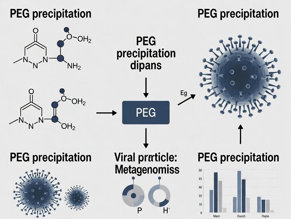
PEG Precipitation for Viral Metagenomics: A Complete Guide for Researchers in Pathogen Discovery and Drug Development
This article provides a comprehensive guide to Polyethylene Glycol (PEG) precipitation for enriching viral particles in metagenomic studies.
PEG Precipitation for Viral Metagenomics: A Complete Guide for Researchers in Pathogen Discovery and Drug Development
Abstract
This article provides a comprehensive guide to Polyethylene Glycol (PEG) precipitation for enriching viral particles in metagenomic studies. Aimed at researchers and biopharma professionals, it covers the foundational principles of viral particle separation, detailed step-by-step protocols for diverse sample types, troubleshooting for common issues like host contamination and low yield, and critical validation strategies comparing PEG to ultracentrifugation and filtration. The goal is to equip scientists with the knowledge to optimize this cost-effective method for uncovering novel viruses, tracking epidemics, and informing therapeutic development.
Viral Metagenomics and PEG Precipitation: Core Principles and Research Applications
Application Notes: Viromics in Pathogen Discovery
Viral metagenomics, or viromics, bypasses traditional culture and PCR-based methods to enable the unbiased discovery of viruses in any sample. Within the thesis framework focusing on PEG precipitation for viral particle enrichment, this approach is critical for surveying viral diversity in clinical and environmental matrices, directly linking to outbreak investigation and novel therapeutic target identification.
Key Applications:
- Outbreak Investigation: Identification of novel viral etiologic agents in unexplained disease clusters.
- Biothreat Surveillance: Monitoring environmental samples for known and emerging viral pathogens.
- Drug & Vaccine Target Discovery: Characterization of viral genomes informs the design of antivirals, monoclonal antibodies, and vaccines.
- Microbiome Research: Defining the viral component (virome) of human and environmental microbiomes and its impact on health and disease.
Quantitative Performance Metrics of Viromics Workflows:
Table 1: Comparison of Viral Nucleic Acid Preparation Methods
| Method | Input Volume | Avg. Viral Recovery Yield* | Host DNA Depletion Efficiency | Suitability for RNA Viruses |
|---|---|---|---|---|
| PEG Precipitation | 50-500 mL | ~40-70% | Moderate | Yes (with carrier) |
| Ultracentrifugation | 10-100 mL | ~60-90% | High | Yes |
| Filtration + Nuclease | 1-50 mL | ~20-50% | Very High | Yes |
| Commercial Kits | 0.1-5 mL | ~10-30% | Variable | Kit-dependent |
*Estimated recovery of spiked control virus particles (e.g., phage ΦX174).
Table 2: Next-Generation Sequencing Platform Suitability for Viromics
| Platform (Example) | Read Length | Output/Flow Cell | Key Advantage for Viromics | Cost per Sample (Approx.) |
|---|---|---|---|---|
| Illumina (NextSeq 2000) | 2x150 bp | 100-400 Gb | High accuracy for strain typing | $800-$1,500 |
| Oxford Nanopore (MinION) | Varies (long) | 10-50 Gb | Real-time, long reads for assembly | $500-$1,000 |
| PacBio (HiFi) | 10-25 kb | 15-50 Gb | Highly accurate long reads | $2,000-$3,500 |
Detailed Protocol: Viral Metagenomics via PEG Precipitation
This protocol details viral particle enrichment using polyethylene glycol (PEG) precipitation, followed by nucleic acid extraction, library preparation, and sequencing.
Part A: PEG Precipitation of Viral Particles from Clarified Sample
- Objective: To concentrate viral particles from a large-volume liquid sample.
- Reagents & Materials: See The Scientist's Toolkit below.
- Procedure:
- Clarification: Centrifuge sample (e.g., sewage, serum) at 10,000 x g for 30 min at 4°C. Filter supernatant through a 0.45 μm PES filter.
- PEG Precipitation: To the filtrate, add NaCl to a final concentration of 0.9 M and PEG 8000 to 10% (w/v). Stir gently at 4°C for 12-16 hours.
- Pellet Recovery: Centrifuge at 10,000 x g for 90 min at 4°C. Discard supernatant.
- Resuspension: Resuspend the invisible pellet in 1/100th of the original volume using 1x PBS or SM Buffer. Incubate on ice for 2 hours with occasional pipetting.
- Clean-up: Centrifuge at 12,000 x g for 10 min to remove debris. Transfer supernatant (containing concentrated virions) to a clean tube.
Part B: Viral Nucleic Acid Extraction & Library Construction
- Objective: To extract total nucleic acid and prepare an NGS library.
- Procedure:
- Digestion: Treat 200 μL of concentrated virus with 5 U of Baseline-ZERO DNase and 2 U of RNase ONE at 37°C for 1 hour to degrade free nucleic acids.
- Inactivation: Add 5 μL of 0.5 M EDTA and heat at 70°C for 10 minutes.
- Extraction: Use a commercial nucleic acid extraction kit with proteinase K treatment. Elute in 30 μL nuclease-free water.
- Amplification: Perform random-primed, multiple displacement amplification (MDA) for DNA viromes or generate cDNA for RNA viromes using random hexamers.
- Library Prep: Fragment amplified DNA (e.g., via ultrasonication), then use a standard NGS library preparation kit (end-repair, A-tailing, adapter ligation). Perform size selection (e.g., 300-700 bp).
- Sequencing: Quantify library by qPCR and sequence on an appropriate platform (e.g., Illumina, 2x150 bp).
Visualizations
Title: PEG Precipitation Viromics Workflow
Title: Bioinformatics Pathogen Discovery Pipeline
The Scientist's Toolkit
Table 3: Key Research Reagent Solutions for PEG Viromics
| Item | Function in Protocol | Example/Note |
|---|---|---|
| PEG 8000 | Precipitates viral particles via volume exclusion and crowding. | Use molecular biology grade. Concentration is sample-dependent (8-16%). |
| DNase I (Baseline-ZERO) | Degrades unprotected host and free DNA post-concentration. | Critical for reducing background. Must be Mg2+/Ca2+ dependent. |
| RNase A/RNase ONE | Degrades unprotected RNA to enrich for encapsulated viral RNA. | Used for RNA viromes or total nucleic acid prep. |
| Proteinase K | Digests viral capsid proteins to release nucleic acids. | Use with SDS in lysis buffer. |
| Random Hexamer Primers | For unbiased reverse transcription of RNA viromes. | Critical for discovering novel viruses with divergent sequences. |
| Phi29 Polymerase | Enzyme for Multiple Displacement Amplification (MDA) of DNA. | Can cause amplification bias; include no-template controls. |
| Size Selection Beads | For NGS library clean-up and selection of optimal insert size. | e.g., SPRIselect beads. Ratios determine size cutoff. |
| Internal Control Virus | Spiked-in process control for yield quantification. | e.g., phage PhiX174 (DNA) or murine norovirus (RNA). |
Within the context of a broader thesis on viral metagenomics, Polyethylene Glycol (PEG) precipitation remains a cornerstone technique for the concentration and purification of viral particles from complex biological and environmental matrices. This method is critical for enabling downstream genomic analyses, including metagenomic sequencing, by increasing viral nucleic acid yield while reducing contaminating host and environmental nucleic acids.
The principle is based on the exclusion of PEG molecules from the solvation shell of viral particles, effectively reducing their solubility and causing aggregation and precipitation. This process is influenced by PEG molecular weight, final concentration, ionic strength, pH, temperature, and incubation time.
Key Advantages for Metagenomics:
- Cost-Effectiveness: Requires minimal specialized equipment.
- Scalability: Easily adapted to process large sample volumes (e.g., seawater, wastewater).
- Broad Specificity: Recovers diverse viral morphologies (enveloped and non-enveloped).
- Compatibility: Precipitated material is suitable for direct nucleic acid extraction or further purification.
Quantitative Performance Data: The following table summarizes key performance metrics from recent studies applying PEG precipitation for viral concentration in metagenomic workflows.
Table 1: Quantitative Performance of PEG Precipitation in Viral Metagenomics Studies
| Sample Matrix | PEG Type & Concentration | Incubation Time/Temp | Avg. Viral Recovery Efficiency* | Key Metric for Metagenomics |
|---|---|---|---|---|
| Wastewater | PEG 8000, 10% w/v | 24h, 4°C | ~40-60% (viral particles) | 5-10x increase in viral read count post-sequencing |
| Sea Water | PEG 6000, 10% w/v + NaCl | Overnight, 4°C | ~30-50% (phage particles) | 3-8x enrichment of viral vs. bacterial sequences |
| Human Serum | PEG 8000, 8.5% w/v | 2h, Room Temp | ~70-80% (enveloped viruses) | Critical for reducing human background DNA (<50% of total reads) |
| Stool Suspension | PEG 6000, 15% w/v | 2h, 4°C | ~60-75% (enteric viruses) | Enabled detection of low-abundance viral species (<0.1% relative abundance) |
| Cell Culture Supernatant | PEG 8000, 10% w/v | Overnight, 4°C | >80% (retroviruses) | Yield sufficient cDNA for library prep from <1 mL sample |
*Recovery efficiency is typically measured via qPCR (specific viruses), plaque assay, or quantitative metagenomics (viral read proportion).
Detailed Experimental Protocols
Protocol 2.1: Standard PEG Precipitation for Viral Metagenomics from Liquid Matrices (e.g., Wastewater, Seawater)
Objective: To concentrate total viral particles from large-volume liquid samples for subsequent viral nucleic acid extraction and metagenomic sequencing.
Materials: See "The Scientist's Toolkit" section.
Procedure:
- Clarification & Pre-filtration: Centrifuge raw sample at 10,000 × g for 30 minutes at 4°C to remove large debris. Filter supernatant sequentially through 5.0 μm and 0.45 μm (or 0.22 μm) pore-size filters. For environmental samples, a 0.22 μm filter may excessively reduce viral titre.
- Nuclease Treatment (Optional but Recommended): Add MgCl₂ to a final concentration of 1-2 mM and DNase I/RNase A to digest free nucleic acids. Incubate for 30-60 minutes at 37°C to specifically enrich for encapsulated viral genomes.
- PEG/NaCl Solution Addition: Add solid NaCl to the filtered sample to a final concentration of 0.5 M (e.g., 29.22 g per liter). Mix until dissolved. Slowly add an appropriate volume of a sterile, autoclaved 50% (w/v) PEG 8000 stock solution to achieve a final concentration of 10% (w/v). Mix thoroughly by gentle inversion.
- Precipitation: Incubate the mixture at 4°C for a minimum of 16 hours (overnight), with gentle agitation if possible.
- Pellet Formation: Centrifuge the mixture at 10,000 × g for 90 minutes at 4°C. Carefully decant the supernatant. A translucent or invisible pellet should be present at the bottom of the tube.
- Pellet Resuspension: Drain the tube completely on clean absorbent paper for 5-10 minutes. Resuspend the viral pellet thoroughly in a suitable, smaller-volume buffer (e.g., SM Buffer, PBS, nuclease-free water) for downstream processing. Volume reduction should be 100-1000x relative to the starting sample.
- Post-PEG Clean-up (Optional): Perform a chloroform:butanol (1:1) treatment to remove residual PEG, which can inhibit downstream enzymatic reactions. Mix resuspended pellet with equal volume of chloroform:butanol, vortex, centrifuge at 10,000 × g for 10 min, and recover the aqueous upper phase containing viruses.
- Proceed to viral nucleic acid extraction and library construction for metagenomic sequencing.
Workflow Diagram:
Diagram Title: Viral Metagenomics PEG Precipitation Workflow
Protocol 2.2: PEG Precipitation for Viral Enrichment from Stool Samples
Objective: To isolate and concentrate viral particles from fecal material for gut virome studies.
Procedure:
- Homogenization: Suspend ~1-5 g of stool in 10-15 mL of SM Buffer or PBS. Vortex vigorously.
- Clarification: Centrifuge at 12,000 × g for 20 minutes at 4°C. Filter supernatant through a 0.45 μm filter.
- PEG Precipitation: To the filtrate, add solid PEG 6000 to a final concentration of 15% (w/v) and NaCl to 0.5 M. Mix and incubate on ice or at 4°C for 2 hours.
- Pellet & Resuspend: Centrifuge at 12,000 × g for 30 minutes at 4°C. Discard supernatant. Resuspend pellet in 1-2 mL of PBS.
- Chloroform Treatment: Add an equal volume of chloroform, vortex for 1 minute, and centrifuge at 10,000 × g for 10 minutes. Recover the aqueous phase.
- Filtration: Pass the aqueous phase through a 0.22 μm filter to remove any remaining bacterial-sized contaminants.
- Proceed to nucleic acid extraction, often incorporating a random amplification step due to low nucleic acid mass.
The Scientist's Toolkit: Key Research Reagent Solutions
Table 2: Essential Materials for PEG Precipitation in Viral Metagenomics
| Reagent/Material | Typical Specification/Concentration | Primary Function in Protocol |
|---|---|---|
| Polyethylene Glycol (PEG) | PEG 6000 or PEG 8000, molecular biology grade. Stock: 50% (w/v) in sterile H₂O. | The precipitating agent. Excludes water, reduces viral solubility. PEG 8000 offers faster precipitation. |
| Sodium Chloride (NaCl) | Molecular biology grade, 5M stock or solid. | Increases ionic strength, enhances virus-PEG interaction, and improves recovery efficiency. |
| Nuclease Enzymes | DNase I (RNase-free) and RNase A, ≥1 U/μL. | Degrades unprotected host and environmental nucleic acids, enriching for encapsulated viral genomes. |
| SM Buffer | 50 mM Tris-HCl (pH 7.5), 100 mM NaCl, 10 mM MgSO₄, 0.01% gelatin. | A common suspension and storage buffer for viral particles, maintaining stability. |
| Sterile Filtration Units | 0.45 μm and 0.22 μm pore size, low protein binding (PES membrane). | Remove bacteria and large debris (0.45μm) or perform final sterilization (0.22μm). |
| High-Speed Centrifuge & Rotors | Capable of ≥10,000 × g with large-volume buckets (e.g., 250mL bottles). | Pellet the PEG-precipitated viral aggregates from large sample volumes. |
| Chloroform & Butanol | Molecular biology grade. | Organic solvents used to remove residual PEG and lipids, clarifying the final viral suspension. |
| Resuspension Buffer | PBS (pH 7.4), SM Buffer, or Nuclease-Free Water. | A small-volume medium to resuspend the concentrated viral pellet for downstream steps. |
Mechanism and Pathway Diagram: The Molecular Logic of PEG Precipitation
The following diagram illustrates the sequential molecular interactions leading to viral precipitation.
Diagram Title: Molecular Mechanism of Viral PEG Precipitation
Application Notes: PEG Precipitation in Viral Metagenomics
Within the broader thesis on advancing viral metagenomics for pathogen discovery and therapeutic development, Polyethylene Glycol (PEG) precipitation remains a cornerstone method for viral particle concentration. Its utility stems from three synergistic advantages that address critical challenges in pre-sequencing sample preparation.
1. Cost-Effectiveness: Compared to ultracentrifugation or commercial spin-column kits, PEG precipitation offers a dramatic reduction in per-sample cost. It requires only basic laboratory centrifuges and inexpensive, shelf-stable reagents, enabling high-throughput processing within constrained budgets. This democratizes access to large-scale virome studies across diverse settings, from academic labs to biopharma R&D.
2. Scalability: The protocol is inherently adaptable to varying input volumes (from milliliters of serum to liters of environmental water) without significant protocol redesign. This linear scalability is crucial for applications ranging from clinical samples to industrial bioreactor monitoring.
3. Broad Viral Recovery: PEG acts as a size-dependent precipitant, non-specifically concentrating a wide spectrum of viral particles (typically >20-30 nm) while excluding most soluble proteins and subcellular debris. This "catch-all" characteristic is vital for unbiased virome profiling, essential for discovering novel viruses and understanding viral community dynamics in drug development contexts (e.g., characterizing viral contaminants in biologics manufacturing).
Quantitative Performance Data Summary
Table 1: Comparative Analysis of Viral Concentration Methods for Metagenomics
| Method | Approx. Cost/Sample (USD) | Processing Time | Typical Viral Recovery Efficiency* | Key Limitation |
|---|---|---|---|---|
| PEG Precipitation | 2 - 10 | 4 - 18 hours (incubation) | 30-70% (varies by matrix/virus) | Co-precipitation of humic substances (environmental samples) |
| Ultracentrifugation | 50 - 200 | 2 - 5 hours (active) | 50-90% | Equipment cost, limited throughput |
| Ultrafiltration | 20 - 100 | 1 - 2 hours | 20-60% | Membrane clogging, shear stress on virions |
| Commercial Kits | 50 - 150 | 1 - 3 hours | 40-80% | High cost, often optimized for specific sample types |
*Recovery is highly dependent on virus type, size, and sample matrix (e.g., stool, seawater, serum). PEG data often reflects a robust average across diverse virion structures.
Detailed Experimental Protocols
Protocol 1: Standard PEG 8000 Precipitation for Diverse Liquid Samples
This protocol is optimized for virome enrichment from stool supernatants, cell culture media, or treated wastewater.
Materials & Reagents:
- Sample (clarified by low-speed centrifugation: 8,000 x g, 10 min)
- Polyethylene Glycol 8000 (PEG 8000)
- Sodium Chloride (NaCl)
- 0.5M EDTA, pH 8.0
- Phosphate-Buffered Saline (PBS), sterile
- Nuclease solution (e.g., Benzonase, Thermolabile Nuclease) to degrade free nucleic acids
Procedure:
- Clarification & Nuclease Treatment: To 10 mL of pre-clarified sample, add 50 µL of 0.5M EDTA and 10 µL of Benzonase (or equivalent). Incubate at 37°C for 30 minutes to digest unprotected nucleic acids.
- PEG/NaCl Solution Preparation: Prepare a 50% (w/v) PEG 8000 and 5M NaCl stock solution in PBS. Filter sterilize (0.22 µm).
- Precipitation: Add the PEG/NaCl stock to the sample to achieve a final concentration of 10% (w/v) PEG and 0.5M NaCl. Mix thoroughly by inversion.
- Incubation: Incubate the mixture at 4°C for a minimum of 12 hours (overnight is optimal).
- Pellet Virions: Centrifuge at 10,000 x g for 60 minutes at 4°C. Carefully decant the supernatant.
- Viral Pellet Resuspension: Resuspend the often invisible pellet in 200 µL of PBS (or suitable buffer for downstream nucleic acid extraction). Let stand on ice for 1-2 hours with occasional gentle pipetting.
- Optional Clean-up: For downstream sequencing, purify the viral nucleic acid using a column-based kit (e.g., DNeasy/RNeasy PowerSoil) or proceed with phenol-chloroform extraction.
Protocol 2: Sequential Filtration-PEG Precipitation for Large-Volume Environmental Samples
Designed for scalable processing of seawater or freshwater for aquatic virome studies.
Procedure:
- Pre-filtration: Sequentially filter water (1-10 L) through a 0.8 µm polycarbonate membrane followed by a 0.22 µm membrane to remove bacteria and large particulates.
- Concentration via Tangential Flow Filtration (TFF): Concentrate the 0.22 µm filtrate to ~100 mL using a 100 kDa TFF system.
- PEG Precipitation: Transfer concentrate to a sterile beaker. Add solid PEG 8000 to 10% (w/v) and NaCl to 0.5M final concentration. Stir gently on a magnetic stirrer at 4°C for 2 hours.
- Collection: Transfer to centrifuge bottles. Pellet at 10,000 x g for 90 minutes at 4°C.
- Resuspension: Resuspend pooled pellets in 2 mL PBS. Centrifuge at 12,000 x g for 5 min to remove residual debris. Transfer supernatant (enriched virions) to a clean tube.
Visualizations
Title: PEG Precipitation Workflow for Viral Metagenomics
Title: Core Advantages and Their Research Impact
The Scientist's Toolkit: Key Research Reagent Solutions
Table 2: Essential Materials for PEG-Based Viral Metagenomics
| Item | Function in Protocol | Key Considerations |
|---|---|---|
| Polyethylene Glycol 8000 (PEG 8000) | Primary precipitating agent; excludes water molecules, forcing viral particle aggregation. | Molecular weight (6000-8000) is standard. Use high-purity grade to avoid contaminants. |
| Benzonase Nuclease | Degrades free DNA/RNA in sample post-clarification, drastically reducing host/non-viral background in sequencing libraries. | Requires Mg²⁺. Thermolabile nuclease is an alternative for heat-inactivation. |
| 0.22 µm PES Membrane Filters | Sterile filtration of PEG stocks and initial removal of bacteria/large particles from samples. | Low protein binding minimizes viral loss during pre-filtration. |
| Phosphate-Buffered Saline (PBS), pH 7.4 | Resuspension buffer for viral pellets; isotonic and compatible with downstream enzymatic steps. | Must be nuclease-free for nucleic acid preservation. |
| DNeasy PowerSoil Pro Kit (or equivalent) | Post-PEG nucleic acid extraction; designed to remove potent PCR inhibitors (humics, salts) common in concentrated samples. | Critical for environmental viromes. Alternative: phenol-chloroform-isoamyl alcohol. |
| DNase/RNase Treatment Reagents (On-column) | Post-extraction treatment to remove any residual external nucleic acids clinging to capsids, ensuring sequenced nucleic acid is encapsidated. | Confirms viral origin of sequenced material. |
| Quantitative PCR (qPCR) Assays for Specific Viruses (e.g., Phage ΦX174) | Spike-in control to quantitatively monitor and optimize viral recovery efficiency through the PEG and extraction process. | Use a non-native virus to the sample as an internal process control. |
Application Notes
Within the framework of PEG precipitation viral metagenomics research, the concentration and unbiased sequencing of viral nucleic acids enable three pivotal applications. Polyethylene glycol (PEG) precipitation provides a cost-effective, scalable method for concentrating diverse viral particles from complex samples prior to nucleic acid extraction and next-generation sequencing (NGS). This approach underpins the following domains:
1. Clinical Diagnostics: Viral metagenomics allows for the agnostic detection of known, variant, and novel viruses in patient samples, overcoming limitations of targeted assays (e.g., PCR, multiplex panels). It is crucial for diagnosing unexplained encephalitis, meningitis, respiratory infections, and febrile illnesses of unknown origin. A 2023 review indicated that clinical metagenomics increased diagnostic yield by ~30-40% in such complex cases compared to conventional testing.
2. Environmental Surveillance: Monitoring wastewater, air, and surfaces for viral pathogens provides early warning for outbreaks and tracks community transmission dynamics. PEG precipitation is ideal for processing large-volume environmental samples. During the COVID-19 pandemic, wastewater surveillance reliably detected SARS-CoV-2 variants 1-2 weeks before clinical case surges, with studies reporting >80% correlation between wastewater viral load trends and hospital admission rates.
3. Novel Virus Identification: This is the discovery frontier. By sequencing all viral nucleic acids in a sample, researchers can identify novel viruses and characterize viral diversity (the virome). This is foundational for pandemic preparedness. Since 2020, initiatives like the Global Virome Project have utilized such methods, leading to a database of over 140,000 newly identified viral sequences from global animal and environmental samples.
Table 1: Quantitative Data Summary for Primary Applications
| Application | Typical Sample Input | Key Performance Metric | Reported Value/Outcome | Common Sequencing Depth |
|---|---|---|---|---|
| Clinical Diagnostics | 200 µL - 1 mL (CSF, plasma, swab) | Diagnostic Yield Increase | ~30-40% over standard care | 5-20 million reads |
| Environmental Surveillance | 10 mL - 1 L (wastewater) | Lead Time for Outbreak Detection | 1-2 weeks ahead of clinical data | 1-5 million reads |
| Novel Virus Identification | 1 mL - 100 mL (tissue, water) | Novel Sequences per Project | >1,000 novel viral contigs per large study | 20-100+ million reads |
Protocols
Protocol 1: PEG Precipitation of Viral Particles from Diverse Sample Types This foundational protocol concentrates viral particles from clinical or environmental matrices.
Materials:
- Sample (e.g., serum, CSF, wastewater filtrate, tissue homogenate supernatant)
- Polyethylene Glycol 8000 (PEG 8000)
- Sodium Chloride (NaCl)
- Nuclease-Free Water
- Centrifuge and fixed-angle rotors
- Phosphate-Buffered Saline (PBS), pH 7.4
Procedure:
- Clarification & Filtration: Centrifuge sample at 10,000 x g for 30 min at 4°C to remove debris. Filter supernatant through a 0.45 µm then a 0.22 µm pore-size filter.
- PEG/NaCl Solution: Prepare a stock solution of 50% (w/v) PEG 8000 and 5 M NaCl in nuclease-free water.
- Precipitation: To the filtered sample, add PEG/NaCl stock to final concentrations of 10% (w/v) PEG and 0.5 M NaCl. Mix thoroughly by inversion.
- Incubation: Incubate the mixture at 4°C for a minimum of 12 hours (or overnight) with gentle agitation.
- Pellet Virus: Centrifuge at 10,000 x g for 90 min at 4°C to pellet viral particles.
- Resuspension: Carefully discard supernatant. Resuspend the invisible pellet in a small volume (e.g., 100-200 µL) of PBS or appropriate buffer for downstream nucleic acid extraction.
- Storage: Process immediately for nucleic acid extraction or store at -80°C.
Protocol 2: Viral Metagenomic Library Preparation from PEG-Precipitated Material Follows nucleic acid extraction (using a kit with DNase/RNase steps to remove host/free nucleic acids).
Materials:
- Extracted viral nucleic acids (DNA and/or RNA)
- Reverse Transcriptase (for RNA viruses)
- Random Hexamer Primers
- DNA Polymerase (with strand-displacement activity)
- Double-Stranded DNA (dsDNA) Fragmentation & Library Prep Kit (e.g., tagmentation-based)
- PCR Purification Kit
- Qubit Fluorometer and TapeStation/Bioanalyzer
Procedure:
- Complementary DNA (cDNA) Synthesis: For RNA viruses, perform reverse transcription using random hexamers to generate cDNA.
- Whole Genome Amplification (Optional but common): Use multiple displacement amplification (MDA) with phi29 polymerase to amplify scant viral DNA/cDNA. Note: This can introduce bias.
- Library Construction: Fragment amplified dsDNA to ~350 bp using mechanical or enzymatic methods. Perform end-repair, A-tailing, and adapter ligation per kit instructions. Alternatively, use a tagmentation-based kit.
- Library Amplification & Clean-up: Amplify the adapter-ligated DNA with 8-12 PCR cycles. Purify the final library using magnetic beads.
- Quality Control: Quantify library concentration (Qubit) and assess size distribution (TapeStation). Pool libraries for sequencing (Illumina NovaSeq/NextSeq recommended for depth).
Visualizations
Diagram 1: Viral Metagenomics Workflow from Sample to Data
Diagram 2: Comparative Application Pathways
The Scientist's Toolkit
Table 2: Essential Research Reagent Solutions
| Reagent/Material | Function in PEG-precipitation Metagenomics | Example Product/Note |
|---|---|---|
| PEG 8000 | Precipitates viral particles by volume exclusion and reducing solubility; core of the concentration step. | Molecular biology grade. Prepare fresh 50% stock. |
| 0.22 µm Sterile Filter | Removes bacteria and large particulates, clarifying sample for viral particle collection. | PES membrane filters are recommended. |
| Nucleic Acid Extraction Kit (with DNase/RNase) | Isolates viral nucleic acids while degrading contaminating free host/bacterial NA. | Kits specifically designed for viral NA (e.g., QIAamp Viral Mini, MagMAX Viral/Pathogen). |
| Random Hexamer Primers | Initiates unbiased reverse transcription of RNA viral genomes and fragmented DNA. | Essential for sequence-agnostic amplification. |
| Phi29 DNA Polymerase | Used in Multiple Displacement Amplification (MDA) to amplify minute amounts of viral DNA/cDNA; can introduce sequence bias. | RepliPhi or GenomiPhi kits. Use with caution and include controls. |
| UltraPure BSA (10 mg/mL) | Added during amplification to stabilize polymerases and sequester inhibitors from complex samples. | Improves yield from inhibition-prone samples (e.g., wastewater). |
| Dual-Indexed Adapter Kit | Allows multiplexing of hundreds of samples in a single NGS run, crucial for surveillance studies. | Illumina TruSeq, Nextera XT, or IDT for Illumina kits. |
| Bioinformatic Pipelines | For raw read processing, host read subtraction, de novo assembly, and viral classification. | FastQC, Trimmomatic, Bowtie2, SPAdes, DIAMOND, Kraken2. |
The efficacy of viral metagenomics, particularly when employing polyethylene glycol (PEG) precipitation for viral particle concentration, is fundamentally dependent on pre-analytical variables. This protocol details the critical considerations for sample type selection and storage conditions to preserve viral nucleic acid integrity and ensure representative sequencing libraries. The context is a broader thesis investigating vironne dynamics across human and environmental matrices using PEG-based enrichment.
Comparative Analysis of Sample Types
Table 1: Characteristics and Pre-Analytical Demands by Sample Type
| Sample Type | Typical Viral Load | Major Inhibitors | Recommended Minimum Volume for PEG Precipitation | Key Stability Concerns |
|---|---|---|---|---|
| Stool | Very High (10^8-10^11 particles/g) | Polysaccharides, bile salts, bacteria, dietary PCR inhibitors. | 0.5 - 1 g | Rapid degradation by nucleases; bacterial overgrowth. |
| Serum/Plasma | Low to Moderate (Variable) | Hemoglobin (in hemolyzed samples), immunoglobulin G, lipids. | 0.5 - 1 mL | Relatively stable for enveloped viruses; freeze-thaw cycles critical. |
| Cerebrospinal Fluid (CSF) | Very Low | Low protein content reduces inhibitors. | 2 - 3 mL | Volume is limiting; extreme sensitivity to contamination. |
| Water (Environmental) | Extremely Low | Humic acids, heavy metals, colloidal particles. | 50 - 1000 mL (pre-concentrated) | Environmental degradation; microbial activity. |
Table 2: Recommended Storage Conditions Prior to PEG Processing
| Sample Type | Optimal Short-Term (<24h) | Optimal Long-Term | Maximum Avoidable Freeze-Thaw Cycles | Stabilization Reagents (if not immediately processing) |
|---|---|---|---|---|
| Stool | 4°C | -80°C (aliquoted) | 1 | RNA/DNA Shield, Stool Transport, and Recovery Buffer. |
| Serum/Plasma | 4°C | -80°C (aliquoted) | 2 | None required if frozen promptly. |
| CSF | 4°C | -80°C (aliquoted) | 1 | None recommended due to low volume; process immediately. |
| Water | 4°C, in the dark | -80°C (after concentration) | 2 | Sulfite-EDTA for RNA viruses; immediate filtration/concentration advised. |
Detailed Protocols for Sample Handling & Storage
Protocol 1: Stool Sample Collection and Pre-PEG Processing
Objective: To collect and stabilize stool samples for viral particle metagenomics. Materials: Sterile collection container with spoon, RNase-free tubes, DNA/RNA Shield or similar, -80°C freezer. Procedure:
- Collect 1-2 g of stool using the spoon integrated into the container lid.
- Immediately suspend the sample in 5-10 mL of commercial DNA/RNA stabilization buffer (e.g., DNA/RNA Shield) at a 1:5 (w/v) ratio. Vortex thoroughly for 2 minutes.
- Aliquot the homogenate into 1-2 mL RNase-free cryovials to avoid repeated freeze-thaw.
- Store at 4°C if processing within 24 hours. For long-term storage, flash-freeze in liquid nitrogen or a -80°C ethanol bath and transfer to a -80°C freezer.
- Prior to PEG precipitation, thaw on ice and clarify by low-speed centrifugation (8,000 x g, 10 min, 4°C). Use supernatant for downstream PEG protocol.
Protocol 2: Serum/Plasma Preparation for Viral Enrichment
Objective: To obtain cell-free serum/plasma suitable for viral concentration. Materials: Blood collection tubes (SST for serum, EDTA/K2EDTA for plasma), centrifuge, RNase-free pipettes and tubes. Procedure:
- For Serum: Allow blood to clot in Serum Separator Tube (SST) for 30 min at room temperature. Centrifuge at 1,500-2,000 x g for 10 min at 4°C. Carefully aspirate the serum layer.
- For Plasma: Centrifuge whole blood in EDTA tube at 1,500-2,000 x g for 10 min at 4°C within 2 hours of collection. Carefully aspirate the plasma layer, avoiding the buffy coat.
- Aliquot 0.5-1 mL of serum/plasma into cryovials. If not processing immediately, flash-freeze and store at -80°C.
- Avoid repeated thawing. Thaw required aliquots on ice immediately before PEG precipitation.
Protocol 3: CSF Handling for Low-Biomass Virome Analysis
Objective: To maximize viral recovery from low-volume CSF samples. Materials: Sterile lumbar puncture kit, low-protein-binding microtubes, clinical centrifuge. Procedure:
- Collect CSF via standard aseptic lumbar puncture. A minimum of 2 mL is recommended for viral metagenomics.
- Centrifuge at 800 x g for 10 min at 4°C to remove cells and debris.
- Aliquot the supernatant into low-protein-binding microtubes (e.g., 0.5 mL per tube). DO NOT ADD STABILIZATION REAGENTS unless empirically validated for downstream PEG/NGS, as they may interfere.
- Flash-freeze aliquots immediately in liquid nitrogen or a -80°C bath. Store at -80°C. Process one aliquot at a time.
Protocol 4: Environmental Water Concentration and Stabilization
Objective: To concentrate viral particles from large water volumes for laboratory processing. Materials: 0.22 µm tangential flow filtration (TFF) system or positive pressure filter, MgCl₂ (1 M), glycine (0.5 M, pH 9.5), -80°C freezer. Procedure:
- Pre-filter water through a 5 µm or 0.45 µm filter to remove large particulates.
- Concentrate viruses from 50-1000 L to ~100 mL using a 0.22 µm TFF system per manufacturer's instructions.
- Further concentrate to a final volume of 5-10 mL using centrifugal ultrafiltration devices (100 kDa MWCO) or secondary TFF.
- Adjust the concentrate to a final concentration of 10 mM MgCl₂ and 50 mM glycine (pH 9.5) to enhance subsequent PEG precipitation efficiency.
- Aliquot, flash-freeze, and store at -80°C.
Visual Workflows
Title: Pre-PEG Sample Handling Decision Workflow
Title: Impact of Pre-Analytical Errors on PEG Workflow
The Scientist's Toolkit: Research Reagent Solutions
Table 3: Essential Materials for Pre-Analytical Sample Preservation
| Item | Function in Pre-PEG Phase | Example Product/Brand |
|---|---|---|
| DNA/RNA Shield | Inactivates nucleases and stabilizes nucleic acids in complex samples like stool. | Zymo Research DNA/RNA Shield |
| RNase-free Tubes & Tips | Prevents introduction of environmental RNases that degrade viral RNA. | Ambion RNase-free, Axygen Maxymum Recovery |
| Low-Protein-Binding Microtubes | Minimizes adsorption of low-abundance viral particles to tube walls, critical for CSF. | Eppendorf LoBind, Thermo Scientific Maxymum Recovery |
| Tangential Flow Filtration (TFF) System | Gentle concentration of viruses from large-volume environmental water samples. | Merck Pellicon, Spectrum Labs KrosFlo |
| Cryogenic Vials & Storage | Reliable -80°C storage with minimal sample degradation or tube cracking. | Corning, Thermo Scientific Nunc |
| MgCl₂ & Glycine Buffer | Pre-treatment for water samples to improve viral recovery during PEG precipitation. | Sigma-Aldrich Molecular Biology Grade |
| Serum Separator Tubes (SST) | Provides clean, cell-free serum for plasma vironne analysis. | BD Vacutainer SST |
| Centrifugal Ultrafilters (100 kDa) | Final concentration step for water and other liquid samples prior to PEG. | Amicon Ultra-15, Pall Macrosep Advance |
Step-by-Step Protocol: Optimizing PEG Precipitation for Viral Metagenomics Sequencing
Application Notes: PEG Precipitation for Viral Metagenomics
In viral metagenomics, the effective concentration of viral particles from diverse environmental or clinical samples is a critical first step. Polyethylene glycol (PEG) precipitation remains a cornerstone method due to its simplicity, scalability, and effectiveness in recovering a broad range of viral sizes and types. The selection between PEG 6000 and PEG 8000, along with the choice and concentration of salts, directly influences viral yield, purity, and subsequent sequencing success. This protocol is contextualized within a thesis exploring the optimization of viral recovery for downstream metagenomic sequencing and biotherapeutic characterization.
PEG 6000 vs. PEG 8000: Key Quantitative Comparisons
Table 1: Comparative Properties of PEG 6000 and PEG 8000 for Viral Precipitation
| Parameter | PEG 6000 | PEG 8000 | Implications for Viral Metagenomics |
|---|---|---|---|
| Average Molecular Weight | 6,000 Da | 8,000 Da | PEG 8000 has longer polymer chains. |
| Typical Final Concentration | 8-10% (w/v) | 6-10% (w/v) | PEG 8000 often requires a slightly lower % for equivalent precipitation efficiency. |
| Precipitation Rate | Faster | Slower | PEG 6000 may precipitate particles more quickly, potentially useful for labile viruses. |
| Stringency & Size Selectivity | Lower | Higher | PEG 8000 may preferentially precipitate larger viral particles; PEG 6000 offers broader size recovery. |
| Co-precipitation of Contaminants | Moderately High | High | PEG 8000 may co-precipitate more humic acids (environmental samples) or host proteins, affecting purity. |
| Common Salt Used | NaCl | NaCl | Both typically used with 0.3-0.5 M final NaCl concentration. |
| Optimal Incubation Time | 1-4 hours (RT or 4°C) | Overnight (4°C) | Overnight incubation with PEG 8000 at 4°C maximizes recovery. |
| Common Resuspension Volume | 100-500 µL (SM Buffer or nuclease-free water) | 100-500 µL (SM Buffer or nuclease-free water) | Dependent on starting sample volume. |
Table 2: Standard Salt Additives in PEG Precipitation
| Salt | Typical Final Concentration | Primary Function |
|---|---|---|
| Sodium Chloride (NaCl) | 0.3 - 0.5 M | Neutralizes surface charge on viral particles, reducing repulsion and enhancing PEG-driven exclusion. |
| Magnesium Chloride (MgCl₂) | 10 - 25 mM | Divalent cations can improve precipitation efficiency for some viral families (e.g., some bacteriophages). |
| Potassium Chloride (KCl) | 0.3 - 0.5 M | Alternative to NaCl, can be used to minimize specific contaminant precipitation. |
Experimental Protocols
Protocol 1: Standard PEG Precipitation for Viral Concentration (Liquid Samples)
Objective: To concentrate viral particles from a liquid supernatant (e.g., cell culture supernatant, environmental water, fecal supernatant) for metagenomic RNA/DNA extraction.
Materials:
- Sample supernatant (clarified by centrifugation at 10,000 x g for 30 min)
- PEG 6000 or PEG 8000 (powder, molecular biology grade)
- 5 M NaCl (filter-sterilized)
- 1 M MgCl₂ (optional, filter-sterilized)
- SM Buffer (50 mM Tris-HCl pH 7.5, 100 mM NaCl, 10 mM MgSO₄)
- Nuclease-free water
- Centrifuge and fixed-angle or swinging-bucket rotor
- Refrigerator (4°C) and/or room temperature incubator
Methodology:
- Sample Clarification: Centrifuge the raw sample at 10,000 x g for 30 minutes at 4°C to remove cells and large debris. Transfer the supernatant to a clean tube.
- PEG/NaCl Addition: To the clarified supernatant, add 1/3 volume of a sterile-filtered PEG/NaCl stock solution. For example, for a 30 mL sample, add 10 mL of stock.
- PEG 6000 Stock: 40% (w/v) PEG 6000, 1.5 M NaCl. (Final: ~10% PEG, 0.375 M NaCl)
- PEG 8000 Stock: 40% (w/v) PEG 8000, 1.5 M NaCl. (Final: ~10% PEG, 0.375 M NaCl)
- Optional: Add MgCl₂ to a final concentration of 10-25 mM from a 1 M stock.
- Mixing: Invert the tube repeatedly (≥50 times) or mix on a rotary mixer for 30 minutes to ensure complete dissolution and even distribution of PEG.
- Incubation: Incubate the mixture to precipitate viral particles.
- For PEG 6000: Incubate at 4°C for 1-4 hours or overnight.
- For PEG 8000: Incubate at 4°C overnight (12-16 hours) for maximum recovery.
- Pellet Formation: Centrifuge at 10,000 x g for 60-90 minutes at 4°C. Carefully decant the supernatant. A small, often invisible, pellet will be present.
- Resuspension: Drain the tube thoroughly on a clean absorbent pad. Resuspend the pellet in a small volume (e.g., 100-500 µL) of SM Buffer or nuclease-free water by gentle pipetting or vortexing. Let stand on ice for 1-2 hours with occasional agitation.
- Optional Clean-up: To remove residual PEG and salts, perform a second, brief clarification spin (5,000 x g, 5 min) and transfer the viral-containing supernatant to a new tube. Proceed to nucleic acid extraction.
Protocol 2: PEG Precipitation for Viral Metagenomics from Complex Matrices (e.g., Stool)
Objective: To isolate and concentrate viral particles from complex, contaminant-rich samples prior to nucleic acid extraction and sequencing.
Materials: All materials from Protocol 1, plus:
- Phosphate-Buffered Saline (PBS)
- Chloroform
- 0.45 µm and 0.22 µm pore-size syringe filters (optional)
- Benzonase Nuclease or DNase I/RNase A (optional)
Methodology:
- Initial Homogenization: Suspend ~1g of stool sample in 10-15 mL of PBS. Vortex vigorously.
- Clarification: Centrifuge at 10,000 x g for 30 min at 4°C. Transfer the supernatant to a new tube.
- Filtration (Optional but Recommended): Sequentially filter the supernatant through 0.45 µm and 0.22 µm filters to remove bacterial-sized particles.
- Treatment with Nucleases (Optional): To reduce free nucleic acid contamination, treat the filtrate with Benzonase (50 U/mL) or a combination of DNase I (5-10 U/mL) and RNase A (5 µg/mL) at 37°C for 1-2 hours. This step enriches for encapsulated viral nucleic acids.
- PEG Precipitation: Follow Protocol 1, Steps 2-6, using PEG 8000 for higher stringency or PEG 6000 for broader recovery. Resuspend in a minimal volume.
- Chloroform Treatment (Optional): Add an equal volume of chloroform to the resuspended pellet, vortex for 30 sec, and centrifuge at 5,000 x g for 5 min. Recover the upper aqueous phase containing viral particles. This step helps remove residual protein/lipid contaminants.
Diagrams
Viral PEG Precipitation Core Workflow
Mechanism of Viral Precipitation by PEG-Salt
The Scientist's Toolkit: Research Reagent Solutions
Table 3: Essential Materials for PEG-Based Viral Metagenomics
| Item | Function & Rationale |
|---|---|
| PEG 6000 & 8000 (Molecular Biology Grade) | Polymers that cause steric exclusion and volume depletion, forcing viral particles out of solution. Choice affects size selectivity and yield. |
| Molecular Biology Grade NaCl & MgCl₂ | Salts shield the negative charges on viral capsids, reducing electrostatic repulsion and allowing PEG-induced aggregation. |
| SM Buffer or PBS | Used for sample dilution, homogenization, and final pellet resuspension. Provides a stable ionic environment for viral integrity. |
| 0.22 µm Pore-Size Sterile Filters | Critical for removing bacterial and eukaryotic cell contaminants from samples prior to precipitation, enriching for viral-sized particles. |
| Benzonase Nuclease | Degrades unprotected (non-encapsidated) DNA and RNA in the sample, dramatically improving the target-to-background ratio for viral metagenomics. |
| Chloroform | An organic solvent used in clean-up steps to denature and remove co-precipitated proteins and lipids, increasing nucleic acid purity. |
| Fixed-Angle High-Speed Centrifuge Rotor | Essential for pelleting the PEG-aggregated viral particles. A fixed-angle rotor is preferred over swinging bucket for tighter pellet formation. |
| Nuclease-Free Microcentrifuge Tubes & Tips | Prevents degradation of purified viral nucleic acids in downstream steps. |
Application Notes
This protocol constitutes the first critical stage in a broader thesis on utilizing polyethylene glycol (PEG) precipitation for viral particle concentration in metagenomics research. The primary objectives of this initial phase are to: 1) liberate viral particles from a complex sample matrix, 2) remove coarse and fine particulate debris that interferes with downstream processing, and 3) enzymatically degrade abundant free host and microbial nucleic acids to enrich for viral genomes. Effective execution of this protocol is foundational for achieving high-purity viral concentrates, which is essential for accurate virome characterization in clinical, environmental, and pharmaceutical drug development contexts.
Nuclease treatment is a pivotal step to selectively degrade unprotected nucleic acids (e.g., from ruptured host cells) while leaving nucleic acids within intact viral capsids intact. The efficacy of this step directly impacts host DNA contamination levels in final sequencing libraries. Based on current literature, the following table summarizes quantitative data on the impact of nuclease treatment on host DNA depletion.
Table 1: Impact of Benzonase Treatment on Host DNA Depletion in Viral Metagenomics Prep
| Sample Type | Treatment Condition | Average Host DNA Reduction | Key Measurement Method | Reference Year |
|---|---|---|---|---|
| Human stool | 50 U/mL Benzonase, 37°C, 30 min | 2.1-log10 reduction | qPCR (16S rRNA gene) | 2023 |
| Marine seawater | 5 U/mL Benzonase & 1 U/mL RNase A, 1 hr | 90% (1-log10) | Fluorometric dsDNA assay | 2024 |
| Mouse fecal homogenate | 100 U/mL Benzonase, 37°C, 45 min | 99.5% reduction | Shotgun sequencing read mapping | 2023 |
| Activated sludge | 50 U/mL Benzonase + Mg2+/Ca2+, 30 min | 15-fold decrease | Host gene copy number (qPCR) | 2022 |
Experimental Protocols
Detailed Protocol: Sample Homogenization, Clarification, and Nuclease Treatment
I. Materials and Reagents
- Sample: (e.g., 10g stool, 50ml seawater, 5g tissue).
- Homogenization Buffer: SM Buffer (100mM NaCl, 10mM MgSO4, 50mM Tris-HCl, pH 7.5) or Phosphate-Buffered Saline (PBS), sterile-filtered (0.22 µm).
- Centrifugation Equipment: Low-speed benchtop centrifuge and high-speed ultracentrifuge or vacuum-driven clarification devices (0.45/0.22 µm).
- Nuclease: Benzonase Nuclease (≥250 U/µL) or equivalent broad-spectrum nuclease.
- Cofactor Solution: 1M Magnesium Chloride (MgCl2) stock.
- Sterile Filtration: 0.22 µm pore-size PES membrane filters.
- Equipment: Vortex mixer, orbital shaker, water bath or incubator set to 37°C.
II. Step-by-Step Procedure
A. Homogenization and Initial Clarification
- Weigh/Measure: Aseptically weigh or measure the sample into a sterile container.
- Add Buffer: Add pre-chilled homogenization buffer at a ratio of 1:5 to 1:10 (w/v or v/v). For solid samples, use a stomacher or sterile filter bag with buffer.
- Homogenize: Homogenize thoroughly for 2-5 minutes until a uniform slurry is achieved. For liquid samples, vortex vigorously.
- Coarse Clarification: Transfer the homogenate to centrifuge tubes. Pellet large debris by centrifugation at 6,000 x g for 15 minutes at 4°C.
- Recovery: Carefully decant or pipette the supernatant into a fresh tube, avoiding the pellet.
B. Fine Clarification (Optional but Recommended)
- Filter: Pass the supernatant sequentially through a 5.0 µm syringe filter (to remove residual fine particles), followed by a 0.45 µm filter. This step removes most bacterial and eukaryotic cells.
- Alternative: High-speed centrifugation at 12,000 x g for 30 minutes at 4°C can be substituted for the 0.45 µm filtration step.
C. Nuclease Treatment to Reduce Host DNA
- Prepare Filtrate: The clarified supernatant (filtrate) is now ready for treatment.
- Add Cofactors: Add MgCl2 to the filtrate to a final concentration of 1-2 mM. This is essential for nuclease activity.
- Add Enzyme: Add Benzonase nuclease to a final concentration of 50-100 U/mL. Mix gently by inversion.
- Incubate: Incubate the mixture at 37°C for 30-60 minutes with gentle agitation (e.g., on an orbital shaker at 150 rpm).
- Terminate Reaction: Place the sample on ice. The nuclease will be inactivated in subsequent steps (e.g., during PEG precipitation or nucleic acid extraction with chaotropic salts/heat).
Note: This treated supernatant is now ready for Protocol Part 2: Viral Particle Concentration via PEG Precipitation.
The Scientist's Toolkit: Research Reagent Solutions
Table 2: Essential Materials for Sample Homogenization & Host DNA Depletion
| Item/Category | Example Product/Tool | Function in Protocol |
|---|---|---|
| Homogenization Buffer | SM Buffer, PBS (0.22 µm filtered) | Maintains viral integrity, provides ionic strength for stability during processing. |
| Broad-Spectrum Nuclease | Benzonase Nuclease, Turbo DNase | Degrades free linear and circular DNA/RNA, dramatically reducing host background. |
| Divalent Cation Solution | 1M Magnesium Chloride (MgCl2) | Essential cofactor for optimal nuclease enzyme activity. |
| Clarification Filters | 5.0 µm & 0.45 µm PES Syringe Filters | Sequentially remove particulate and cellular debris while allowing virions to pass. |
| Centrifugation Tubes | Conical polypropylene tubes (50mL) | Withstand forces during clarification spins. |
Visualizations
Title: Workflow for Sample Prep & Host DNA Reduction
Title: Nuclease Mechanism: Degrading Free Host Nucleic Acids
Within the context of a thesis on PEG precipitation for viral metagenomics, the optimization of polyethylene glycol (PEG) incubation parameters is critical for the efficient recovery of diverse viral particles from complex samples. This protocol details the standardized parameters for PEG incubation—time, temperature, and concentration—and the subsequent pellet formation step, which collectively influence viral yield, purity, and the representativeness of downstream metagenomic analyses.
The efficacy of PEG precipitation is governed by three interdependent variables. The following table synthesizes current recommendations from recent literature for the precipitation of broad viral communities from environmental and clinical matrices.
Table 1: Optimized PEG Incubation Parameters for Viral Metagenomics
| Parameter | Typical Range for Viral Metagenomics | Recommended Optimal Value (Starting Point) | Key Considerations & Rationale |
|---|---|---|---|
| PEG Type & Molecular Weight | PEG 6000 - PEG 8000 | PEG 8000 | Higher MW (PEG 8000) offers more consistent precipitation of diverse virion sizes. |
| Final PEG Concentration (w/v) | 8% - 15% | 10% | 10% balances high recovery of small viruses with reduced co-precipitation of contaminants. |
| NaCl Concentration | 0.2 M - 0.5 M | 0.3 M | Provides necessary ionic strength to shield virion surface charge, aiding aggregation. |
| Incubation Temperature | 4°C - 25°C | 4°C | Enhances PEG exclusion effect, increases virion stability, and reduces enzymatic degradation. |
| Incubation Time | 1 hour - Overnight (12-18 hrs) | Overnight (12-16 hrs) | Longer incubation maximizes recovery, especially for low-abundance or smaller virions. |
| Centrifugation Force & Time | 8,000 - 12,000 x g for 30-90 min | 10,000 x g for 60 min at 4°C | Sufficient to pellet aggregated virions while minimizing compaction of non-viral debris. |
Detailed Experimental Protocol
Reagent Preparation
- PEG-NaCl Stock Solution (10% PEG 8000, 0.3 M NaCl): Dissolve 100 g of PEG 8000 and 17.53 g of NaCl in ~800 mL of molecular biology-grade water with stirring. Bring final volume to 1 L. Filter sterilize (0.22 µm pore size) and store at 4°C.
- SM Buffer or PBS: For pellet resuspension.
Step-by-Step Protocol for PEG Precipitation and Pellet Formation
Input: Clarified and concentrated sample (e.g., from filtration/ultracentrifugation) in a volume of 1-50 mL. Output: Viral pellet ready for nucleic acid extraction or resuspension.
- Sample Conditioning: Ensure the sample is in a low-protein buffer (e.g., Tris-EDTA, PBS). High organic content may require pre-dilution.
- PEG Addition: In a sterile, conical-bottom centrifuge tube (e.g., 50 mL), add 1 volume of cold PEG-NaCl Stock Solution to 4 volumes of sample (e.g., 4 mL sample + 1 mL PEG stock for a final concentration of ~10% PEG, 0.25 M NaCl). For precise optimization, adjust stock concentration to achieve desired final values (see Table 1).
- Mixing: Invert tube gently but thoroughly 10-15 times to mix completely. Do not vortex, to avoid shearing viral particles.
- Incubation: Place the tube on a rocking mixer or static rack at 4°C for 12-16 hours (overnight).
- Pellet Formation (Centrifugation): a. Pre-cool a fixed-angle or swinging-bucket rotor to 4°C. b. Centrifuge the incubated sample at 10,000 x g for 60 minutes at 4°C. c. Post-centrifugation, carefully decant and discard the supernatant without disturbing the often glassy/translucent pellet. A brief, low-speed spin (e.g., 500 x g for 1 min) can collect residual liquid for complete removal.
- Pellet Drainage: Invert the tube on a clean absorbent pad for 2-5 minutes to drain residual PEG.
- Pellet Resuspension: Resuspend the viral pellet in a suitable, smaller-volume buffer (e.g., SM Buffer, 0.1X PBS, or nuclease-free water) for downstream processing. Use 0.5-1% of the original sample volume. Allow pellet to soak for 30-60 minutes on ice, then pipette mix gently. Do not vortex.
Critical Validation Step
Assess precipitation efficiency and purity via:
- Quantitative PCR (qPCR): Using a viral target (e.g., phage φX174 spike-in control) to calculate recovery yield.
- Metagenomic Sequencing Library Concentration: Measure post-extraction DNA/ cDNA library yield as a proxy for total recovered virions.
Visualizations
PEG Precipitation Experimental Workflow
Title: Viral PEG Precipitation and Pellet Formation Workflow
Parameter Interdependence Logic
Title: Logic of PEG Parameter Optimization for Metagenomics
The Scientist's Toolkit: Research Reagent Solutions
Table 2: Essential Materials for PEG Precipitation in Viral Metagenomics
| Item | Specification/Example | Function in Protocol |
|---|---|---|
| PEG 8000 | Molecular Biology Grade, Powder | The primary precipitating agent; excludes virions from solution, driving aggregation. |
| NaCl | Molecular Biology Grade, Powder | Provides ionic strength to neutralize surface charges on virions, facilitating PEG-driven aggregation. |
| Nuclease-Free Water | 0.22 µm filtered, DEPC-treated or equivalent | Used for buffer preparation to prevent degradation of viral nucleic acids. |
| Conical Centrifuge Tubes | Polypropylene, Sterile, 15 mL or 50 mL | Vessels for incubation and pellet formation during centrifugation. |
| Fixed-Angle Centrifuge Rotor | Pre-coolable to 4°C (e.g., for 50 mL tubes) | Provides the high, consistent g-force required for efficient pelleting of PEG-aggregated virions. |
| Resuspension Buffer | SM Buffer (50 mM Tris, 10 mM MgSO₄, 100 mM NaCl, pH 7.5) or 0.1X PBS | A stable, compatible medium for resuspending the viral pellet post-centrifugation. |
| External Process Control | Known-titer non-enveloped virus (e.g., bacteriophage φX174) | Spiked into sample to quantitatively monitor and optimize precipitation recovery efficiency. |
| 0.22 µm Sterilizing Filter | PES or cellulose acetate membrane | For sterilizing PEG/NaCl stock solutions to prevent contamination of metagenomic samples. |
In viral metagenomics research for pathogen discovery and drug development, polyethylene glycol (PEG) precipitation is a cornerstone method for concentrating dilute viral particles from complex samples like seawater, serum, or stool. The subsequent critical step—extracting viral nucleic acids from the PEG-precipitated pellet—directly influences downstream sequencing success. This application note provides a current, practical guide for researchers selecting and using extraction kits optimized for viral DNA, RNA, or total nucleic acid (TNA) from PEG-concentrated material, within the context of a thesis focused on enhancing viral recovery for metagenomic sequencing.
The Core Challenge: PEG Carryover and Inhibitor Co-precipitation
PEG precipitation efficiently concentrates viruses but also co-precipitates humic acids, salts, and polysaccharides. Residual PEG itself is a potent inhibitor of downstream enzymatic reactions like reverse transcription and PCR. The chosen extraction kit must robustly remove these inhibitors while maximizing yield of often low-abundance viral nucleic acids.
Kit Selection: DNA, RNA, or Total Nucleic Acid (TNA)
Table 1: Comparison of Nucleic Acid Extraction Kit Types for Post-PEG Processing
| Kit Type | Target | Typical Technology | Best For Post-PEG Because... | Potential Drawback |
|---|---|---|---|---|
| Viral RNA Kits | RNA | Silica-membrane/bead (spin column/magnetic) | Optimized lysis for RNA viruses; includes RNA-specific carriers. | Misses DNA viruses; DNA contamination possible. |
| Viral DNA Kits | DNA | Silica-membrane/bead (spin column/magnetic) | Efficient elution of high-molecular-weight dsDNA. | Misses RNA viruses. |
| Total Nucleic Acid (TNA) Kits | DNA & RNA | Silica-membrane/bead, often with selective elution | Captures entire virome; no pre-selection bias. | RNA may be less stable during co-extraction; may require DNase/RNase treatment post-extraction. |
| Paramagnetic Particle (PMP) Kits | DNA & RNA | Magnetic silica particles in liquid handling | High-throughput, automatable; efficient inhibitor removal. | Higher initial equipment cost; protocol optimization needed. |
Quantitative Data Summary:
- Inhibitor Removal: PMP and column-based kits with inhibitor-removal wash buffers can reduce PEG carryover to <0.001% (v/v), critical for sequencing library prep.
- Yield Variance: For identical post-PEG pellets, TNA kits recover 80-95% of viral RNA compared to dedicated RNA kits, but DNA recovery can be 5-15% higher in dedicated DNA kits.
- Input Volume: Most commercial kits are optimized for 100-200 µL input. Post-PEG pellets must be thoroughly resuspended in kit lysis buffer, often requiring volume adjustment.
Detailed Experimental Protocols
Protocol 1: Resuspension and Processing of PEG Pellet for Extraction
Materials: PEG pellet, appropriate kit lysis buffer, sterile syringe (for homogenization), benchtop microcentrifuge.
- Centrifuge the PEG-containing sample at 10,000 x g for 30 min at 4°C. Decant supernatant completely.
- Add kit-specific lysis/binding buffer directly to the visible pellet (e.g., 100-200 µL). For tough pellets, use a sterile syringe to gently aspirate and expel the mixture 10-15 times.
- Incubate at room temperature for 5-10 min to ensure complete dissociation.
- If the kit protocol includes a proteinase K step, add it now and incubate at 56°C for 10-15 min.
- Proceed immediately to the chosen kit's standard binding, wash, and elution steps.
Protocol 2: Total Nucleic Acid Extraction Using a Silica-Column Kit (Post-PEG)
Featured Kit: QIAamp MinElute Virus Spin Kit (adapted for TNA).
- Resuspend PEG pellet in 100 µL PBS. Add 100 µL kit Buffer AVL (containing carrier RNA) and vortex for 15 sec.
- Incubate at room temp (15-25°C) for 10 min.
- Add 200 µL ethanol (96-100%), vortex, and pulse-spin.
- Apply entire mixture to the QIAamp MinElute column. Centrifuge at 6000 x g for 1 min. Discard flow-through.
- Wash with 500 µL Buffer AW1. Centrifuge. Discard flow-through.
- Wash with 500 µL Buffer AW2. Centrifuge. Discard flow-through.
- Centrifuge at full speed (20,000 x g) for 3 min to dry membrane.
- Elute DNA/RNA in 20-50 µL Buffer AVE or nuclease-free water. Incubate on column for 5 min before final centrifugation.
Protocol 3: Sequential DNase/RNase Treatment for TNA Eluates
Purpose: To obtain pure viral RNA or DNA from a TNA extract for specific applications.
- Split the TNA eluate (e.g., 50 µL) into two 25 µL aliquots.
- For Viral DNA: Add 5 µL DNase I, RNase-free, to one aliquot. Incubate 37°C for 30 min. Inactivate per enzyme protocol. The resulting solution is enriched for RNA.
- For Viral RNA: Add 5 µL RNase A to the other aliquot. Incubate 37°C for 30 min. Inactivate per enzyme protocol. The resulting solution is enriched for DNA.
- Purify each treated aliquot using a clean-up column if necessary.
Visualizing the Decision and Workflow
Title: Kit Selection Workflow for Post-PEG Viral Nucleic Acid Extraction
Title: Core Steps of Column-Based Nucleic Acid Extraction
The Scientist's Toolkit: Key Research Reagent Solutions
Table 2: Essential Materials for Post-PEG Viral Nucleic Acid Extraction
| Item | Function in Protocol | Example Product/Brand |
|---|---|---|
| Viral Lysis/Binding Buffer | Contains chaotropic salts (guanidinium) to denature proteins, inactivate nucleases, and promote nucleic acid binding to silica. | Buffer AVL (QIAGEN), Lysis Buffer (Zymo Research) |
| Nucleic Acid Carrier | Enhances recovery of low-concentration viral nucleic acids by providing a matrix for efficient binding. | Poly-A RNA, tRNA, or glycogen |
| Silica-Based Purification Matrix | The solid phase that selectively binds nucleic acids under high-salt conditions. | Silica membrane spin columns, magnetic silica beads |
| Inhibitor Removal Wash Buffer | High-salt/ethanol buffers that remove co-precipitated contaminants and residual PEG without eluting nucleic acids. | Buffer AW1/AW2 (QIAGEN) |
| Nuclease-Free Elution Buffer | Low-ionic-strength solution (TE or water) to release purified nucleic acids from the silica matrix. | Buffer AVE, Nuclease-Free Water |
| DNase I, RNase-free | Digests DNA in TNA eluates to isolate pure viral RNA for RNA-seq or RT-qPCR. | Turbo DNase (Thermo Fisher) |
| RNase A | Digests RNA in TNA eluates to isolate pure viral DNA for DNA-seq. | RNase A (QIAGEN) |
| Automated Nucleic Acid Extractor | For high-throughput processing using magnetic bead technology, ensuring consistency. | KingFisher (Thermo Fisher), QIAcube (QIAGEN) |
Library Preparation and Sequencing Considerations for PEG-Precipitated Viromes
Within the broader thesis on PEG-precipitation-based viral metagenomics, the steps following viral particle concentration are critical. Library preparation and sequencing dictate the depth, bias, and ultimate biological insight derived from virome samples. This application note details current protocols and considerations for converting PEG-precipitated viral nucleic acids into sequencing-ready libraries, emphasizing strategies for the often minute yields and diverse nucleic acid types (dsDNA, ssDNA, RNA) found in viromes.
Key Quantitative Considerations for PEG Virome Libraries
The following table summarizes core quantitative parameters that must be optimized during library preparation for PEG-precipitated viromes.
Table 1: Quantitative Benchmarks & Decisions for Virome Library Prep
| Parameter | Typical Range/Options | Consideration for PEG Viromes |
|---|---|---|
| Input Nucleic Acid Mass | 0.1 pg – 10 ng | Yields post-PEG are often sub-nanogram. Ultra-low input (picogram) protocols are essential. |
| Amplification Method | Multiple Displacement Amplification (MDA), Linker Amplification PCR, Tagmentation-based PCR | MDA introduces severe bias for ssDNA/dsDNA viruses. PCR-based methods are preferred but require careful cycle optimization to minimize chimeras. |
| PCR Cycle Number | 10 – 35 cycles | Minimize cycles (<20) to reduce duplicate reads and bias. Use qPCR to determine minimal sufficient cycles. |
| Library Size Selection Range | 300 – 700 bp (standard), >1kbp (long-read) | Removing host sub-100bp fragments is crucial. Size selection post-amplification improves library quality. |
| Sequencing Depth | 5 – 100 million reads per sample | Depth depends on community complexity. 20-50M paired-end reads often sufficient for viral community profiling. |
| Read Length & Type | 2x150 bp (Illumina), 2x250 bp, Long-read (PacBio, Nanopore) | Longer reads improve viral genome assembly. Paired-end reads are standard for Illumina platforms. |
Detailed Protocols
Protocol 1: Low-Input dsDNA Virome Library Preparation (Post-PEG & DNase Treatment)
This protocol is designed for viromes where dsDNA has been extracted from viral capsids following PEG precipitation and DNase treatment to remove free nucleic acids.
Materials:
- Purified viral dsDNA (in low TE buffer, pH 8.0).
- NEBNext Ultra II FS DNA Library Prep Kit (or similar ultra-low input kit).
- AMPure XP beads.
- Size Selection Beads (e.g., SPRSelect).
- PCR machine with heated lid.
- Qubit fluorometer and Bioanalyzer/TapeStation.
Procedure:
- DNA Repair and End-Prep: Combine up to 10 ng of viral dsDNA with End Prep Enzyme Mix. Incubate at 20°C for 15 minutes, then 65°C for 15 minutes.
- Adapter Ligation: Dilute NEBNext Adapter (1:20). Add Blunt/TA Ligase and diluted adapter to the reaction. Incubate at 20°C for 15 minutes. Purify with 0.9x AMPure XP beads.
- Adapter Clean-up & Size Selection: Elute in 15 µL. Perform a dual-sided size selection with SPRSelect beads to remove adapter dimers and fragments <150bp. Follow manufacturer's ratio guidelines.
- PCR Amplification: Amplify the size-selected DNA using limited-cycle PCR (e.g., 12 cycles). Use indexed primers for multiplexing. Purify with 0.9x AMPure XP beads.
- Library QC: Assess concentration via Qubit dsDNA HS assay and profile via Bioanalyzer High Sensitivity DNA chip. A peak should be visible in the target size range (e.g., 350-700 bp).
Protocol 2: Whole-Viral-RNA (ViRNAome) Library Prep from PEG Pellet
This protocol addresses the preparation of RNA viromes from the same PEG-precipitated material, focusing on small inputs.
Materials:
- PEG-precipitated viral particles, resuspended.
- DNase I (RNase-free).
- RNase inhibitor.
- Zymo-Seq RiboFree Total RNA Library Kit (or similar ribodepletion-capable kit).
- AMPure XP RNAClean beads.
Procedure:
- Nucleic Acid Extraction & DNase: Extract total nucleic acid from resuspended PEG pellet using a column-based kit with an on-column DNase I digestion step to remove contaminating DNA.
- Ribodepletion: Treat the eluted RNA with a probe-based ribodepletion kit designed for low input (e.g., Zymo-Seq). This is critical to deplete ribosomal RNA from co-concentrated cellular debris.
- First-Strand Synthesis: Using random hexamers, synthesize first-strand cDNA with reverse transcriptase.
- Second-Strand Synthesis & Library Construction: Generate dsDNA using a Second Strand Synthesis module. Then proceed with standard end-prep, adapter ligation, and low-cycle PCR amplification as in Protocol 1, Step 4.
- Library QC: Assess library using Qubit HS and Bioanalyzer. Expect a broader size distribution due to fragmented RNA.
The Scientist's Toolkit: Essential Research Reagent Solutions
Table 2: Key Reagents for PEG-Virome Library Construction
| Reagent / Kit | Primary Function | Critical Consideration for Viromes |
|---|---|---|
| AMPure & SPRSelect Beads | Nucleic acid purification and size selection. | SPRSelect allows precise removal of host-derived small fragments and adapter dimers. |
| NEBNext Ultra II FS | Ultra-low input DNA library prep. | "FS" (Fragmentation, Size Selection) module is omitted; input is already fragmented viral DNA. |
| SMARTer Stranded RNA Kits | Low-input RNA-Seq with strand specificity. | Maintains strand info to identify sense/antisense viral RNA. |
| Zymo-Seq RiboFree Kit | Total RNA library prep with ribodepletion. | Effective ribodepletion on low-input samples is vital for RNA virome discovery. |
| Qubit dsDNA HS / RNA HS Assay | Accurate quantification of dilute nucleic acids. | Fluorometric specificity is superior to UV absorbance for contaminated, low-concentration samples. |
| Phi29 Polymerase (MDA) | Whole-genome amplification from low inputs. | Use with extreme caution; major bias against ssDNA viruses and over-amplification of contaminant dsDNA. |
| Bioanalyzer/TapeStation | Fragment size distribution analysis. | Essential for diagnosing adapter contamination and verifying successful size selection. |
Visualization of Workflows and Considerations
PEG Virome Library Prep Decision Workflow
Virome Library Prep Decision & Bias Pathway
1. Introduction: Framing within PEG Precipitation Viral Metagenomics Research
The overarching thesis of this research program posits that optimized polyethylene glycol (PEG) precipitation is a critical, frontline concentration technique for enabling comprehensive viral metagenomics from complex, low-biomass samples. This application note demonstrates its utility in two transformative fields: Wastewater-Based Epidemiology (WBE) for population-level public health surveillance and the analysis of Unbiased Clinical Fevers for pathogen discovery in individual patients. Both applications rely on capturing the complete "virome" without a priori assumptions about the causative agent.
2. Case Study 1: WBE for SARS-CoV-2 Variant Surveillance
- Objective: Quantify SARS-CoV-2 viral load and determine variant lineage prevalence from municipal wastewater.
- Core Protocol: PEG Precipitation for Wastewater Solids.
- Sample Collection: Collect 24-hour composite wastewater influent (200-500 mL). Transport on ice and process within 24 hours.
- Solid Separation: Centrifuge at 4,500 x g for 30 minutes at 4°C. Retain the solid pellet.
- Viral Elution: Resuspend pellet in 10-15 mL of 10% beef extract (pH 9.5). Vortex vigorously for 2 minutes.
- PEG Precipitation: Adjust pH to 7.0-7.5. Add NaCl to 0.3 M and PEG-8000 to 10% (w/v). Incubate overnight at 4°C with gentle mixing.
- Viral Pellet: Centrifuge at 10,000 x g for 90 minutes at 4°C. Discard supernatant.
- Nucleic Acid Extraction: Resuspend pellet in nuclease-free water. Extract total nucleic acids using a silica-membrane-based kit with carrier RNA.
- Metagenomics: Perform RT-qPCR for quantification and whole-genome sequencing via amplicon or shotgun metagenomic approaches.
Table 1: Quantitative Data from a Representative WBE Study
| Metric | Raw Influent | After PEG Concentration | Fold-Change |
|---|---|---|---|
| SARS-CoV-2 RNA (GC/L) | 5.2 x 10^5 | 3.1 x 10^7 | ~60x |
| Sequencing Depth (M reads) | 5 | 50 | 10x |
| Variants Detected | 2 (Major only) | 8 (Incl. sub-lineages) | 4x |
3. Case Study 2: Unbiased Metagenomics for Clinical Fever of Unknown Origin (FUO)
- Objective: Identify causative viral pathogen in plasma from an immunocompromised patient with persistent fever, negative in standard panels.
- Core Protocol: PEG Precipitation of Plasma for Viral Metagenomics.
- Sample Preparation: Clarify 1 mL of patient plasma by centrifugation at 12,000 x g for 10 minutes to remove cellular debris.
- Nuclease Treatment: Treat supernatant with a cocktail of Benzonase and RNase A (37°C, 1 hour) to degrade free nucleic acids not protected within viral capsids.
- PEG Precipitation: Add PEG-8000 to 8% (w/v) and NaCl to 0.3 M. Incubate on ice for 2 hours.
- Viral Recovery: Centrifuge at 16,000 x g for 90 minutes at 4°C. Resuspend pellet in 100 µL of PBS.
- Nucleic Acid Extraction & Library Prep: Extract DNA/RNA. Synthesize cDNA, amplify via SISPA (Sequence-Independent Single Primer Amplification), and prepare sequencing library.
- Bioinformatics: Analyze reads via pipeline: quality trim -> subtract human host reads (against hg38) -> de novo assembly -> BLAST against viral databases.
Table 2: Metagenomic Sequencing Results from FUO Case Study
| Analysis Step | Read Count | Percentage | Key Finding |
|---|---|---|---|
| Total Sequenced Reads | 20,000,000 | 100% | - |
| Human (Host) Reads | 18,500,000 | 92.5% | Removed |
| Microbial/Viral Reads | 1,500,000 | 7.5% | Analyzed |
| Identified Pathogen | 1,250,000 | 83% of viral reads | Human Pegivirus 2 |
WBE Sample Processing and Analysis Workflow
Unbiased Clinical Fever Diagnostic Pathway
4. The Scientist's Toolkit: Key Research Reagent Solutions
Table 3: Essential Materials for PEG Precipitation Viral Metagenomics
| Reagent/Material | Function | Key Consideration |
|---|---|---|
| PEG-8000 | Precipitates viral particles by excluding solvent volume; core concentrating agent. | Molecular weight (6k-8k Da) is optimal for virus precipitation. Concentration (8-10%) is sample-dependent. |
| Carrier RNA | Co-precipitates with and protects low-abundance viral nucleic acid during extraction, dramatically improving yield. | Essential for low-biomass samples (e.g., wastewater, plasma). |
| Benzonase / RNase A | Degrades unprotected host and bacterial nucleic acids, enriching for encapsidated viral sequences. | Critical for reducing host background in metagenomic sequencing. |
| Beef Extract (Glycine Buffer) | Elutes viruses adsorbed to wastewater solids by competing for binding sites. | pH adjustment (to 9.5) is crucial for efficient elution. |
| Silica-Membrane Nucleic Acid Kit | Purifies total nucleic acids (DNA & RNA) from complex concentrates. | Must include a robust inhibitor removal step for wastewater samples. |
| SISPA Primers | Enables sequence-independent amplification of the entire viral nucleic acid population for NGS. | Allows detection of novel/divergent viruses with no prior sequence knowledge. |
Solving Common PEG Precipitation Challenges: Yield, Purity, and Bias
Application Notes
The efficient concentration of viral particles from complex sample matrices is a critical, yet often limiting, step in viral metagenomics and subsequent drug discovery pipelines. Within the broader thesis on advancing PEG precipitation for environmental and clinical virome studies, this protocol addresses the common challenge of low viral yield. Polyethylene glycol (PEG) precipitation is a cornerstone technique due to its cost-effectiveness and scalability, but its efficacy is highly dependent on three interdependent parameters: PEG concentration, solution pH, and incubation time. This document provides optimized, sample-specific protocols to maximize viral recovery and purity for downstream Next-Generation Sequencing (NGS) applications.
Key Findings from Recent Optimization Studies:
- PEG Concentration: Low molecular weight PEG (e.g., PEG 6000-8000) is standard. Optimal concentration is sample-dependent; high organic content (e.g., feces) often requires higher PEG concentrations (e.g., 10-15% w/v) to efficiently precipitate small virions, while cleaner samples like cell culture supernatant may be effectively processed at 8-10%.
- pH: The isoelectric point of viral capsids varies. Adjusting pH away from the sample's native state (often to a slightly acidic range, pH ~5.5-6.5) can reduce virion solubility and enhance precipitation efficiency without compromising integrity.
- Incubation Time: Extended incubation at 4°C (e.g., 8-24 hours) maximizes recovery but increases co-precipitation of non-viral nucleic acids. Shorter times (2-4 hours) may favor purity in sample types prone to humic acid or protein contamination.
Table 1: Optimized PEG Precipitation Parameters for Specific Sample Types
| Sample Type | Recommended PEG 8000 (% w/v) | Optimal pH Range | Incubation Time at 4°C | Key Rationale & Notes |
|---|---|---|---|---|
| Fecal Suspension | 12 - 15% | 5.5 - 6.2 | 12 - 18 hrs | High debris & organic matter; lower pH reduces contaminant solubility. Pre-clearing via low-speed centrifugation is essential. |
| Sea/Environmental Water | 10% | 6.0 - 6.5 (in 0.5M NaCl) | 8 - 12 hrs | Large volume processing; added salt (NaCl) improves virus recovery. Tween-80 (0.001%) can reduce virus adsorption to walls. |
| Cell Culture Supernatant | 8 - 10% | 7.0 - 7.4 | 4 - 8 hrs | Relatively clean matrix; neutral pH maintains envelope integrity for many viruses. Can use PEG 6000. |
| Serum/Plasma | 8 - 10% | 6.5 - 7.0 | Overnight (16-24 hrs) | High protein content; longer incubation maximizes recovery of low-abundance virions. Consider pre-treatment with nucleases. |
| Urine | 10 - 12% | 6.0 - 6.8 | 12 - 16 hrs | Variable salt & urea content; mid-range parameters provide robust capture. Centrifugation at >20,000 x g is critical for pellet formation. |
Table 2: Impact of Parameter Adjustment on Yield and Purity Outcomes
| Parameter Change | Expected Impact on Viral Yield | Expected Impact on Purity (for NGS) | Recommended Use Case |
|---|---|---|---|
| Increase PEG % | ↑ (to a plateau) | ↓ (more co-precipitation) | Samples with very small virions (<50 nm) or high organic content. |
| Decrease pH | ↑ (if away from virion pI) | Varies (can precipitate humics) | Environmental samples; to reduce solubility of contaminants. |
| Increase Incubation Time | ↑ | ↓ | Concentrating low-titer viruses where recovery is the priority. |
| Add Salt (e.g., 0.5M NaCl) | ↑↑ | ↓ | Large-volume water samples; enhances virus aggregation. |
| Add Carrier (e.g., Glycogen) | ↑ (for recovery) | ↓ | Critical for visualizing pellet; use molecular biology grade. |
Experimental Protocols
Protocol 1: Optimized PEG Precipitation for Complex Samples (e.g., Fecal, Soil Slurry)
- Objective: Maximize viral particle recovery from samples with high particulate and organic content.
- Materials: See "The Scientist's Toolkit" below.
- Method:
- Homogenization & Clarification: Suspend 1g sample in 10mL SM Buffer or PBS. Vortex vigorously for 5 min. Centrifuge at 10,000 x g for 20 min at 4°C. Filter supernatant through a 0.45μm PES membrane filter.
- Nuclease Treatment: Add MgCl₂ (to 10mM) and DNase I/RNase A (1-5 μg/mL each). Incubate at 37°C for 60 min to degrade free nucleic acids.
- PEG Precipitation: Transfer clarified supernatant to a fresh tube. Adjust pH to 5.8 using acetic acid (1M). Add solid NaCl to 0.5M final concentration. Dissolve completely.
- Add solid PEG 8000 to a final concentration of 13% (w/v). Dissolve by gentle inversion at room temperature.
- Incubation: Incubate at 4°C for a minimum of 16 hours (overnight) with gentle agitation (e.g., on a rocker).
- Pellet Formation: Centrifuge at 15,000 x g for 90 min at 4°C. Carefully decant the supernatant.
- Viral Pellet Resuspension: Resuspend the often invisible pellet in 100-200 μL of nuclease-free buffer (e.g., 0.1X SM Buffer or PBS). Incubate on ice for 2-4 hours with periodic gentle pipetting.
- Optional Clean-up: For downstream NGS, purify resuspended virus using a 0.22μm centrifugal filter.
Protocol 2: Rapid PEG Precipitation for Clean Matrices (e.g., Cell Culture Supernatant)
- Objective: Efficient, rapid concentration while maintaining viral infectivity and purity.
- Method:
- Clarify supernatant by centrifugation at 5,000 x g for 15 min at 4°C.
- To the cleared supernatant, add solid NaCl to 0.4M and solid PEG 8000 to 9% (w/v). Dissolve by gentle inversion.
- Incubate on ice or at 4°C for 4-6 hours (no agitation required).
- Pellet by centrifugation at 12,000 x g for 60 min at 4°C.
- Resuspend pellet in a suitable volume (e.g., 1/100th of original volume) of desired assay buffer.
Mandatory Visualization
Title: Workflow for Optimized Viral Concentration via PEG
Title: Thesis Context of PEG Parameter Optimization
The Scientist's Toolkit: Key Research Reagent Solutions
| Item | Function & Rationale in Protocol |
|---|---|
| PEG 8000 (Polyethylene Glycol) | A neutral, water-soluble polymer that excludes virions from solution, reducing their solubility and inducing aggregation/precipitation. Molecular weight 8000 is optimal for precipitating a broad range of virus sizes. |
| SM Buffer (100mM NaCl, 8mM MgSO₄, 50mM Tris-Cl pH 7.5) | A common viral storage and suspension buffer. Mg²⁺ ions help stabilize viral capsids. Used for sample homogenization and final pellet resuspension. |
| Nuclease Cocktail (DNase I & RNase A) | Degrades unprotected host and environmental nucleic acids outside of viral capsids. Critical for reducing contaminating non-viral genetic material in metagenomic preps. |
| Molecular Biology Grade Glycogen (or Linear Polyacrylamide) | An inert, co-precipitating carrier. Aids in visualizing the often-invisible viral pellet and improves recovery by providing a physical matrix for precipitation. |
| Polyethersulfone (PES) Syringe Filters (0.45μm & 0.22μm) | For initial clarification (0.45μm) and final sterile filtration (0.22μm) of viral concentrates. PES is low-protein-binding, maximizing virus recovery. |
| High-Speed Centrifuge & Fixed-Angle Rotor | Essential for pelleting PEG-aggregated virions. Requires forces >12,000 x g. A fixed-angle rotor yields a more compact pellet than a swinging bucket. |
| pH Meter & Adjustment Solutions (e.g., HCl, Acetic Acid) | Precise pH adjustment is critical for optimization. Using weak acids (e.g., acetic) for lowering pH is often gentler on viral structures than strong acids. |
| Ultra-pure NaCl | Added to increase ionic strength, which shields negative charges on virus particles and reduces electrostatic repulsion, enhancing PEG-driven precipitation efficiency. |
Metagenomic analysis of viral particles, concentrated via Polyethylene Glycol (PEG) precipitation from complex samples like serum or environmental sources, is a cornerstone of virome discovery and characterization. However, a persistent challenge is the high level of contaminating host and free-floating environmental nucleic acids, which can obscure viral signals, consume sequencing depth, and complicate bioinformatic analysis. This application note details enhanced strategies, integrated post-PEG precipitation, to deplete this contamination through optimized nuclease treatment and tangential flow filtration (TFF) steps, thereby enriching for viral nucleic acids and improving the sensitivity and specificity of downstream metagenomic sequencing.
Table 1: Comparative Efficacy of Host Nucleic Acid Depletion Strategies Post-PEG Precipitation
| Strategy | Target Contaminant | Typical Log10 Reduction in Host DNA/RNA | Yield Impact on Viral Nucleic Acids | Key Limitation |
|---|---|---|---|---|
| Benchmark: DNase I + RNase A Treatment | Free & loosely bound DNA/RNA | 1.5 - 2.5 | Moderate loss (15-30%) | Inefficient against protected nucleic acids (e.g., in vesicles, complexes). |
| Enhanced: Benzonase + TURBO DNase | All forms of DNA & RNA (single/double-stranded) | 3.0 - 4.0 | Higher loss (25-40%) | Requires careful inactivation/removal; more costly. |
| Benchmark: 0.22µm Syringe Filtration | Large cellular debris & bacteria | >6.0 (for cells) | High loss of large viruses (>0.2µm) | Does not remove subcellular particles/nanovesicles. |
| Enhanced: 100kDa TFF (Hollow Fiber) | Host organelles, vesicles, proteins | 2.0 - 3.0 (for vesicle-associated NA) | Minimal loss for standard viruses | Requires specialized equipment; optimization needed. |
| Combined: Enhanced Nuclease + 100kDa TFF | Free, vesicular, and complexed NA | 4.5 - 6.0+ | Acceptable loss (30-50%) for pure virome | Multi-step protocol; cumulative viral particle loss. |
Table 2: Metagenomic Sequencing Outcomes Before and After Protocol Enhancement
| Metric | PEG Precipitation Only (Control) | PEG + Enhanced Nuclease + TFF |
|---|---|---|
| % Host-derived reads | 85 - 99.5% | 5 - 25% |
| % Viral-like reads | 0.5 - 12% | 40 - 85% |
| Number of viral species detected | Low (5-15) | High (20-100+) |
| Sequencing depth required for 10x viral coverage | Very High (>50M reads) | Moderate (5-20M reads) |
Detailed Experimental Protocols
Protocol 3.1: Enhanced Dual-Nuclease Treatment Post-PEG Resuspension
Objective: To degrade all forms of contaminating nucleic acids (dsDNA, ssDNA, RNA) co-precipitated with viral particles.
Materials:
- Resuspended PEG pellet in 1x PBS or TM buffer.
- Benzonase Nuclease (≥250 units/µL).
- TURBO DNase (2 units/µL).
- MgCl₂ (1M stock).
- CaCl₂ (0.5M stock).
- EDTA (0.5M stock, pH 8.0).
- Thermonixer.
Procedure:
- Prepare Nuclease Master Mix: For 1 mL of resuspended pellet, combine:
- 10 µL of 1M MgCl₂ (Final: 10 mM)
- 2 µL of 0.5M CaCl₂ (Final: 1 mM)
- 5 µL Benzonase (Final: ~1.25 units/µL)
- 5 µL TURBO DNase (Final: 0.01 units/µL)
- Incubation: Add the master mix to the sample. Mix thoroughly by gentle inversion.
- Incubate at 37°C for 60 minutes in a thermomixer with gentle agitation (300 rpm).
- Inactivation: Add EDTA to a final concentration of 5-10 mM (e.g., 10-20 µL of 0.5M EDTA per mL) to chelate Mg²⁺/Ca²⁺ and halt nuclease activity.
- Proceed immediately to the TFF purification step (Protocol 3.2) to remove nucleases, digestion products, and EDTA.
Protocol 3.2: Tangential Flow Filtration (TFF) for Virion Purification
Objective: To separate intact viral particles from nuclease enzymes, digested nucleotides, and sub-100kDa contaminants.
Materials:
- Nuclease-treated sample.
- Benchtop TFF system with a 100 kDa molecular weight cut-off (MWCO) hollow fiber filter.
- Purification Buffer: 1x PBS, pH 7.4, 0.01% Tween-80 (optional, reduces adhesion).
- Peristaltic pump or syringe driver.
- Centrifugal concentrators (100kDa MWCO) for final volume reduction.
Procedure:
- System Preparation: Flush and prime the TFF system and 100kDa filter with >100 mL of Purification Buffer.
- Sample Dilution: Dilute the nuclease-treated sample 1:5 in Purification Buffer to reduce viscosity.
- Diafiltration: Load the sample into the TFF system. Perform diafiltration with 10-15 volume exchanges of Purification Buffer. This continuously washes away small molecules while retaining viral particles (>100kDa).
- Concentration: After diafiltration, concentrate the retentate to a manageable volume (e.g., 1-2 mL) using the TFF system.
- Final Recovery: Recover the concentrated viral retentate. If further volume reduction is needed, use a centrifugal concentrator (100kDa MWCO) at 4,000 x g at 4°C.
- The purified viral concentrate is now ready for nucleic acid extraction and library preparation for metagenomic sequencing.
Visualizations
Diagram 1: Workflow for Enhanced Host NA Depletion
Diagram 2: Contaminant Challenges & Strategic Solutions
The Scientist's Toolkit: Key Research Reagent Solutions
Table 3: Essential Materials for Enhanced Host Nucleic Acid Depletion
| Item (Vendor Examples) | Function in Protocol | Critical Specification/Note |
|---|---|---|
| PEG 8000 (e.g., MilliporeSigma) | Initial viral precipitation from large volumes. | Use high-purity grade. Concentration (8-10%) and salt conditions are sample-dependent. |
| Benzonase Nuclease (Merck Millipore) | Degrades all forms of DNA and RNA (linear, circular, chromosomal). | High specific activity (≥250 U/µL). Requires Mg²⁺. Key for degrading vesicle-released NA. |
| TURBO DNase (Thermo Fisher) | Highly potent double-stranded DNA degradation. | Effective in diverse buffers. Used in combination with Benzonase for comprehensive coverage. |
| 100kDa MWCO Hollow Fiber Filter (Repligen) | Tangential flow filtration device for virion purification. | Hollow fiber format minimizes clogging. 100kDa cutoff retains most viral particles. |
| Mini TFF System (Repligen) | Benchtop instrument for controlled diafiltration/concentration. | Enables precise buffer exchange and volume reduction with minimal shear stress. |
| RNase/DNase-Inhibiting Buffers (e.g., Zymo) | For nucleic acid extraction post-purification. | Prevents residual nuclease activity from degrading viral genomes during extraction. |
| Metagenomic Library Prep Kit (e.g., Illumina DNA Prep) | Sequencing library construction from purified viral NA. | Select kits with high sensitivity for low-input, fragmented DNA/RNA. |
Within a broader thesis on PEG precipitation for viral particle concentration in metagenomic studies, the carryover of polyethylene glycol (PEG) and associated salts represents a critical, often overlooked, bottleneck. While PEG precipitation is a cost-effective and scalable method for concentrating viral particles from diverse matrices, residual PEG can severely inhibit downstream enzymatic reactions essential for sequencing library preparation, including reverse transcription, PCR amplification, and ligation. This application note details the mechanisms of inhibition, provides quantitative data on tolerance thresholds, and outlines validated protocols for effective resuspension and clean-up to ensure high-quality viral metagenomic data.
Mechanisms of PEG-Induced Inhibition
Residual PEG interferes with downstream molecular biology via multiple mechanisms:
- Macromolecular Crowding: PEG alters solution thermodynamics, stabilizing secondary DNA structures and non-specifically inhibiting enzyme activity by altering solvent properties.
- Direct Enzyme Inhibition: PEG can denature or sequester enzymes, directly reducing the efficiency of polymerases and restriction endonucleases.
- Nucleic Acid Binding: PEG may co-precipitate with nucleic acids, physically blocking primer binding sites and polymerase processivity.
Quantitative Data on Downstream Inhibition Thresholds
The following table summarizes published tolerance levels for key downstream reactions.
Table 1: Threshold Concentrations of PEG 8000 for Inhibition of Key Enzymatic Reactions
| Downstream Reaction | Critical Inhibitor | Reported Tolerance Threshold (w/v%) | Observed Effect |
|---|---|---|---|
| Reverse Transcription | PEG 8000 | >0.25% | >50% drop in cDNA yield |
| PCR Amplification | PEG 8000 | >0.5% | Significant reduction in amplicon yield and specificity |
| DNA Ligation | PEG 8000 + Salt | >0.1% | >80% reduction in ligation efficiency |
| NGS Library Prep | PEG 8000 | >0.25% | Severe bias, low library complexity, and high duplication rates |
Table 2: Efficiency of Clean-Up Protocols for PEG Removal
| Clean-Up Method | Protocol | Estimated PEG Removal Efficiency | Nucleic Acid Recovery Yield | Suitability for Viral Metagenomics |
|---|---|---|---|---|
| Chloroform:Benzene Extraction | Protocol A (Below) | >99% | 60-80% | Excellent for DNA; caution with RNA integrity |
| Silica Membrane Spin Column | Standard kit protocol | >95% | 70-90% | Good; potential bias against small fragments |
| Alcohol Re-precipitation | 0.7 Vol Ethanol, 0.3M NaOAc | ~90% | 50-70% | Moderate; carries over salts, requires optimization |
| Dialysis (100kDa MWCO) | 4°C, 2hrs in TE buffer | >99% | >95% | Excellent for intact viral particles pre-lysis |
Detailed Experimental Protocols
Protocol A: Chloroform:Benzene Extraction for Complete PEG Removal
- Principle: PEG is soluble in organic phases (chloroform, benzene), while nucleic acids remain in the aqueous phase.
- Materials: Chloroform, Benzene (ACS grade), Phase Lock Gel Heavy tubes (optional), TE buffer (pH 8.0).
- Procedure:
- Resuspend the PEG pellet in 200 µL of TE buffer (pH 8.0) instead of water to stabilize nucleic acids.
- Add an equal volume (200 µL) of a 1:1 mixture of Chloroform:Benzene. Vortex vigorously for 30 seconds.
- Centrifuge at 12,000 × g for 5 minutes at room temperature to separate phases. (Using Phase Lock Gel tubes simplifies recovery).
- Carefully transfer the top aqueous phase to a new tube.
- Repeat the extraction once more with a fresh 200 µL of Chloroform:Benzene.
- Perform a final extraction with 200 µL of chloroform only to remove residual benzene.
- Recover the aqueous phase and proceed with ethanol precipitation or column clean-up to concentrate the nucleic acids.
Protocol B: Optimized Resuspension and Dilution to Mitigate Inhibition
- Principle: Diluting residual PEG below the inhibition threshold for subsequent reactions.
- Materials: Molecular biology grade water, TE buffer, qPCR/PCR reagents.
- Procedure:
- Resuspend the final PEG pellet in a larger than typical volume (e.g., 50-100 µL) of nuclease-free water or low-EDTA TE buffer.
- Quantify nucleic acid concentration.
- Critical Step: Perform a pilot qPCR inhibition assay. Serially dilute the template (1:5, 1:10, 1:20) in water. A significant decrease in Cq with dilution indicates PEG/salt inhibition.
- Based on the pilot, dilute all samples to a PEG concentration empirically determined to be below 0.1% (v/v) for the downstream reaction master mix. Calculate using estimated carryover from the initial pellet.
Visualization of Workflows and Relationships
Title: Decision Workflow for Managing PEG Carryover Post-Precipitation
Title: Mechanisms of PEG Inhibition Impacting Metagenomic Data Quality
The Scientist's Toolkit: Key Research Reagent Solutions
Table 3: Essential Materials for PEG Precipitation Clean-Up
| Reagent/Material | Function & Rationale |
|---|---|
| PEG 8000 (in NaCl/MgCl₂) | Precipitant. High molecular weight (8000) offers optimal virus pelleting with slower kinetics, reducing co-precipitation of contaminants. |
| Chloroform:Benzene (1:1) | Organic extraction solvent. Effectively partitions PEG away from the aqueous nucleic acid phase. Benzene is highly toxic; use in fume hood with proper PPE. |
| Phase Lock Gel Heavy Tubes | Facilitates clean separation of organic and aqueous phases, maximizing recovery and minimizing cross-contamination during extraction. |
| Silica Membrane Clean-Up Kits (e.g., MinElute) | Rapid removal of PEG, salts, and inhibitors. Select kits with high (>70%) recovery for small DNA/RNA fragments typical in viromes. |
| Dialysis Membranes (100kDa MWCO) | Gentle removal of PEG and buffer exchange for intact viral capsids prior to DNA/RNA extraction, preserving particle integrity. |
| Inhibition Spike-in Control (qPCR) | Synthetic non-competitive internal control added to lysis buffer to detect inhibition in downstream qPCR, distinguishing failed extraction from true negative. |
| Glycogen (Molecular Grade) | Carrier to improve visibility and recovery of nucleic acid pellets during alcohol precipitation steps post-clean-up. |
Application Notes and Protocols
1. Context within PEG Precipitation Viral Metagenomics Thesis Polyethylene glycol (PEG) precipitation is a cornerstone method for concentrating viral particles from diverse sample types (e.g., seawater, wastewater, clinical samples) for downstream viral metagenomics (virome) analysis. A core thesis in this field posits that standard PEG protocols, optimized for total viral yield, introduce systematic biases in viral community representation. A primary source of bias is the differential recovery of enveloped viruses (possessing a lipid bilayer) versus non-enveloped viruses (comprising only a protein capsid). This bias directly impacts the ecological and clinical interpretations derived from virome data. These notes detail the experimental evidence and protocols for quantifying and addressing this bias.
2. Quantitative Data on Recovery Bias
Table 1: Comparative Recovery Efficiency of Model Viruses via Standard PEG Precipitation
| Virus Model | Envelope Status | Genome Type | Average Recovery Efficiency (%) (Mean ± SD) | Key Factor Influencing Loss |
|---|---|---|---|---|
| Phi6 (Pseudomonas phage) | Enveloped | dsRNA | 15.2 ± 4.1 | Envelope destabilization by PEG; co-precipitation with organic matter |
| Murine Hepatitis Virus (MHV) | Enveloped | (+)ssRNA | 22.7 ± 5.8 | Envelope fragility during pelleting; aggregation |
| MS2 (Escherichia phage) | Non-enveloped | (+)ssRNA | 68.5 ± 7.3 | High stability; efficient precipitation |
| T4 (Escherichia phage) | Non-enveloped | dsDNA | 72.1 ± 8.4 | Large size; efficient precipitation |
| Porcine Parvovirus (PPV) | Non-enveloped | ssDNA | 58.3 ± 6.9 | Smaller size may reduce precipitation efficiency slightly |
Table 2: Impact of Protocol Modifications on Enveloped Virus Recovery
| Protocol Modification | Target | Enveloped Virus Recovery (% Change vs. Std. Protocol) | Non-Enveloped Virus Recovery (% Change vs. Std. Protocol) | Notes |
|---|---|---|---|---|
| Reduced PEG 8000 Concentration (8% w/v) | Gentler precipitation | +85% | -22% | Increases supernatant loss of small, non-enveloped viruses. |
| Addition of 0.5M NaCl before PEG | Reduces organics binding | +40% | +5% | Improves specificity of viral precipitation. |
| Use of a Glycogen Carrier (50 µg/mL) | Enhances pellet formation | +55% | +10% | Especially beneficial for low-volume samples. |
| Ultracentrifugation on 20% Sucrose Cushion | Avoids hard pelleting | +120% | +2% | Preserves envelope integrity; requires specialized equipment. |
| Sequential Filtration (0.45µm + 0.22µm) + PEG on Filtrate | Removes bacteria/debris | +30% | +25% | Redes inhibitors but may filter large viruses. |
3. Detailed Experimental Protocols
Protocol 3.1: Standard PEG Precipitation for Virome Sample Preparation Objective: Concentrate viral particles from a liquid sample. Reagents: PEG 8000 (10% w/v final), NaCl (0.5M final), Phosphate Buffered Saline (PBS, pH 7.4), sterile nuclease-free water. Procedure:
- Clarify 100 mL of sample by centrifugation at 10,000 x g for 30 min at 4°C.
- Filter supernatant through a 0.45 µm PES membrane filter.
- To the filtrate, add NaCl to a final concentration of 0.5 M. Mix thoroughly.
- Add solid PEG 8000 to a final concentration of 10% (w/v). Stir gently on a magnetic stirrer at 4°C for 1-2 hours until fully dissolved.
- Incubate the mixture overnight at 4°C without stirring.
- Pellet the precipitated material by centrifugation at 10,000 x g for 60 min at 4°C.
- Carefully decant the supernatant. Resuspend the pellet in 1 mL of PBS (pH 7.4). Transfer to a 1.5 mL microcentrifuge tube.
- Proceed to nucleic acid extraction or store at -80°C.
Protocol 3.2: Evaluating Recovery Bias Using Spike-in Controls Objective: Quantify differential recovery of enveloped vs. non-enveloped viruses. Reagents: Model viruses (e.g., enveloped Phi6, non-enveloped MS2), qPCR/RTPCR reagents, sample matrix (e.g., sterile PBS or environmental background). Procedure:
- Spike-in: Aliquot a known quantity (e.g., 1 x 10^8 genome copies) of each model virus into identical volumes of sample matrix (e.g., 50 mL).
- Processing: Subject each spiked sample to the standard PEG precipitation protocol (3.1).
- Nucleic Acid Extraction: Extract nucleic acids from the final resuspended pellet and from an equivalent volume of the original spiked sample (pre-precipitation control) using a viral NA kit.
- Quantification: Perform absolute quantification (qPCR/RTPCR) for each virus in both the pre-precipitation control and the post-precipitation pellet. Use standard curves of known copy number.
- Calculation: Recovery Efficiency (%) = (Post-precipitation copy number / Pre-precipitation copy number) * 100. Compare efficiencies between virus types.
Protocol 3.3: Modified PEG Protocol for Enhanced Enveloped Virus Recovery Objective: Concentrate viral particles while minimizing enveloped virus loss. Reagents: PEG 8000, NaCl, Glycogen (molecular biology grade), PBS, Sucrose (20% w/v in PBS). Procedure:
- Perform steps 1-2 from Protocol 3.1.
- Add NaCl to 0.5M and Glycogen to 50 µg/mL final concentration. Mix.
- Add PEG 8000 to a final concentration of 8% (w/v). Stir and incubate as in Protocol 3.1, steps 4-5.
- Instead of a standard pelleting spin, underlay the incubated mixture with 5 mL of a 20% sucrose cushion (in PBS) in a ultracentrifuge tube.
- Centrifuge at 100,000 x g for 2 hours at 4°C in a swinging-bucket rotor.
- Carefully aspirate and discard the supernatant. The viral concentrate will be in the sucrose cushion and pellet.
- Carefully collect the sucrose cushion and pellet, combine, and dialyze against PBS or dilute in a small volume for downstream processing.
4. Visualization of Workflows and Concepts
Title: Virome Prep Workflow & Bias Outcome
Title: Mechanism of Enveloped Virus Loss in PEG Prep
5. The Scientist's Toolkit: Key Research Reagent Solutions
| Item | Function in Experiment | Key Consideration |
|---|---|---|
| PEG 8000 | Induces viral precipitation by volume exclusion and macromolecular crowding. Molecular weight 8000 affects precipitation efficiency. | Concentration is critical (8-10%). Higher conc. increases yield but may increase bias. |
| Model Viruses (Spike-ins) | Internal controls to quantify recovery efficiency and bias (e.g., Phi6, MS2, MHV). | Must be non-infectious to lab personnel and not native to sample. Quantify via qPCR. |
| Molecular Grade Glycogen | An inert carrier to improve visibility and recovery of small pellet during centrifugation. | Use nuclease-free, PCR-grade to avoid contamination of virome libraries. |
| Sucrose Cushion (20%) | Provides a dense barrier during ultracentrifugation, preventing hard pelleting and preserving fragile envelopes. | Requires ultracentrifuge and appropriate rotor. Must be prepared sterilely. |
| Nuclease-Free PBS | Resuspension and dilution buffer that maintains viral stability without degrading nucleic acids. | Essential for preventing RNA/DNA degradation before extraction. |
| PES Membrane Filters (0.45/0.22µm) | Clarify samples by removing bacteria and large particulates that can co-precipitate with viruses. | Low protein binding minimizes viral loss. Sequential filtering improves clarity. |
| Viral Nucleic Acid Extraction Kit | Isolates total viral RNA/DNA from concentrated samples, often with DNase/RNase steps to remove host nucleic acids. | Choose broad-spectrum kits validated for diverse virus types and low input. |
| Absolute qPCR Quantification Standards | Generate standard curves from plasmids or synthetic genes to calculate absolute copy numbers of spike-in viruses. | Critical for accurate recovery efficiency calculations. |
This document details critical quality control (QC) checkpoints for monitoring efficiency within a research pipeline focused on PEG precipitation and metagenomic analysis of viral particles (virome). The methodologies are framed within a broader thesis investigating environmental or clinical viromes, where the integrity of viral nucleic acid recovery—from concentration through library preparation—is paramount for accurate downstream sequencing and analysis. Implementing qPCR, Transmission Electron Microscopy (TEM), and spike-in controls provides orthogonal validation of physical recovery, structural integrity, and quantitative accuracy.
Application Notes & Protocols
Quantitative PCR (qPCR) for Viral Recovery & Inhibition Assessment
Application Note: qPCR is employed to quantify total viral recovery after PEG precipitation and to detect the presence of PCR inhibitors co-concentrated during sample processing. It serves as a primary checkpoint for nucleic acid extraction efficiency.
Protocol: SYBR Green-based qPCR for a Conserved Viral Gene Region
- Template Preparation: Use 2 µL of extracted viral DNA/RNA (converted to cDNA if needed) from the PEG-precipitated sample. Include a standard curve using a linearized plasmid containing the target amplicon (e.g., a fragment of the phage T3 or φX174 genome for spiking control) at known concentrations (e.g., 10^1 to 10^7 copies/µL).
- Reaction Setup (20 µL total volume):
- 10 µL of 2X SYBR Green Master Mix.
- 0.8 µL each of forward and reverse primer (10 µM stock).
- 2 µL of template DNA.
- 6.4 µL of nuclease-free water.
- Thermocycling Conditions (on a standard real-time PCR system):
- Step 1: 95°C for 5 min (initial denaturation).
- Step 2: 40 cycles of:
- 95°C for 15 sec (denaturation).
- 60°C for 30 sec (annealing/extension, with fluorescence acquisition).
- Step 3: Melt curve analysis: 65°C to 95°C, increment 0.5°C.
- Data Analysis: Quantify viral copy number in the sample by interpolation from the standard curve. Compare to input/spike-in values to calculate percent recovery.
Transmission Electron Microscopy (TEM) for Particle Integrity
Application Note: TEM provides visual confirmation of successful viral particle concentration and assesses their physical integrity (intact vs. damaged capsids) post-PEG precipitation and purification, which is crucial for interpreting metagenomic data.
Protocol: Negative Stain TEM for Viral Particles
- Sample Preparation: Concentrate the PEG-precipitated viral suspension further via ultracentrifugation (e.g., 150,000 x g, 4°C, 3 hours). Resuspend pellet in 50 µL of 0.1 µm-filtered 1X ammonium acetate buffer (pH 7.0).
- Grid Preparation:
- Glow-discharge a carbon-coated Formvar grid (300–400 mesh) for 30 seconds to increase hydrophilicity.
- Piper 5–10 µL of the sample onto the grid and allow to adsorb for 2 minutes.
- Wick away excess liquid with filter paper.
- Staining:
- Immediately apply a drop of 2% uranyl acetate (pH 4.0) to the grid for 60 seconds.
- Wick away the stain and allow the grid to air-dry completely.
- Imaging: Examine the grid using a TEM operated at 80–100 kV. Capture images at various magnifications (e.g., 25,000x to 100,000x) to visualize particle morphology and distribution.
Synthetic Spike-In Controls for Metagenomic Sequencing
Application Note: Adding known quantities of non-native viral particles or nucleic acids (spike-ins) prior to sample processing allows for absolute quantification and benchmarking of every step in the workflow, from PEG precipitation and nucleic acid extraction to library preparation and sequencing.
Protocol: Implementation of Whole-Virus & Nucleic Acid Spike-Ins
- Spike-In Selection: Use a combination of:
- Whole-Virus Spike: A culturable virus not expected in the sample (e.g., processovirus [Murine Norovirus] for fecal samples, or藻病毒 [Phage ϕ6] for aquatic samples). Add a known PFU count prior to PEG precipitation.
- External RNA/DNA Controls (ERC): Synthetic, non-homologous DNA/RNA sequences (e.g., from the ERCC RNA Spike-In Mix) added after nucleic acid extraction but before library prep.
- Spiking Workflow:
- To the raw sample, add a precise volume of the whole-virus spike to achieve a known concentration (e.g., 10^5 PFU/mL).
- Process the sample through the entire PEG precipitation and nucleic acid extraction protocol.
- Quantify the recovery of the whole-virus spike via targeted qPCR (as in 2.1).
- After final nucleic acid elution, add a defined attomole amount of the ERC sequences.
- Proceed with metagenomic library preparation and sequencing.
- Bioinformatic Analysis: Map sequencing reads to the reference genomes/sequences of the spike-ins. Calculate recovery rates based on expected versus observed read counts, normalizing for length and input amount.
Data Presentation: Quantitative Benchmarks
Table 1: Expected QC Metrics for PEG Precipitation Virome Workflow
| Checkpoint | Method | Target Metric | Acceptance Range | Purpose |
|---|---|---|---|---|
| Particle Recovery | qPCR for whole-virus spike-in | % Recovery of input spike | 10–50% (matrix-dependent) | Quantifies precipitation & extraction efficiency |
| Inhibition Check | qPCR with dilution series | ∆Cq between neat & 1:10 dilution | < 1.5 Cq | Identifies residual PCR inhibitors |
| Particle Integrity | TEM | % Intact viral capsids | >70% visually intact | Confirms physical preservation during concentration |
| Sequencing Calibration | Bioinformatic recovery of ERCs | Linear log10(observed/expected) reads | R² > 0.95 | Controls for library prep bias & enables absolute quantification |
| Library Complexity | Sequencing (pre-QC) | Unique read ratio | > 70% non-duplicate reads | Indicates sufficient input material & low amplification bias |
The Scientist's Toolkit: Research Reagent Solutions
Table 2: Essential Materials for Virome QC Workflow
| Item | Function | Example Product/Catalog |
|---|---|---|
| PEG 8000 | Precipitates viral particles from large-volume, low-concentration samples. | Polyethylene Glycol 8000, molecular biology grade. |
| Nuclease-Free Water | Prevents nucleic acid degradation in buffers and eluates. | 0.1 µm-filtered, PCR-grade water. |
| Whole-Virus Spike | Provides a process control for physical recovery steps (precipitation, extraction). | Murine Norovirus (ATCC VR-1937) or Phage ϕ6 (ATCC 21781-B1). |
| External RNA/DNA Controls (ERC) | Synthetic sequences for benchmarking library prep and sequencing. | ERCC RNA Spike-In Mix (Thermo Fisher 4456740) or Sequins synthetic standards. |
| SYBR Green Master Mix | For quantitative PCR assessment of recovery and inhibition. | PowerUp SYBR Green Master Mix (Applied Biosystems). |
| Uranyl Acetate (2%) | Negative stain for TEM visualization of viral particles. | Aqueous uranyl acetate solution, filtered. |
| Viral Metagenomics Kit | Optimized for maximum yield from low-input, diverse viral nucleic acids. | QIAamp Viral RNA Mini Kit (Qiagen) or NEBNext Microbiome DNA Enrichment Kit. |
Visualization: Experimental Workflow & Logical Relationships
Diagram 1: Integrated QC Workflow for Viral Metagenomics
Diagram 2: Decision Logic for QC Checkpoint Failure
Benchmarking PEG Precipitation: Performance vs. Ultracentrifugation and Filtration
Within the context of metagenomics research of viral particles, the efficient and unbiased concentration of viruses from diverse sample matrices (e.g., seawater, wastewater, serum) is a critical first step. Two prevalent physical-chemical methods dominate: Polyethylene Glycol (PEG) Precipitation and Ultracentrifugation. This application note provides a detailed, head-to-head comparison of these techniques, focusing on their impact on downstream metagenomic analysis, including viral recovery, nucleic acid yield, and community representation.
Table 1: Core Performance Metrics Comparison
| Parameter | PEG Precipitation | Ultracentrifugation | Notes for Metagenomics |
|---|---|---|---|
| Typical Viral Recovery Yield | 50-80% | 70-95% | Ultracentrifugation generally offers higher recovery of diverse virions. |
| Concentration Factor | 50-200x | 100-10,000x | Ultracentrifugation allows for higher concentration from large volumes. |
| Processing Time | 4-24 hours (passive) | 2-6 hours (active) | PEG is longer but mostly hands-off; UC is faster but requires dedicated equipment. |
| Sample Volume Flexibility | High (mL to L) | Limited by rotor capacity (mL) | PEG is more adaptable to very large environmental sample volumes. |
| Co-precipitation of Contaminants | High (proteins, cellular debris) | Moderate (dependent on gradient purity) | High contaminant load in PEG can inhibit downstream enzymatic steps. |
| Capital Cost | Very Low | Very High | Requires ultracentrifuge and rotors. |
| Operational Cost per Sample | Low | Moderate | UC costs include rotor maintenance, tubes, and potential gradient media. |
| Impact on Viral Integrity | Moderate (pH/Osmotic stress) | Low (when optimized) | Better integrity from UC may improve DNA/RNA extraction efficiency. |
| Suitability for Sensitive Virions | Low (e.g., enveloped viruses) | High (with gentle gradients) | UC with sucrose cushions preserves fragile viral structures. |
| Throughput Potential | High (batch processing) | Low to Medium | Multiple samples require multiple runs or large rotors. |
Table 2: Downstream Metagenomic Sequencing Outcomes
| Outcome Metric | PEG Precipitation | Ultracentrifugation | Rationale |
|---|---|---|---|
| Host/Non-Viral Nucleic Acid Background | High | Lower | UC better separates viruses from free nucleic acids and cellular particles. |
| Representation of Viral Community Diversity | Potential bias against low-abundance/small virions | More comprehensive recovery | Higher purity and recovery in UC reduces bias. |
| Nucleic Acid Yield (Total) | Often higher | Can be lower | PEG co-precipitates non-viral nucleic acids, inflating yield. |
| Nucleic Acid Purity (A260/A280) | Lower (~1.4-1.7) | Higher (~1.8-2.0) | Protein/polysaccharide contamination common in PEG preps. |
| Sequencing Library Preparation Success Rate | Variable; may require cleanup | Consistently high | Purer input material leads to more efficient adapter ligation/amplification. |
Detailed Experimental Protocols
Protocol 3.1: PEG Precipitation for Viral Concentration (for Metagenomics)
Principle: Viruses are precipitated via a crowding agent (PEG) and salts, followed by low-speed centrifugation.
Materials: See "Scientist's Toolkit" below. Procedure:
- Clarification: Centrifuge raw sample (e.g., 100 mL wastewater) at 10,000 × g for 30 min at 4°C. Retain supernatant.
- Nuclease Treatment (Optional but Recommended): Add Benzonase (50 U/mL) and MgCl2 (1 mM) to supernatant. Incubate 30-60 min at 37°C to degrade free nucleic acids.
- PEG/NaCl Addition: To the supernatant, add NaCl (final 0.5 M) and PEG 8000 (final 10% w/v). Dissolve gently by stirring or inversion.
- Precipitation: Incubate at 4°C for a minimum of 4 hours (overnight incubation often improves yield).
- Pellet Collection: Centrifuge at 10,000 × g for 60 min at 4°C. Carefully decant supernatant.
- Pellet Resuspension: Resuspend the often invisible pellet in a small volume (e.g., 500 µL - 1 mL) of SM Buffer or molecular-grade PBS. Resuspend gently overnight at 4°C or for several hours on a rocking platform.
- Clarification (Optional): Centrifuge at 5,000 × g for 5 min to remove large aggregates. The supernatant contains concentrated viruses. Proceed to nucleic acid extraction.
Protocol 3.2: Ultracentrifugation via Sucrose Cushion
Principle: Viruses are pelleted through a dense sucrose layer via high g-force, separating them from soluble contaminants.
Materials: See "Scientist's Toolkit" below. Procedure:
- Clarification & Filtration: Clarify sample as in 3.1, Step 1. Optionally, filter through a 0.45 µm or 0.22 µm PES filter to remove bacteria.
- Nuclease Treatment: As in 3.1, Step 2, to reduce background DNA/RNA.
- Cushion Preparation: Underlay 3-5 mL of clarified sample with 1-2 mL of 20% (w/v) sucrose cushion (in TNE or PBS) in a polypropylene ultracentrifuge tube (e.g., for SW41 Ti rotor).
- Ultracentrifugation: Centrifuge at 150,000 × g (e.g., ~35,000 rpm in SW41 Ti) for 2-3 hours at 4°C.
- Viral Pellet Collection: Carefully aspirate the supernatant down to just above the pellet. The viral pellet may be visible as a translucent speck. Gently wash the pellet with 500 µL of PBS or buffer.
- Resuspension: Completely resuspend the pellet in 100-200 µL of appropriate buffer by gentle pipetting or overnight incubation at 4°C. Avoid vortexing.
- Storage: Use immediately for nucleic acid extraction or store at -80°C.
Visualization: Workflow & Decision Pathway
Decision Workflow for Viral Concentration Methods
Viral Metagenomics Processing Workflow
The Scientist's Toolkit
Table 3: Essential Research Reagent Solutions
| Item | Function in Protocol | Key Considerations for Metagenomics |
|---|---|---|
| PEG 8000 | Crowding agent to precipitate viral particles. | Molecular biology grade. Concentration (usually 10-15%) and incubation time affect yield and contamination. |
| Sucrose (Optima Grade) | Forms density cushion/gradient for ultracentrifugation. | High purity minimizes inhibition in downstream enzymatic reactions. |
| Benzonase Nuclease | Degrades unprotected (non-encapsidated) DNA and RNA. | Critical for reducing background host nucleic acids. Requires Mg²⁺. |
| SM Buffer (100 mM NaCl, 8 mM MgSO₄, 50 mM Tris-Cl, pH 7.5) | Common resuspension/storage buffer for viral particles. | Maintains viral stability. Mg²⁺ helps preserve virion integrity. |
| Polypropylene Ultracentrifuge Tubes | Hold sample during high-speed centrifugation. | Must be compatible with rotor (e.g., open-top, thin-wall). Leak-proof sealing is critical. |
| 0.22 µm PES Syringe Filters | Remove bacteria-sized particles prior to UC. | Low protein binding minimizes viral loss. PES preferred over nitrocellulose. |
| DNase I & RNase A | Used during nucleic acid extraction to further remove external genomes. | Different from Benzonase; targets specific nucleic acid types post-concentration. |
| Proteinase K | Digests viral capsids during nucleic acid extraction. | Essential for efficient release of viral genomes, especially for robust capsids. |
| PBS (Molecular Biology Grade) | Dilution, washing, and resuspension buffer. | Calcium- and magnesium-free versions prevent aggregation of some viruses. |
Application Notes on PEG Precipitation in Viral Metagenomics
Within the broader thesis on optimizing viral metagenomics for pathogen discovery and biotherapeutic development, the concentration of viral particles from complex samples is a critical first step. Polyethylene glycol (PEG) precipitation remains a cornerstone method due to its balance of efficiency, cost, and accessibility. These application notes provide a comparative analysis of PEG precipitation against common alternative concentration methods and detail a standardized protocol for its implementation in a research pipeline.
Comparative Metrics of Viral Concentration Methods
The selection of a concentration method directly impacts downstream metagenomic sequencing results, influencing the diversity and abundance of recovered viral genomes. The following table summarizes key performance metrics for four common techniques.
Table 1: Comparative Analysis of Viral Particle Concentration Methods
| Metric | PEG Precipitation | Ultracentrifugation | Ultrafiltration | Anion-Exchange Chromatography |
|---|---|---|---|---|
| Viral Recovery Efficiency | Moderate-High (~40-70%)*; varies by virus type and sample matrix. | Very High (~60-90%); considered a "gold standard." | Moderate (~30-60%); prone to filter clogging and particle adhesion. | High (~50-80%); selective for enveloped viruses at specific pH. |
| Operational Cost | Very Low (cents per sample). Reagents only. | Very High (equipment cost >$50k, significant maintenance). | Moderate (cost of centrifugal devices). | High (cost of resins and columns). |
| Processing Time | Slow (Overnight incubation + 1-2 hours hands-on). | Moderate-Slow (2-4 hours hands-on + run time). | Fast (30-60 minutes hands-on). | Moderate (1-2 hours hands-on). |
| Required Expertise | Low (Standard biochemistry lab skills). | High (Specialized equipment training, biosafety for pelleting). | Low-Medium (Technique-sensitive to avoid filter damage). | Medium (Understanding of buffer exchange and column operation). |
| Sample Throughput | High (Easily scalable to dozens of samples). | Low (Limited by rotor capacity). | Medium (Limited by device availability). | Low-Medium (Column dependent). |
| Co-precipitation of Contaminants | High (Proteins, nucleic acids, particulates). | Medium (Cellular debris if pre-cleared). | Low-Medium (Some soluble contaminants). | Low (High purity; buffer-dependent). |
*Efficiency is highly protocol-dependent. Optimized PEG/NaCl concentrations and incubation times can improve recovery.
Protocol: PEG Precipitation of Viral Particles from Complex Aqueous Samples
This protocol is optimized for concentrating viral particles from cell-free supernatants, seawater, or stool filtrates for downstream nucleic acid extraction and metagenomic sequencing.
I. Materials and Reagent Setup
The Scientist's Toolkit: Essential Research Reagents
| Item | Function in Protocol |
|---|---|
| PEG 8000 | High molecular weight polymer that excludes viral particles, causing their precipitation via volume exclusion and macromolecular crowding. |
| Sodium Chloride (NaCl) | Provides ionic strength to neutralize surface charges on viral particles, enhancing PEG-driven aggregation and precipitation. |
| Nuclease-Free Water | Used to resuspend the viral pellet to preserve nucleic acid integrity for downstream applications. |
| 0.22µm or 0.45µm PES Syringe Filter | Pre-clearing step to remove bacteria and large particulates, preventing their co-precipitation. |
| Proteinase K (optional) | Post-resuspension treatment to degrade co-precipitated proteins and improve nucleic acid purity. |
| DNase I / RNase A (optional) | Treatment of resuspended pellet to digest free nucleic acids not contained within viral capsids. |
II. Step-by-Step Procedure
Sample Pre-Clearing:
- Centrifuge the raw sample (e.g., fecal suspension, culture supernatant) at 10,000 x g for 30 minutes at 4°C to pellet cellular debris.
- Carefully filter the supernatant through a 0.22µm or 0.45µm pore-size polyethersulfone (PES) membrane filter using a syringe.
PEG/NaCl Solution Addition:
- To the cleared filtrate, add solid PEG 8000 and NaCl to final concentrations of 10% (w/v) and 0.5 M, respectively. Example: For 10 mL of filtrate, add 1.0g PEG 8000 and 0.292g NaCl.
- Place the mixture on a magnetic stir plate or rotary mixer at 4°C until the PEG and salt are completely dissolved (approximately 1 hour).
Incubation for Precipitation:
- Transfer the solution to a sealed tube and incubate at 4°C for a minimum of 12 hours (overnight). Gentle end-over-end mixing during incubation can enhance recovery.
Pellet Recovery:
- Centrifuge the incubated sample at 10,000 x g for 90 minutes at 4°C.
- Carefully decant the supernatant. A small, often invisible, pellet will be present.
- Drain the tube on clean absorbent paper for 5-10 minutes.
Viral Pellet Resuspension:
- Resuspend the pellet in 50-200 µL of nuclease-free water or preferred buffer (e.g., SM Buffer) by vigorous pipetting. Let stand on ice for 30-60 minutes with intermittent pipetting.
- Optional: Add Proteinase K (to 1 mg/mL) and incubate at 56°C for 30 minutes to digest proteins. Follow with a DNase/RNase treatment step if intent is to sequence encapsulated genomes only.
Downstream Processing:
- The resuspended viral concentrate is now ready for nucleic acid extraction using a commercial kit (e.g., QIAamp Viral RNA Mini Kit) or phenol-chloroform-based methods, followed by library preparation for metagenomic sequencing.
Visualizations
PEG Precipitation Viral Metagenomics Workflow
Decision Guide for Viral Concentration Method
Application Notes
Within the broader thesis on PEG precipitation for viral particle concentration in metagenomics, a key limitation is co-precipitation of contaminating soluble proteins, nucleic acids, and exosomes, which can inhibit downstream sequencing and analysis. This note details the application of two methodological hybrids designed to enhance purity post-PEG concentration. By integrating a size-based spin filter polish or a multimodal chromatographic capture step, researchers can significantly reduce contaminants, leading to higher-quality viral nucleic acid yields for metagenomic sequencing.
Key Research Reagent Solutions
| Item | Function in Protocol |
|---|---|
| PEG 8000 | Induces viral particle precipitation via volume exclusion and macromolecular crowding. |
| NaCl (0.5M final) | Provides ionic strength to reduce charge repulsion, enhancing PEG precipitation efficiency. |
| 0.22µm Spin Filter (PES membrane) | Post-PEG clarification device to remove large aggregates and potential bacterial cells. |
| 100kDa MWCO Spin Filter | Size-exclusion polish to remove sub-100kDa soluble proteins and small contaminants. |
| Capto Core 700 | Multimodal chromatography resin with a porous shell. Captures impurities (proteins, nucleic acids, exosomes) while allowing intact viral particles to flow through. |
| Phosphate Buffered Saline (PBS) | Standard buffering and equilibration solution for filters and columns. |
| Proteinase K & DNase/RNase | Enzymatic treatment post-purification to digest residual protein/nucleic acid contaminants. |
| Nucleic Acid Extraction Kit | For final isolation of viral DNA/RNA from purified viral particle preparation. |
Quantitative Data Summary
Table 1: Comparison of Purity and Yield Metrics from Hybrid Methods vs. Standard PEG
| Method | Avg. Viral Recovery¹ (%) | Host Protein Removal² (%) | dsDNA Contaminant Reduction³ (log10) | Suitability for RNA Viromes |
|---|---|---|---|---|
| PEG Only (Standard) | 100 (Baseline) | 70-85 | 1.0-1.5 | Moderate |
| PEG + 100kDa Spin Polish | 85-92 | 95-98 | 1.8-2.2 | High (Gentler on envelopes) |
| PEG + Capto Core 700 | 78-88 | >99 | 2.5-3.5+ | Moderate (Flow-through is pH sensitive) |
¹Measured via qPCR for a spiked control virus (e.g., PhiX174). ²Measured via BCA or Bradford assay relative to starting material. ³Measured via fluorometric assay (e.g., Qubit) on purified flow-through.
Table 2: Recommended Method Selection Guide Based on Sample Type
| Sample Input | Primary Contaminant Concern | Recommended Hybrid | Key Consideration |
|---|---|---|---|
| Complex Feces/Gut | Soluble protein, dietary particles | PEG + 100kDa Spin Polish | Robust, handles particulates well. |
| Cell Culture Supernatant | Fetal bovine serum proteins, exosomes | PEG + Capto Core 700 | Superior for exosome depletion. |
| Marine/Water | Dissolved organic matter, humics | PEG + 100kDa Spin Polish | Large volume processing compatibility. |
| Tissue Homogenate | Cellular debris, genomic DNA | Sequential: PEG → 0.22µm → Capto Core 700 | Maximizes nucleic acid purity for sequencing. |
Detailed Experimental Protocols
Protocol A: PEG Precipitation with 100kDa Spin Filter Polish
Objective: To concentrate viral particles from a clarified environmental or biological sample and subsequently remove sub-100kDa contaminants. Workflow: Clarification → PEG Precipitation → Resuspension → Size-Polish Filtration.
- Sample Clarification: Centrifuge raw sample (e.g., 10 mL fecal slurry, seawater) at 10,000 x g for 30 min at 4°C. Filter supernatant through a 0.22µm PES membrane syringe filter.
- PEG Precipitation: To the clarified filtrate, add NaCl to 0.5 M final concentration and PEG 8000 to 10% (w/v). Dissolve by gentle inversion. Incubate overnight at 4°C.
- PEG Pellet Collection: Centrifuge at 12,000 x g for 90 min at 4°C. Discard supernatant. Resuspend the visible pellet in 500 µL of 1x PBS.
- 100kDa Spin Polish: Transfer the resuspended PEG pellet to a 100kDa molecular weight cut-off (MWCO) centrifugal filter device. Centrifuge at 4,000 x g for 15-20 min at 4°C until ~50-100 µL retentate remains.
- Retentate Recovery: Invert the filter device and place it in a clean collection tube. Centrifuge at 1,000 x g for 2 min to recover the purified viral concentrate (~100 µL). Proceed to nucleic acid extraction.
Protocol B: PEG Precipitation Followed by Capto Core 700 Chromatography
Objective: To achieve ultra-pure viral particle preparations by removing impurities via core-shell multimodal chromatography after PEG concentration. Workflow: Clarification → PEG Precipitation → Resuspension → Capto Core 700 Flow-Through Purification.
- Steps 1-3: Identical to Protocol A (Clarification, PEG Precipitation, Resuspension in 500 µL PBS).
- Column Preparation: Pack a 1 mL column (e.g., in a syringe or commercial housing) with Capto Core 700 resin. Equilibrate with at least 5 column volumes (CV) of 1x PBS, pH 7.4.
- Sample Application & Purification: Apply the resuspended PEG pellet directly to the top of the equilibrated column. Collect the unbound flow-through fraction immediately. Wash the column with 2 CV of PBS, collecting the wash with the initial flow-through.
- Concentration: The combined flow-through/wash fraction (now depleted of impurities) can be concentrated using a 100kDa spin filter (as in Protocol A, Step 4) if needed.
- Elution of Impurities (Optional): To regenerate the column or analyze captured contaminants, elute with 3 CV of 1 M NaCl or a stepped pH gradient. The viral particles of interest are in the initial flow-through.
Visualizations
Decision Workflow for PEG Hybrid Purification Methods
Capto Core 700 Impurity Capture Mechanism
Within viral metagenomics (virome) studies utilizing polyethylene glycol (PEG) precipitation for viral particle enrichment, robust validation of sequencing results is critical. This document details application notes and protocols for correlating metagenomic data with clinical parameters and confirming viral findings through orthogonal enrichment techniques, ensuring the biological and clinical relevance of identified viral signatures.
Protocol: Correlating Virome Data with Clinical Parameters
Objective: To statistically associate viral abundance metrics (from PEG-precipitated samples) with patient clinical data.
Materials:
- Processed metagenomic sequencing data (viral read counts or normalized abundances).
- Annotated clinical dataset (e.g., disease severity scores, cytokine levels, liver/kidney function markers, patient outcomes).
- Statistical software (R, Python with pandas/scipy/statsmodels).
Procedure:
- Data Matrix Preparation:
- Create a sample-by-virus matrix of normalized read counts (e.g., Reads Per Million (RPM) or proportions).
- Merge this matrix with the corresponding clinical data matrix, using de-identified sample IDs as the key.
Correlation & Association Testing:
- For continuous clinical variables (e.g., ALT levels, viral load), perform Spearman or Pearson correlation analysis for each significantly detected virus.
- For categorical clinical variables (e.g., disease severity group: mild/moderate/severe), conduct non-parametric tests (Kruskal-Wallis test for >2 groups, Mann-Whitney U test for 2 groups) on viral abundance across groups.
Regression Modeling:
- Employ multivariate regression models (e.g., negative binomial regression for count data) to assess the relationship between viral abundance and clinical outcomes, adjusting for covariates like age, sex, and co-infections.
Visualization & Interpretation:
- Generate heatmaps of significant correlations.
- Plot boxplots of viral abundance stratified by clinical categories.
Data Presentation: Table 1: Example Correlation of Detected Viral Sequences with Clinical Markers (Simulated Data)
| Virus (Genus) | Mean RPM (PEG) | Clinical Marker | Correlation Coefficient (ρ) | P-value | Adjusted P-value (FDR) |
|---|---|---|---|---|---|
| Anellovirus | 45,200 | Serum Creatinine | 0.78 | 0.002 | 0.015 |
| Mastadenovirus | 3,150 | CRP (mg/L) | 0.65 | 0.012 | 0.043 |
| Pegivirus | 890 | ALT (U/L) | -0.41 | 0.085 | 0.210 |
Protocol: Orthogonal Confirmation via Alternative Enrichment
Objective: To confirm viral sequences detected via PEG precipitation using an independent, non-precipitation-based enrichment method (e.g., nuclease treatment).
Materials:
- Same original biological sample aliquots used for PEG precipitation.
- Baseline: Benzonase Nuclease or DNase I/RNase A.
- Filtration units (0.22 µm or 0.45 µm).
- Nucleic acid extraction kits.
- Qubit fluorometer, PCR reagents.
Procedure: A. Nuclease-Based Enrichment Workflow:
- Clarification & Filtration: Centrifuge sample (e.g., plasma, stool supernatant) at 5,000 x g for 10 min. Pass supernatant through a 0.45 µm filter.
- Nuclease Treatment: Divide filtered sample into two aliquots (+/- nuclease).
- To the treatment aliquot, add MgCl₂ (final 2mM) and Benzonase (50 U/mL) or DNase I/RNase A mix.
- Incubate at 37°C for 1-2 hours.
- Enzyme Inactivation: Add EDTA (final 10mM) to chelate Mg²⁺ and inactivate nucleases.
- Nucleic Acid Extraction: Proceed with total nucleic acid extraction from both treated and untreated aliquots using a silica-membrane or magnetic bead-based kit.
- Downstream Analysis: Perform reverse transcription (for RNA viruses), library preparation, and sequencing identical to the PEG protocol. Alternatively, use targeted PCR/qPCR for specific viruses of interest.
B. Data Comparison:
- Compare the relative abundance and diversity of viral genomes between PEG and nuclease-treated samples.
- A true viral signal should be resistant to nuclease treatment but enriched by PEG. Host or free nucleic acid background should be depleted in both.
Data Presentation: Table 2: Comparison of Viral Read Counts: PEG vs. Nuclease Enrichment Methods
| Sample ID | Target Virus (PCR-based) | PEG Prep (RPM) | Nuclease Prep (RPM) | Fold-Change (PEG/Nuclease) |
|---|---|---|---|---|
| PT-01 | Human betaherpesvirus 5 | 12,500 | 10,800 | 1.16 |
| PT-01 | Mammalian orthoreovirus | 8,900 | 9,200 | 0.97 |
| PT-02 | Human alphatorquevirus | 105,000 | 92,000 | 1.14 |
| Background Marker | Human GAPDH gene | 150 | 5 | 30.00 |
Visualizations
Diagram 1: Validation Workflow for PEG Virome Studies (95 chars)
Diagram 2: Nuclease Confirmation Protocol Flow (89 chars)
The Scientist's Toolkit
Table 3: Key Research Reagent Solutions for Validation
| Item | Function in Validation | Example/Note |
|---|---|---|
| PEG 8000 | Primary viral concentration; forms a mesh to precipitate virus particles. | Use consistent concentration (e.g., 8-10%) and salt conditions. |
| Benzonase Nuclease | Degrades unprotected DNA/RNA; confirms encapsidated viral nucleic acid. | Requires Mg²⁺; inactivate with EDTA post-treatment. |
| DNase I & RNase A Mix | Alternative to Benzonase for degrading free host nucleic acids. | Can be used sequentially or as a mix. |
| Nucleic Acid Extraction Kit | Isolate total nucleic acid post-enrichment for downstream analysis. | Choose kits optimized for low-input/viral nucleic acid. |
| Spike-in Control (e.g., Phage) | External control for process efficiency and normalization. | Added pre-processing to monitor enrichment yield. |
| Human Microbiome Standards | Positive control communities to assess method bias and sensitivity. | e.g., ZymoBIOMICS Spike-in Control. |
| Statistical Software Packages | Perform correlation, differential abundance, and multivariate analysis. | R (phyloseq, DESeq2), Python (SciPy, statsmodels). |
In the context of PEG precipitation for viral particle metagenomics research, selecting the appropriate methodology is a critical determinant of success. This decision is influenced by three primary constraints: available sample volume, budgetary allocations, and specific research goals—whether they are broad viral surveys, targeted pathogen discovery, or quantitative virome profiling. These constraints guide the choice between ultracentrifugation, filtration, precipitation, and commercially available kits. This application note provides a structured framework, current experimental protocols, and reagent solutions to optimize experimental design and outcomes.
Decision Framework Table: Method Selection Based on Constraints
The following table synthesizes quantitative data and key characteristics of common viral concentration and nucleic acid extraction methods relevant to viromics.
Table 1: Viral Metagenomics Preparation Method Comparison
| Method | Typical Sample Input Volume | Estimated Cost per Sample (USD) | Time Requirement | Primary Research Goal Suitability | Key Advantages | Key Limitations |
|---|---|---|---|---|---|---|
| PEG Precipitation | 50 mL - 10 L | $5 - $20 | 12-24 hrs (O/N incubation) | Broad viral surveys, large-scale environmental studies | High volume capacity, low cost, simple, concentrates diverse viruses | Co-precipitation of contaminants, moderate purity, lengthy |
| Ultracentrifugation | 1 mL - 100 mL | $50 - $200 (incl. amortization) | 4-6 hrs | High-purity virome, virus quantitation, structural studies | Excellent purity and recovery, gold standard | High equipment cost, technical skill, lower throughput |
| Filtration (0.45/0.22 µm) + Centrifugal Concentration | 10 mL - 1 L | $20 - $100 | 2-4 hrs | Pathogen discovery in clinical/environmental samples | Rapid, removes large contaminants, scalable | Filter clogging, potential virus adhesion/loss |
| Commercial Spin-Column Kits | 0.1 mL - 1 mL | $30 - $150 | 1-2 hrs | Fast pathogen detection, small-volume clinical samples | High purity, rapid, reproducible, minimal equipment | High per-sample cost, small input volume, may bias recovery |
| FeCl3 Flocculation | 250 mL - 5 L | $2 - $10 | 4-6 hrs | Large-scale environmental virome concentration | Very low cost, effective for large volumes | Chemical interference, requires cleanup, less common |
Detailed Experimental Protocols
Protocol 2.1: Standard PEG 8000 Precipitation for Diverse Sample Types
Application: Concentrating viral particles from large volumes of stool, seawater, or wastewater supernatant.
Materials:
- Sample supernatant (clarified by centrifugation at 10,000 x g, 30 min, 4°C)
- PEG 8000 Solution (50% w/v in sterile molecular-grade water)
- NaCl (5 M stock solution)
- Nuclease-free PBS or SM Buffer
- Centrifuge and fixed-angle rotor (e.g., for 50 mL conical tubes)
Procedure:
- To clarified sample supernatant, add NaCl to a final concentration of 0.5 M (e.g., 1.17 mL of 5 M NaCl per 10 mL sample).
- Add PEG 8000 to a final concentration of 10% w/v (e.g., 2.5 mL of 50% PEG per 10 mL sample).
- Mix thoroughly by inversion. Incubate at 4°C for a minimum of 12 hours (overnight).
- Pellet precipitated material by centrifugation at 10,000 x g for 90 minutes at 4°C.
- Carefully decant supernatant. Invert tube on clean absorbent paper for 5 minutes.
- Resuspend the often invisible pellet in 100-500 µL of PBS or SM Buffer (volume depends on initial sample size). Let stand on ice for 1-2 hours with periodic gentle pipetting.
- Optional: Perform a brief, low-speed spin (5,000 x g, 5 min) to remove any insoluble debris. The supernatant contains concentrated virions.
Protocol 2.2: Nucleic Acid Extraction from PEG-Precipitated Viral Concentrate
Application: Preparing viral metagenomic DNA/RNA for sequencing.
Materials:
- PEG-concentrated viral suspension (from Protocol 2.1)
- DNase I (RNase-free) and RNase A
- Proteinase K
- Commercial nucleic acid extraction kit (e.g., QIAamp Viral RNA Mini Kit, DNeasy PowerSoil Kit for challenging samples)
- Thermal shaker or water bath
Procedure:
- Nuclease Treatment (Optional but Recommended): Treat ~200 µL of viral concentrate with 1-2 µL of DNase I (and RNase A if only DNA is desired) to degrade free nucleic acids not protected by a capsid. Incubate at 37°C for 30-60 min.
- Viral Lysis: Add Proteinase K and lysis buffer (from kit or e.g., 40 µL of 10% SDS, 20 µL of 0.5 M EDTA, pH 8.0). Incubate at 56°C for 60-90 min.
- Nucleic Acid Purification: Follow manufacturer instructions for your selected commercial kit. For broad virome analysis, use kits that co-extract DNA and RNA. Elute in a small volume (e.g., 30-50 µL) of nuclease-free water or TE buffer.
- Quality Assessment: Quantify yield using a fluorescence-based assay (e.g., Qubit). Assess fragment size distribution via Bioanalyzer or TapeStation.
Visualized Workflows and Pathways
Diagram 1: Method Selection Decision Tree (Max Width: 760px)
Diagram 2: PEG Precipitation & Metagenomics Workflow (Max Width: 760px)
The Scientist's Toolkit: Key Research Reagent Solutions
Table 2: Essential Materials for PEG-Based Viral Metagenomics
| Reagent/Material | Function & Rationale | Example Product/Supplier |
|---|---|---|
| Polyethylene Glycol 8000 (PEG 8000) | Induces viral particle precipitation by excluding volume and reducing solubility; the workhorse for large-volume concentration. | Sigma-Aldrich 89510 |
| SM Buffer (NaCl, MgSO₄, Tris, Gelatin) | Ideal resuspension and storage buffer for concentrated virions; maintains phage stability. | Commonly prepared in-lab per Sambrook & Russel. |
| DNase I (RNase-free) | Degrades unprotected host and environmental DNA post-concentration, enriching for capsid-protected viral nucleic acids. | Thermo Fisher EN0521 |
| Proteinase K | Digests viral capsid proteins and contaminating nucleases during lysis, releasing and protecting nucleic acids. | Qiagen 19131 |
| All-in-One Viral DNA/RNA Extraction Kit | Simultaneously purifies viral DNA and RNA for holistic virome analysis, maximizing discovery potential. | Zymo Quick-DNA/RNA Viral MagBead |
| Qubit dsDNA HS / RNA HS Assay Kits | Fluorometric quantification of low-yield nucleic acids with high specificity, superior to UV absorbance for metagenomic prep. | Invitrogen Q32851 / Q32855 |
| Dual Indexing Primers for Library Prep | Enables high-throughput multiplexing of virome samples for cost-effective next-generation sequencing (NGS). | Illumina Nextera XT Index Kit |
| Bioanalyzer High Sensitivity DNA/RNA Chips | Critical quality control to assess nucleic acid fragment size distribution and detect contaminants before sequencing. | Agilent 5067-4626 / 5067-5579 |
Conclusion
PEG precipitation remains a foundational, accessible, and highly effective method for viral particle enrichment in metagenomic studies, particularly valuable for large-scale screening and resource-limited settings. By understanding its principles (Intent 1), meticulously applying and adapting protocols (Intent 2), proactively troubleshooting biases and contaminants (Intent 3), and rigorously validating its performance against gold-standard methods (Intent 4), researchers can reliably harness its power. Future directions include developing standardized, automated protocols, creating tailored PEG formulations for specific viral groups, and integrating PEG precipitation seamlessly with long-read sequencing and single-virus genomics. This will further solidify its role in accelerating pathogen discovery, outbreak response, and the development of novel antiviral therapeutics and vaccines.