Phage Display Technology: From Antibody Discovery to Next-Generation Therapeutics and Diagnostics
This article provides a comprehensive overview of phage display technology, detailing its core principles and evolution.
Phage Display Technology: From Antibody Discovery to Next-Generation Therapeutics and Diagnostics
Abstract
This article provides a comprehensive overview of phage display technology, detailing its core principles and evolution. It explores its pivotal applications in antibody discovery, peptide engineering, and vaccine development. The piece addresses common experimental challenges, optimization strategies, and compares phage display to alternative display platforms. Aimed at researchers and drug developers, the guide synthesizes current trends and projects the future clinical impact of this transformative biotechnological tool.
What is Phage Display? Core Principles and Historical Breakthroughs
Phage display is a molecular biology technique that physically links a protein or peptide's genetic code (genotype) with its expressed binding property (phenotype). This is achieved by fusing the DNA encoding the protein of interest to a gene encoding a viral coat protein of a bacteriophage. The resulting fusion protein is displayed on the phage surface while its genetic material resides within. This fusion enables the rapid screening of vast libraries (10^7–10^11 variants) for binders against any target of interest, driving advances in therapeutics, diagnostics, and basic research. This document provides application notes and detailed protocols within the broader thesis context of advancing phage display technology.
Key Quantitative Data in Phage Display
Table 1: Common Phage Display Systems and Their Characteristics
| Phage System (Coat Protein) | Display Valency | Typical Library Size | Primary Applications |
|---|---|---|---|
| M13 pIII (Gene 3) | Low (≤5 copies) | 10^9 – 10^11 | High-affinity scFv/peptide selection, maturation |
| M13 pVIII (Gene 8) | High (∼2700 copies) | 10^7 – 10^9 | Epitope mapping, lower-affinity peptide selection |
| T7 (Gene 10 capsid) | High (10-415 copies) | 10^7 – 10^11 | cDNA expression, rapid in vitro biopanning |
Table 2: Typical Output Metrics from a Phage Display Selection (Biopanning) Campaign
| Panning Round | Phage Input (PFU) | Phage Recovery (PFU) | Enrichment Ratio (Output/Input) | Indicator of Success |
|---|---|---|---|---|
| 1 | 10^11 | 10^3 – 10^5 | 10^-8 – 10^-6 | Low recovery expected |
| 2 | 10^11 | 10^4 – 10^6 | 10^-7 – 10^-5 | 10-100x increase |
| 3 | 10^11 | 10^5 – 10^7 | 10^-6 – 10^-4 | 100-1000x increase vs Round 1 |
| 4 | 10^11 | 10^6 – 10^8 | 10^-5 – 10^-3 | Plateau suggests convergence |
Protocols
Protocol 1: Solid-Phase Biopanning Against an Immobilized Protein Target
Objective: To select phage-displayed peptides or antibodies that bind to a purified target protein coated on a microtiter plate well.
Materials (Research Reagent Solutions Toolkit):
- M13 Phage Display Library: A diverse collection of phage clones, each displaying a unique peptide or antibody fragment (e.g., scFv).
- Target Antigen: Purified protein of interest.
- Coating Buffer: 50 mM Sodium Carbonate/Bicarbonate, pH 9.6.
- Blocking Buffer: PBS (Phosphate Buffered Saline) containing 2-5% (w/v) Bovine Serum Albumin (BSA) or non-fat dry milk.
- Washing Buffer (PBS-T): PBS containing 0.1% (v/v) Tween-20.
- Elution Buffer: 0.1 M Glycine-HCl, pH 2.2, neutralized immediately with 1 M Tris-HCl, pH 9.1.
- E. coli Host Strain: Log-phase E. coli ER2738 or similar F+ strain for M13 phage infection.
- LB Media & Agar: For bacterial culture and titration.
- PEG/NaCl Solution: 20% Polyethylene Glycol 8000, 2.5 M NaCl for phage precipitation.
Methodology:
- Coating: Dilute target antigen to 10-100 µg/mL in coating buffer. Add 100 µL per well to a 96-well immunosorbent plate. Incubate overnight at 4°C.
- Blocking: Aspirate coating solution. Add 200 µL of blocking buffer per well. Incubate at 37°C for 1-2 hours.
- Phage Binding: Dilute the phage library (~10^11 PFU) in blocking buffer. Add 100 µL to the blocked well. Incubate at room temperature for 1-2 hours with gentle agitation.
- Washing: Aspirate unbound phage. Wash wells 10 times with PBS-T using a multichannel pipette, ensuring thorough removal of non-specifically bound phage.
- Elution: Add 100 µL of elution buffer to the well. Incubate for 10 minutes at RT with agitation to dissociate bound phage. Immediately transfer the eluate to a tube containing 15 µL of 1 M Tris-HCl (pH 9.1) for neutralization.
- Amplification: Infect 5 mL of log-phase E. coli with the eluted phage. Grow, then pellet cells. Precipitate amplified phage from the supernatant using PEG/NaCl. Resuspend pellet in PBS.
- Titration: Perform serial dilutions of input, wash, and output phage. Infect E. coli, plate on LB/IPTG/X-gal agar, and count plaques to determine phage titer (PFU/mL) and calculate enrichment.
- Repeat: Use amplified output as input for the next round of panning. Typically, 3-4 rounds are performed.
Protocol 2: Phage ELISA for Screening Individual Clones
Objective: To identify and confirm target-binding clones from post-panning populations.
Materials:
- Individual Phage Clones: Isolated from the final panning output.
- HRP-conjugated Anti-M13 Antibody: For detecting bound phage.
- TMB Substrate Solution: 3,3',5,5'-Tetramethylbenzidine.
- Stop Solution: 1 M H2SO4.
Methodology:
- Clone Amplification: Pick individual plaques into E. coli culture tubes. Grow, and PEG-precipitate phage from the supernatant to produce monoclonal phage stocks.
- ELISA Setup: Coat a plate with target antigen and a non-target control protein as in Protocol 1, Steps 1-2.
- Binding: Add 10^10 PFU of each monoclonal phage stock in blocking buffer to target and control wells. Incubate for 1 hour.
- Detection: Wash wells. Add HRP-conjugated anti-M13 antibody. Incubate for 1 hour.
- Development & Readout: Wash wells, add TMB substrate. After color development, stop reaction with H2SO4. Measure absorbance at 450 nm. Clones showing strong signal on target but not control are positive hits.
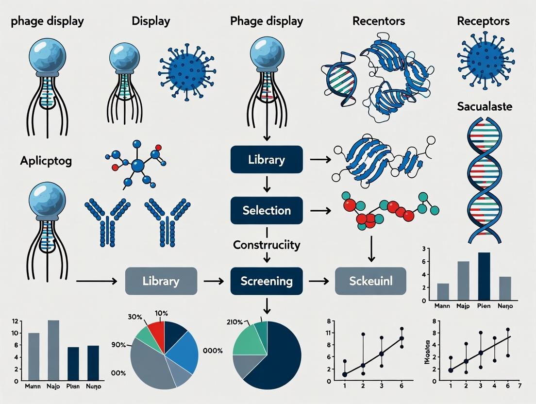
Visualizations
Phage Display Biopanning Workflow
Fusion of Genotype and Phenotype
The Scientist's Toolkit: Essential Research Reagent Solutions
Table 3: Key Reagents for Phage Display Experiments
| Reagent / Material | Function / Purpose |
|---|---|
| M13KE or similar Phage Vector | Engineered bacteriophage genome for cloning foreign DNA in-frame with coat protein genes (gIII or gVIII). |
| E. coli ER2738 | An F-plus pilus expressing bacterial host strain essential for M13 phage infection and propagation. |
| PEG/NaCl Precipitation Solution | Used to concentrate and purify phage particles from bacterial culture supernatants. |
| HRP/Anti-M13 Antibody | Horseradish peroxidase-conjugated antibody specific to the M13 phage major coat protein for immunoassays (ELISA). |
| IPTG/X-gal | Used in LB agar for blue/white screening of phage plaques when using vectors with a LacZα insert. |
| Protease Inhibitors (e.g., PMSF) | Added during phage purification to prevent degradation of displayed proteins. |
| Streptavidin-coated Magnetic Beads | For solution-phase biopanning using biotinylated target molecules and magnetic separation. |
| Next-Generation Sequencing (NGS) Reagents | For deep sequencing of phage library pools pre- and post-selection to analyze diversity and enrichment. |
This application note details the historical development of phage display technology, from its conceptual inception to its recognition with the Nobel Prize, framed within the thesis that this platform is foundational to modern biotherapeutics and diagnostic research. The protocol sections provide actionable methodologies for key experiments that have defined the field.
Historical Development & Quantitative Milestones
Table 1: Key Historical Milestones and Quantitative Impact
| Year | Key Event/Publication | Primary Contributor(s) | Key Quantitative Outcome/Impact |
|---|---|---|---|
| 1985 | First demonstration of peptide display on filamentous phage | George P. Smith | Showed foreign peptides (6-aa) could be fused to pIII and recovered using affinity purification. |
| 1990 | First antibody fragment (scFv) display on phage | McCafferty et al. | Demonstrated functional antibody fragments could be displayed, founding phage antibody technology. |
| 1991 | First in vitro selection from large libraries termed "biopanning" | Scott, Smith | Defined the iterative selection process; library size ~10^8 clones. |
| 1994 | First human therapeutic antibody (Adalimumab) developed using phage display (approved 2002) | Cambridge Antibody Technology | Affinity in nM range; annual peak sales >$20B. |
| 2018 | Nobel Prize in Chemistry awarded for phage display | George P. Smith, Sir Gregory P. Winter | Recognized the technology's transformative role in drug development. |
| 2023 (Approx.) | Over 100 phage-derived therapeutics in clinical development | Various | 14+ approved drugs, including monoclonal antibodies and peptides. |
Detailed Experimental Protocols
Protocol 1: Original Peptide Display & Biopanning (Smith, 1985)
Objective: To clone and display a foreign peptide on the surface of filamentous phage via fusion to the minor coat protein pIII and recover it via binding to a solid-phase antibody.
Materials: See "Research Reagent Solutions" below.
Method:
- Genetic Fusion: Insert oligonucleotide encoding the target peptide into the EcoRI site of plasmid vector fUSE5, creating an in-frame fusion with gene III.
- Phage Propagation: Transform the ligation into an E. coli host strain (e.g., K91). Grow transformed cells in LB + tetracycline to log phase. Superinfect with helper phage (e.g., M13KO7) to supply wild-type proteins for phage assembly.
- Phage Precipitation: Culture overnight. Precipitate phage particles from supernatant using PEG/NaCl (4% PEG-8000, 0.5 M NaCl). Resuspend pellet in Tris-buffered saline (TBS).
- Affinity Selection (Biopanning): a. Coat a polystyrene Petri dish with 1 mL of purified antibody (10-100 µg/mL in bicarbonate buffer) overnight at 4°C. b. Block dish with 2% BSA in TBS for 2 hours. c. Incubate dish with ~10^11 phage particles in 1 mL of TBS + 1% BSA for 1-4 hours. d. Wash dish 10x with TBS-Tween 20 (0.1%) to remove non-specifically bound phage. e. Elute bound phage by incubating with 1 mL of elution buffer (0.1 M HCl, pH adjusted with glycine to 2.2) for 10 min. Neutralize immediately with 2M Tris base.
- Amplification: Infect log-phase E. coli with eluted phage. Propagate for subsequent rounds of panning (typically 3-4 rounds).
- Analysis: Plate infected bacteria to obtain single clones. Sequence DNA from individual plaques/colonies to determine displayed peptide sequence.
Protocol 2: Phage Display Antibody Library Screening (McCafferty et al., 1990)
Objective: To isolate antigen-specific single-chain Fv (scFv) fragments from a phage display library.
Method:
- Library Construction: Amplify VH and VL gene repertoires from immune or naïve B-cell mRNA via RT-PCR. Assemble into scFv format (VH-linker-VL) via splice-by-overlap-extension PCR. Clone into phagemid vector (e.g., pHEN2) downstream of a pelB signal sequence and fused to gene III.
- Phage Rescue: Transform library into E. coli TG1. Grow a portion of the library in 2xTY medium with ampicillin and glucose to OD600 ~0.5. Infect with helper phage (M13KO7) for 1 hour. Centrifuge, resuspend in 2xTY with ampicillin and kanamycin (no glucose) to induce expression. Culture overnight at 30°C.
- Phage Purification: Precipitate phage from culture supernatant with PEG/NaCl as in Protocol 1.
- Antigen Panning: a. Coat immunotube or magnetic beads with purified antigen (5-50 µg/mL). b. Block with 2% MPBS (skim milk powder in PBS). c. Incubate with purified phage library in 2% MPBS for 1-2 hours. d. Wash with PBS-Tween (0.1%), increasing stringency (Tween concentration, wash number) over selection rounds. e. Elute with 100 mM triethylamine or via protease cleavage site (e.g., 3C protease) if included in the vector. f. Neutralize and amplify eluted phage by infecting log-phase TG1 cells.
- Clone Characterization: After 3-4 rounds, pick individual colonies for monoclonal phage ELISA. Express soluble scFv by omitting helper phage infection and inducing with IPTG. Test for antigen binding via ELISA or SPR.
Research Reagent Solutions
Table 2: Essential Reagents for Phage Display
| Reagent | Function & Key Detail |
|---|---|
| Filamentous Phage Vector (fUSE5) | Gene III fusion vector; contains phage origin for packaging and antibiotic resistance. |
| Phagemid Vector (e.g., pHEN2) | Plasmid with phage origin, antibiotic resistance, and in-frame cloning site for fusions to truncated gene III; requires helper phage. |
| E. coli Host Strain (e.g., K91, TG1) | F+ pilus expressing strain for phage infection; often suppressor strain for amber stop codon read-through in phagemid systems. |
| Helper Phage (e.g., M13KO7) | Provides wild-type phage proteins in trans to package phagemid DNA; carries kanamycin resistance. |
| PEG/NaCl Solution | Polyethylene glycol (PEG-8000) and high-salt solution for precipitating and concentrating phage particles. |
| Coating Antigen/Antibody | Purified target for panning; immobilized on polystyrene plates, immunotubes, or magnetic beads. |
| Blocking Agent (e.g., BSA, Skim Milk) | Reduces non-specific binding of phage during selection steps. |
| Wash Buffer with Detergent (e.g., PBS + 0.1% Tween 20) | Removes weakly bound phage; increasing Tween % increases selection stringency. |
| Elution Buffer (e.g., Triethylamine, Low pH Glycine) | Disrupts antigen-antibody binding to recover specifically bound phage for amplification. |
Visualizations
Title: Timeline of Phage Display Key Milestones
Title: Phage Display Biopanning Workflow
Within the broader thesis on the Applications of Phage Display Technology, the M13 filamentous bacteriophage stands as the foundational workhorse. Its unique molecular biology—non-lytic replication, repetitive coat structure, and ssDNA genome—makes it uniquely suited for the display of peptide and protein libraries. This application note details its biology, key protocols, and reagent toolkit essential for researchers and drug development professionals.
Molecular Biology & Relevance to Phage Display
M13 is a rod-shaped, F-pili specific phage infecting E. coli. Its ~6.4 kb single-stranded DNA genome encodes 11 proteins. Five coat proteins are critical for display:
- pVIII: Major coat protein (~2700 copies). Used for displaying short peptides.
- pIII: Minor coat protein (3-5 copies at one tip). Used for displaying large proteins (e.g., antibodies, scaffolds).
- pVI, pVII, pIX: Other minor proteins, also used for display.
The phage is secreted from infected cells without lysis, allowing continuous production and easy purification of displayed polypeptides.
Key Quantitative Data
Table 1: M13 Bacteriophage Structural & Genomic Data
| Parameter | Value | Significance for Phage Display |
|---|---|---|
| Genome Type | Single-stranded DNA (ssDNA) | Simplifies DNA manipulation and library construction. |
| Genome Size | ~6407 nucleotides | Compact, well-characterized sequence. |
| Virion Length | ~880 nm | Large surface area for display. |
| Major Coat Protein (pVIII) Copies | ~2700 | High-valency display of short peptides. |
| Minor Coat Protein (pIII) Copies | 3-5 | Low-valency display for high-affinity selection. |
| Infection Specificity | F-pili of E. coli | Requires F+ or F' strains (e.g., TG1, XL1-Blue). |
| Replication Cycle | Non-lytic, secretory | Host cell remains viable; phage harvested from supernatant. |
Table 2: Common Phagemid vs. Helper Phage System Output
| Component | Typical Titer/Quantity | Function |
|---|---|---|
| Phagemid Vector (e.g., pComb3) | Library size: 10^9 - 10^11 CFU | Carries gene for antibody fragment (scFv/Fab) fused to pIII/pVIII and antibiotic resistance. |
| Helper Phage (e.g., M13K07) | ~10^12 PFU/mL in stock | Supplies all phage proteins for replication and assembly; has a packaging signal defect. |
| Phage Particle Output (after rescue) | 10^10 - 10^13 CFU/mL | Infectious particles displaying the library member. |
Core Protocols
Protocol 1: Rescue of Phage Display Library from a Phagemid System
Objective: To produce infectious phage particles displaying antibody fragments (or other proteins) from a cloned phagemid library.
Materials:
- E. coli strain (e.g., TG1) harboring the phagemid library.
- Helper phage (e.g., M13K07, VCSM13).
- 2x YT media with appropriate antibiotics (e.g., ampicillin, tetracycline).
- PEG/NaCl solution (20% PEG-8000, 2.5 M NaCl).
Procedure:
- Inoculate 10 mL of 2x YT with antibiotic selecting for the phagemid. Grow overnight at 37°C, 220 rpm.
- Subculture 1 mL of overnight culture into 50 mL of fresh 2x YT (with antibiotic) to an OD600 of ~0.1. Grow to OD600 = 0.4-0.6 (mid-log phase).
- Add helper phage at a multiplicity of infection (MOI) of 10-20 (e.g., for 5x10^8 cells in 50 mL, add 5-10x10^9 pfu helper phage). Incubate 30 min at 37°C without shaking, then 30 min with shaking.
- Centrifuge cells (3000 x g, 10 min). Resuspend pellet in 100 mL of 2x YT containing ampicillin (100 µg/mL) and kanamycin (50 µg/mL) to select for cells containing both phagemid and helper phage.
- Incubate overnight (~16-20 hrs) at 30°C, 220 rpm. Note: Lower temperature improves display.
- Centrifuge culture (10,000 x g, 15 min, 4°C). Transfer supernatant to a fresh tube.
- Precipitate phage by adding 1/5 volume of PEG/NaCl solution. Mix and incubate on ice for ≥1 hour.
- Centrifuge (10,000 x g, 30 min, 4°C). Discard supernatant.
- Resuspend phage pellet in 1-2 mL of PBS or TBS. Centrifuge briefly to remove debris.
- Titer the phage stock (see Protocol 2) and store at 4°C for short-term or -80°C with 15% glycerol for long-term.
Protocol 2: Titering M13 Phage by Plaque or Transduction Assay
Objective: To determine the concentration of infectious phage particles (CFU/mL).
Procedure (Transduction/Colony Forming Units - Standard for Display Phage):
- Prepare serial dilutions (10^-8 to 10^-12) of the phage stock in sterile LB or PBS.
- Mix 10 µL of each dilution with 100 µL of mid-log phase E. coli TG1 cells (OD600 ~0.5).
- Incubate 30 min at 37°C without shaking.
- Plate each mixture onto 2x YT agar plates containing the antibiotic that selects for the phagemid (e.g., ampicillin).
- Incubate plates overnight at 37°C.
- Count colonies and calculate titer: Titer (CFU/mL) = (Colonies counted x Dilution Factor x 100) / 0.01 mL.
The Scientist's Toolkit: Key Reagent Solutions
Table 3: Essential Research Reagents for M13 Phage Display
| Reagent | Function & Explanation |
|---|---|
| F+ E. coli Strains (TG1, XL1-Blue) | Essential host strains expressing the F-pilus for M13 infection and phage propagation. |
| Helper Phage (M13K07, VCSM13) | Genetically modified M13 phage with a defective origin; supplies all structural/ replication proteins in trans to package a phagemid. |
| Phagemid Vectors (pComb3, pHEN1) | Plasmid containing phage origin, antibiotic resistance, and cloning site for fusion to pIII/pVIII gene fragment. Core of display library construction. |
| PEG/NaCl Solution | Polyethylene glycol precipitates phage particles from culture supernatant for concentration and purification. |
| Blocking Agents (BSA, Skim Milk) | Used to block non-specific binding sites during panning/biopanning selection steps. |
| Elution Buffers (Glycine-HCl pH 2.2, Triethylamine) | Acidic or basic conditions disrupt phage-target binding for recovery of bound phage during panning. |
| Trypsin/Protease | Used for specific elution by cleaving a designed site between displayed protein and phage coat protein. |
Visualized Workflows & Pathways
Diagram Title: M13 Phage Display Panning Cycle Workflow
Diagram Title: M13 Genome Key Genes and Virion Structure
Within the broader thesis on the Applications of Phage Display Technology Research, the iterative cycle of Phage Vector, Library Construction, and Affinity Selection constitutes the foundational engine. This technology enables the high-throughput screening of up to (10^{11}) unique peptides or antibodies for binding to a target of interest, revolutionizing drug discovery, epitope mapping, and protein engineering. This document provides detailed application notes and protocols for these core components.
Core Components: Application Notes
Phage Display Vectors
Phage vectors are engineered bacteriophages (commonly M13, fd, or T7) that genetically fuse the DNA encoding a peptide/protein of interest to a gene for a viral coat protein (pIII or pVIII), resulting in display on the virion surface.
Key Research Reagent Solutions:
| Reagent/Solution | Function & Explanation |
|---|---|
| M13KE Vector (or similar) | A common, genetically stable phage genome for fusing inserts to the pIII protein. Allows for antibiotic selection and phage propagation. |
| Helper Phage (e.g., M13K07) | Provides wild-type coat proteins in trans for the packaging of phagemid particles during library amplification. Essential for phagemid systems. |
| PEG/NaCl Solution | Polyethylene glycol (PEG) precipitates phage particles from solution for concentration and purification post-infection or panning. |
| E. coli ER2738 | An F-pilus expressing, tetracycline-sensitive bacterial strain specifically optimized for the infection and propagation of M13 phage libraries. |
Peptide/Protein Library
Libraries are constructed by cloning degenerate oligonucleotides or gene fragments into the phage vector. Diversity is a critical parameter.
Table 1: Common Library Types and Characteristics
| Library Type | Typical Diversity | Insert Size | Common Display Format | Primary Application |
|---|---|---|---|---|
| Linear Peptide | (10^9) - (10^{11}) | 7-12 aa | pIII or pVIII | Epitope mapping, finding short binders |
| Constrained Peptide (e.g., Cys-loop) | (10^8) - (10^{10}) | 7-10 aa | pIII | Finding higher-affinity, structured peptides |
| scFv Antibody | (10^8) - (10^{10}) | ~750 bp | pIII | Therapeutic & diagnostic antibody discovery |
| Fab Fragment | (10^8) - (10^{10}) | ~1.3 kbp | pIII (fusion to heavy chain) | Stable antibody fragment discovery |
| Domain Library (e.g., DARPin) | (10^9) - (10^{11}) | ~330 bp | pIII | Alternative scaffold binders |
Affinity Selection (Panning)
Panning is the iterative process of isolating target-binding phage from the vast excess of non-binders.
Table 2: Quantitative Panning Metrics for Monitoring Progress
| Panning Round | Input Phage (cfu) | Eluted Phage (cfu) | Output/Input Ratio | Enrichment Indicator |
|---|---|---|---|---|
| 1 | (1.0 \times 10^{12}) | (5.0 \times 10^{3}) | (5.0 \times 10^{-9}) | Baseline |
| 2 | (5.0 \times 10^{11}) | (2.5 \times 10^{5}) | (5.0 \times 10^{-7}) | ~100x enrichment |
| 3 | (2.5 \times 10^{11}) | (1.0 \times 10^{7}) | (4.0 \times 10^{-5}) | ~80x further enrichment |
| 4 | (1.0 \times 10^{11}) | (5.0 \times 10^{8}) | (5.0 \times 10^{-3}) | ~125x further enrichment |
cfu: colony-forming units.
Detailed Experimental Protocols
Protocol 1: Solid-Phase Panning Against Immobilized Protein Target
Materials:
- Target protein in coating buffer (e.g., 100 mM NaHCO₃, pH 8.6).
- Blocking buffer: 5% (w/v) non-fat dry milk in PBS or 3% BSA in PBS.
- Phage library in blocking buffer.
- Washing buffer: PBS with 0.1% (v/v) Tween-20 (PBS-T) and PBS alone.
- Elution buffer: 0.1 M Glycine-HCl (pH 2.2) or 100 mM Triethylamine.
- Neutralization buffer: 1 M Tris-HCl (pH 9.1).
- Log-phase E. coli ER2738 culture.
Methodology:
- Coating: Add 100 µL of target protein (10-100 µg/mL in coating buffer) to a well of an immunoassay plate. Incubate overnight at 4°C.
- Blocking: Aspirate coating solution. Add 300 µL of blocking buffer. Incubate at room temperature (RT) for 1-2 hours.
- Binding: Aspirate blocking buffer. Add 100 µL of pre-blocked phage library ((10^{11}) - (10^{12}) cfu in blocking buffer). Incubate at RT for 1-2 hours with gentle agitation.
- Washing: Aspirate phage solution. Perform 10 rapid washes with PBS-T (first round). Increase stringency in subsequent rounds (e.g., 15-20 washes with 0.5% Tween-20).
- Elution: For acidic elution, add 100 µL of 0.1 M Glycine-HCl (pH 2.2). Incubate at RT for 10 min with agitation. Transfer eluate to a tube containing 15 µL of neutralization buffer.
- Amplification: Mix eluted phage with 20 mL of log-phase ER2738 cells. Incubate 30 min at 37°C with shaking. Transfer to larger culture volume with helper phage (if using phagemid) and antibiotic. Propagate overnight. PEG-precipitate phage for the next round.
- Titration: Serially dilute input, wash flow-through, and eluted phage, infect ER2738, and plate on selective media to calculate titers (Table 2).
Protocol 2: Phage Rescue & Amplification from Selected Clones
Materials:
- E. coli ER2738 culture.
- LB medium with appropriate antibiotic (e.g., Tetracycline for M13KE).
- PEG/NaCl Solution: 20% PEG-8000, 2.5 M NaCl.
- TBS: 50 mM Tris-HCl, 150 mM NaCl, pH 7.5.
Methodology:
- Pick individual bacterial colonies from titering plates into 1 mL of LB with antibiotic in a 96-deep well block.
- Incubate at 37°C with shaking (~900 rpm) for 4-5 hours.
- Add helper phage (if needed) at high MOI (~20), incubate 30 min statically.
- Transfer culture to a larger volume or add antibiotic for selection of infected cells. Incubate overnight at 37°C with shaking.
- Centrifuge the next day (3,300 x g, 15 min). Transfer supernatant to a new tube.
- Precipitate phage by adding 1/5 volume of PEG/NaCl. Incubate on ice for 1 hour.
- Centrifuge (12,000 x g, 15 min, 4°C). Discard supernatant. Resuspend pellet in 100 µL TBS. This is the monoclonal phage stock for ELISA.
Mandatory Visualizations
Panning Cycle Workflow
Target Binding and Recovery Steps
Application Notes
Within the broader thesis on the applications of phage display technology in therapeutic and diagnostic research, three key advantages establish its dominance in library screening: Rapid Screening, Direct Genetic Linkage, and Robustness. These attributes collectively accelerate the path from target identification to lead candidate validation, making it indispensable for researchers and drug development professionals.
Rapid Screening is enabled by the ability to perform iterative biopanning cycles against immobilized targets in vitro, bypassing complex cellular systems. This in vitro selection typically completes 3-5 rounds of enrichment within 1-2 weeks, drastically shortening the timeline compared to in vivo methods like hybridoma technology for antibody discovery. Recent studies highlight the use of next-generation sequencing (NGS) post-panning to analyze entire selection outputs, identifying high-binders within a single, deep-sequenced round, further compressing timelines.
Direct Genetic Linkage is the foundational principle of phage display, where the physical connection between the displayed phenotype (protein/peptide) and its encapsulated genotype (DNA) is preserved. This allows for immediate identification of binding sequences through Sanger or NGS of phage DNA from selected clones, eliminating the need for separate cloning and expression steps during early screening. Modern workflows integrate NGS analysis with bioinformatic clustering to deconvolute enriched families, directly linking sequence to function.
Robustness refers to the inherent stability of the filamentous phage particle (e.g., M13) and the resilience of the screening process. Phages are resistant to a range of pH conditions (pH 3-11), temperatures, and denaturing agents, allowing for stringent off-rate selection through aggressive washing and competitive elution. This robustness enables the selection of high-affinity, stable binders. Furthermore, the bacterial production system is scalable and cost-effective, ensuring reproducible generation of diverse libraries (>10^9-11 unique members).
Table 1: Comparison of Key Phage Display Screening Parameters with Hybridoma Technology
| Parameter | Phage Display (Peptide/Antibody) | Hybridoma Technology (mAb) |
|---|---|---|
| Library Diversity | 10^9 - 10^11 independent clones | ~10^4 - 10^6 splenocytes |
| Screening Cycle Time | 1-2 weeks (3-5 panning rounds) | 4-8 weeks (cell culture & fusion) |
| Primary Hit Identification | Direct sequencing post-panning | ELISA screening of supernatants |
| Affinity Range (Kd) | nM - μM (primary hits); pM after maturation | nM - pM (post-selection) |
| Key Strength | In vitro control, genotype-phenotype link | Native mammalian folding & glycosylation |
| Common Elution Methods | Acidic pH (Glycine-HCl), competitive ligand, protease | Not applicable (cell-based) |
Table 2: Impact of NGS Integration on Phage Display Output Metrics
| Analysis Method | Sequencing Depth per Sample | Time from Panning to Hit List | Key Deliverable |
|---|---|---|---|
| Traditional (Sanger) | 96 - 384 clones | 1-2 weeks | Individual high-frequency sequences |
| NGS-Enhanced | 10^5 - 10^7 reads | 3-5 days (post-DNA prep) | Enriched family clusters, consensus sequences, binding motifs |
Experimental Protocols
Protocol 1: Standard Biopanning for Antigen-Specific scFv Selection
Objective: To isolate single-chain variable fragments (scFvs) binding to a purified, immobilized target antigen over 3-4 rounds of selection.
Materials (See Toolkit Section)
- Phage display library (e.g., human scFv library in phagemid vector).
- Target antigen in coating buffer (PBS or carbonate-bicarbonate, pH 9.6).
- Blocking buffer: 5% (w/v) non-fat dry milk in PBS.
- Washing buffers: PBS-T (PBS + 0.1% Tween-20) and PBS.
- Elution buffers: 0.1 M Glycine-HCl (pH 2.2) and 1 M Tris-HCl (pH 9.1) for neutralization.
- E. coli strain for infection (e.g., TG1 or XL1-Blue).
- Tetracycline or carbenicillin for selection, M13KO7 helper phage.
Methodology:
- Coating: Coat immunotube or 96-well plate with 4 mL or 100 µL of target antigen (10-100 µg/mL in coating buffer). Incubate overnight at 4°C.
- Blocking: Aspirate coating solution, wash twice with PBS. Add 5% milk/PBS blocking buffer (4 mL or 300 µL). Incubate at 37°C for 2 hours.
- Phage Binding: Blocked phage library (pre-blocked in milk/PBS for 30 min) is added to the coated, washed tube/well. Incubate for 2 hours at room temperature with gentle rotation/tapping.
- Stringent Washing: For Round 1, perform 10 washes with PBS-T followed by 10 washes with PBS. Increase Tween-20 concentration (up to 0.5%) and wash count (e.g., 20x) in subsequent rounds.
- Elution: For acidic elution, add 1 mL or 100 µL of 0.1 M Glycine-HCl (pH 2.2). Incubate for 10 min with agitation. Immediately transfer eluate to a tube containing 0.5 mL or 75 µL of 1 M Tris-HCl (pH 9.1) for neutralization.
- Amplification: Infect 10 mL of log-phase E. coli (OD600 ~0.5) with the eluted phage. Add M13KO7 helper phage (MOI >20) and incubate 1 hour at 37°C without shaking. Plate on large selective agar plates (e.g., with tetracycline) or grow in selective liquid culture for phage precipitation (PEG/NaCl).
- Titering: Titre input, unbound, and eluted phage after each round to monitor enrichment.
- Repeat: Perform 3-4 rounds of panning. Subject phage output from rounds 3 and 4 to NGS or proceed with monoclonal phage ELISA.
Protocol 2: Competitive Elution for High-Affinity Binder Selection
Objective: To selectively elute phages displaying binders with the highest affinity or those binding to a specific epitope using a soluble competitive ligand.
Materials: Includes all from Protocol 1, plus purified soluble competitor (target antigen or known antibody for the epitope of interest).
Methodology:
- Follow steps 1-4 of Protocol 1 for coating, blocking, binding, and washing.
- Competitive Elution: Instead of acidic elution, add a high-concentration solution of the soluble competitor (e.g., 1-10 µM in blocking buffer) to the washed tube/well.
- Incubate for 1-2 hours at room temperature or overnight at 4°C with gentle agitation.
- Collect the eluate containing phages displaced by the competitor.
- Proceed with amplification and titering (Steps 6-7, Protocol 1). This method directly selects for binders to the specific epitope recognized by the competitor.
Protocol 3: NGS Sample Preparation from Phage Pool
Objective: To prepare the phage display library panning output for high-throughput sequencing to analyze enrichment.
Materials: QIAprep Spin M13 Kit (or equivalent), PCR primers flanking the variable region, high-fidelity DNA polymerase, NGS library preparation kit (e.g., Illumina Nextera).
Methodology:
- Phage DNA Extraction: Purify single-stranded DNA (ssDNA) from 1 mL of precipitated phage pool (post-round 3/4) using the QIAprep Spin M13 Kit according to the manufacturer's protocol. Elute in 50 µL nuclease-free water.
- Amplification of Variable Insert: Set up a PCR reaction using 5 µL of eluted ssDNA as template with primers containing overhangs compatible with your NGS platform indexes. Use a high-fidelity polymerase (15-20 cycles).
- Purify Amplicon: Clean up the PCR product using a PCR purification kit. Quantify by spectrophotometry (Nanodrop) or fluorometry (Qubit).
- NGS Library Preparation: Follow the specific protocol for your sequencing platform (e.g., Illumina). Typically, this involves a second, limited-cycle PCR to attach full adapter indices.
- Sequencing: Pool libraries and sequence on an Illumina MiSeq or NovaSeq platform with paired-end reads of sufficient length to cover the variable region.
Visualizations
Title: Phage Display Screening & Hit Isolation Workflow
Title: Genotype-Phenotype Link in Phage Display
The Scientist's Toolkit
Table 3: Key Research Reagent Solutions for Phage Display Screening
| Item | Function / Rationale |
|---|---|
| Phagemid Vector (e.g., pComb3X) | Plasmid containing phage origin, antibiotic resistance, and cloning site for peptide/antibody fragment insertion. |
| Helper Phage (e.g., M13KO7) | Provides all viral proteins for phage assembly; superinfects bacteria containing phagemid to produce virions. |
| E. coli F+ Strain (e.g., TG1) | Essential host with F-pili for phage infection and propagation. |
| Immunotubes / Streptavidin Beads | Solid support for immobilizing target antigen (protein or biotinylated molecules). |
| PEG/NaCl Precipitation Solution | Standard method for concentrating and purifying phage particles from bacterial supernatant. |
| Anti-M13 Antibody (HRP conjugated) | For detection of phage binding in monoclonal phage ELISA. |
| QIAprep Spin M13 Kit | Optimized for rapid purification of single-stranded phage DNA for sequencing. |
| NGS Library Prep Kit | For preparing amplicons from the variable region of enriched phage pools for deep sequencing analysis. |
How Phage Display is Used: Key Methodologies and Transformative Applications
Within the broader thesis on the Applications of Phage Display Technology Research, the construction of high-quality, diverse libraries is the foundational step. This process determines the success of subsequent selection campaigns for identifying novel binders, modulators, and therapeutics. This application note details protocols and design principles for constructing peptide, single-chain variable fragment (scFv), antigen-binding fragment (Fab), and protein domain libraries.
Library Design Principles and Quantitative Benchmarks
Table 1: Key Design Parameters for Different Library Types
| Library Type | Typical Size (Clones) | Diversity Source | Framework/Backbone | Key Design Considerations |
|---|---|---|---|---|
| Peptide | 10⁸ – 10¹¹ | Randomized linear or constrained loops (e.g., Cys-Cys) | Fusion protein (pIII, pVIII) | Length (6-15 aa), randomization strategy (NNK vs. NNS), context for constraint. |
| scFv | 10⁸ – 10¹⁰ | CDR-H3 & CDR-L3, often all CDRs | Stable human frameworks (e.g., VH3-23/Vκ1-39) | CDR length distribution, tailormade vs. naive diversity, stability selection. |
| Fab | 10⁸ – 10¹⁰ | CDR-H3 & CDR-L3, often all CDRs | Paired heavy & light chain frameworks | Proper heavy-light pairing, efficient cloning of two chains, Fab display efficiency. |
| Protein Domain | 10⁷ – 10⁹ | Surface residues, loops, or entire sequence | Stable scaffold (e.g., FN3, DARPins, A-domain) | Minimizing structural perturbation, maintaining scaffold integrity, focusing diversity on functional surfaces. |
Table 2: Common Mutagenesis Strategies and Outcomes
| Strategy | Method | Theoretical Diversity | Practical Library Size | Best For |
|---|---|---|---|---|
| Oligonucleotide-Directed | Kunkel mutagenesis, PCR assembly | Very High (10¹²+) | Limited by transformation (~10¹⁰) | All library types, precise control. |
| Error-Prone PCR | PCR with Mn²⁺, unbalanced dNTPs | Moderate, random | 10⁶ – 10⁸ | Affinity maturation, introducing low-level diversity across gene. |
| DNA Shuffling | Fragmentation & reassembly | High, recombination-based | 10⁷ – 10⁹ | Diversifying homologous sequences, family shuffling. |
| Codon-Based Degeneracy | TRIM, NNK/NNS codons | Defined by codon scheme | Defined by transformation | Focused diversity at specific positions. |
Experimental Protocols
Protocol 1: Construction of a Naive Human scFv Library Using Kunkel Mutagenesis
Objective: Generate a large (>10⁹ member) scFv library with diversity incorporated into all six Complementarity-Determining Regions (CDRs).
Materials:
- Template: M13 phagemid vector containing scFv gene with amber stop codon in CDR regions, in E. coli CJ236 (dut⁻ ung⁻).
- Oligonucleotides: Degenerate oligonucleotides designed to replace CDRs with NNK/TRIMM mixtures.
- Enzymes: T7 DNA Polymerase, T4 DNA Ligase, T4 Polynucleotide Kinase.
- Host Strain: Electrocompetent E. coli SS320 (or TG1) for high-efficiency transformation.
Method:
- Prepare Uracil-Containing Single-Stranded DNA (ssDNA): Infect CJ236 harboring the template phagemid with helper phage (e.g., M13K07). Precipitate phage particles from supernatant, then purify ssDNA via phenol/chloroform extraction and ethanol precipitation.
- Phosphorylate Oligonucleotides: Pool all mutagenic oligos. Phosphorylate using T4 PNK and ATP.
- Annealing: Mix ~1 µg uracil-ssDNA template with 5-10 fold molar excess of phosphorylated oligo pool. Anneal in a thermal cycler (90°C for 2 min, ramp to 25°C over 45 min).
- Synthesis & Ligation: Add dNTPs, ATP, T7 DNA polymerase, and T4 DNA ligase. Incubate on ice for 5 min, room temp for 5 min, then 37°C for 90 min. This extends the oligos and ligates the nicks, creating closed circular heteroduplex DNA.
- Template Degradation & Transformation: Digest the product with Exonuclease III or DpnI to degrade the uracil-containing template strand. Desalt the synthesized dsDNA. Electroporate ~100 ng DNA into 1 mL highly electrocompetent SS320 cells (≥10¹⁰ cfu/µg). Recover cells in SOC medium for 1 hour.
- Library Amplification & Storage: Plate serial dilutions to determine library size. Amplify the library in liquid culture, add helper phage to produce phage particles, and precipitate/physical storage at -80°C in glycerol.
Protocol 2: Construction of a Constrained Peptide Library on the pVIII Coat Protein
Objective: Display a CX₇C (where X is any amino acid) peptide library on the major coat protein pVIII for high-valency display, useful for selecting high-affinity binders.
Materials:
- Vector: Phage vector (e.g., fUSE5) allowing peptide fusion to pVIII.
- Oligonucleotide: Degenerate oligonucleotide encoding 5'-(AGC) Cys (NNK)₇ Cys (TCA)-3'.
- Enzymes: Restriction enzymes for the chosen cloning site.
Method:
- Vector Preparation: Digest the phage vector with appropriate restriction enzymes to linearize. Gel purify the linearized backbone.
- Oligo Annealing: Synthesize complementary oligos. Anneal them to form double-stranded DNA with compatible sticky ends.
- Ligation: Ligate the annealed oligo duplex into the prepared vector at a high insert:vector molar ratio (~10:1) to maximize diversity.
- Electroporation: Electroporate the ligation mixture into electrocompetent E. coli (e.g., ER2738). Perform multiple electroporations to achieve >10⁹ independent transformants.
- Phage Amplification: Pool all transformations, add helper phage if necessary (for type 8+8 systems), and grow overnight. PEG precipitate phage from the supernatant to create the crude library stock for subsequent panning.
Protocol 3: Fab Library Cloning by Restriction-Based Assembly
Objective: Clone a diversified Fd fragment (VH-CH1) and light chain (VL-CL) into a phagemid vector for Fab display.
Method:
- Separate PCR Amplification: Amplify the diversified Fd fragment (using primers adding e.g., SfiI/NotI sites) and the diversified light chain (using primers adding e.g., XbaI/SacI sites).
- Sequential Cloning: First, digest the Fd fragment and phagemid vector with SfiI and NotI. Ligate and transform. Isolate the resulting vector pool. Second, digest the light chain fragment and the intermediate vector pool with XbaI and SacI. Ligate and transform.
- Library Production: The final phagemid encodes the heavy chain fused to pIII and the light chain as a soluble partner. Transform the final ligation into E. coli via electroporation, amplify, and rescue with helper phage to produce Fab-displaying phage.
Visualizations
Diagram 1: scFv Library Construction via Kunkel Mutagenesis
Diagram 2: Fab Phagemid Vector & Display Logic
The Scientist's Toolkit
Table 3: Key Research Reagent Solutions for Phage Library Construction
| Reagent / Material | Function & Rationale |
|---|---|
| Helper Phage (e.g., M13K07, VCSM13) | Provides all wild-type phage proteins in trans during rescue to package the phagemid DNA into infectious particles. Essential for display valency control. |
| Electrocompetent E. coli (e.g., SS320, TG1) | High-efficiency transformation cells (>10¹⁰ cfu/µg) are critical for achieving large library sizes. SS320 is often used for its high conjugation efficiency. |
| dut⁻ ung⁻ E. coli Strain (CJ236) | Essential for Kunkel mutagenesis. Lacks dUTPase and uracil deglycosylase, allowing incorporation of uracil into phage DNA for template strand degradation. |
| T7 DNA Polymerase | Highly processive enzyme used in Kunkel synthesis for efficient extension of the annealed oligonucleotide around the entire template. |
| NNK Degenerate Codon Oligos | Oligonucleotides with NNK (N=A/T/G/C; K=G/T) degeneracy encode all 20 amino acids with only one stop codon (TAG), optimizing diversity representation. |
| Phagemid Vector (e.g., pComb3, pHEN) | Plasmid containing phage origin of replication, antibiotic resistance, and gene for fusion protein (pIII or pVIII). Allows efficient DNA manipulation and monovalent display. |
| PEG/NaCl Precipitation Solution | Standard method for concentrating and purifying phage particles from bacterial culture supernatants, removing contaminants and cell debris. |
Within the broader thesis on Applications of Phage Display Technology Research, biopanning represents the cornerstone experimental technique for isolating high-affinity peptide or antibody ligands against a target of interest. This protocol details the iterative affinity selection process used to screen phage display libraries, enabling the discovery of binders for therapeutic, diagnostic, and research applications.
Key Research Reagent Solutions
| Reagent/Material | Function in Biopanning |
|---|---|
| Phage Display Library | A diverse collection of filamentous phage (e.g., M13) each displaying a unique peptide or protein variant on its surface coat protein (pIII or pVIII). Provides the genetic diversity for selection. |
| Immobilized Target | The molecule of interest (e.g., protein, enzyme, cell receptor) immobilized on a solid support (e.g., immunotube, magnetic bead, column resin). Serves as the bait for affinity selection. |
| Blocking Buffer | A solution of irrelevant proteins (e.g., BSA, skim milk) used to coat non-specific binding sites on the immobilization surface and the phage, reducing background noise. |
| Elution Buffer | A solution designed to disrupt phage-target binding. Common agents include low-pH glycine buffer, high-pH triethylamine, or a molar excess of soluble target for competitive elution. |
| E. coli Host Strain | Typically an F+ strain (e.g., ER2738) susceptible to M13 phage infection. Used to amplify eluted phage for subsequent rounds of panning, linking phenotype to genotype. |
Experimental Protocol: Standard Solid-Phase Panning
Objective: To isolate specific phage clones binding to a purified protein target immobilized on a plastic surface.
Day 1: Target Immobilization & Blocking
- Coating: Dilute the purified target protein to 10-100 µg/mL in an appropriate coating buffer (e.g., 0.1 M NaHCO₃, pH 8.6). Add 1-4 mL to a polystyrene immunotube or 96-well plate. Incubate overnight at 4°C or for 2 hours at 37°C.
- Washing: Remove the coating solution. Wash the tube three times with Tris-Buffered Saline (TBS), pH 7.5.
- Blocking: Fill the tube with 4 mL of blocking buffer (e.g., 5% w/v non-fat dry milk or 3-5% BSA in TBS). Incubate at 37°C for 1-2 hours to block non-specific sites.
Day 1: Phage Library Binding & Washing
- Preparation: Dilute the phage display library (typically 10¹¹ - 10¹³ pfu) in 1 mL of blocking buffer.
- Binding: Remove the blocking buffer from the immunotube. Add the diluted phage library. Incubate for 1-2 hours at room temperature with gentle rocking to allow binding.
- Stringent Washes: Remove unbound phage by performing a series of washes. For the first round, perform 10 gentle washes with TBST (TBS + 0.1% Tween-20). For subsequent rounds, increase stringency by increasing wash number (e.g., 20 washes) and/or switching to TBS alone for final washes.
Day 1: Phage Elution
- Elution: To recover specifically bound phage, choose one method:
- Acidic Elution: Add 1 mL of 0.2 M glycine-HCl (pH 2.2) with 1 mg/mL BSA. Incubate for 10 minutes at RT with rocking. Immediately neutralize with 150 µL of 1 M Tris-HCl (pH 9.1).
- Competitive Elution: Add 1 mL of a high-concentration solution of soluble target (1 mg/mL) and incubate for 1 hour at RT to competitively displace bound phage.
Day 1: Phage Amplification & Purification
- Infection: Mix the eluted phage (neutralized if acidic) with 20 mL of mid-log phase E. coli host culture (OD₆₀₀ ≈ 0.5). Incubate for 30 minutes at 37°C with shaking to allow infection.
- Outgrowth & Phage Production: Transfer the entire culture to 500 mL of selective LB medium (with appropriate antibiotic). Grow for 4-6 hours at 37°C with vigorous shaking.
- Phage Precipitation: Centrifuge the culture at 10,000 x g for 15 min to pellet cells. Precipitate phage from the supernatant by adding 1/6 volume of 20% PEG-8000 / 2.5 M NaCl. Incubate overnight at 4°C or for 1 hour on ice.
- Phage Pellet: Centrifuge at 10,000 x g for 30 min at 4°C. Resuspend the phage pellet in 1-2 mL of TBS. Microcentrifuge briefly to remove residual debris. Store amplified phage output at 4°C for the next panning round.
Subsequent Rounds (Days 2-4)
- Repeat steps 4-11 for a total of 3-5 rounds, using the amplified phage output from the previous round as the input for the next. Increase wash stringency each round.
Day 4-5: Clone Analysis
- After the final round, plate infected bacteria from the elution on selective agar plates to obtain single colonies.
- Pick individual clones for monoclonal phage ELISA to confirm target-specific binding.
- Sequence the DNA of positive clones to identify the displayed peptide or protein sequence.
| Panning Round | Input Phage (pfu) | Eluted Phage (pfu) | Recovery Rate (%) | Wash Stringency (TBST Washes) |
|---|---|---|---|---|
| Round 1 | 1.0 x 10¹² | 1.0 x 10⁶ | 1.0 x 10⁻⁴ | 10 |
| Round 2 | 1.0 x 10¹¹ | 5.0 x 10⁶ | 5.0 x 10⁻³ | 15 |
| Round 3 | 1.0 x 10¹¹ | 2.0 x 10⁷ | 2.0 x 10⁻² | 20 |
| Round 4 | 1.0 x 10¹¹ | 5.0 x 10⁷ | 5.0 x 10⁻² | 20 + 5 TBS |
Visualization of Protocols and Workflows
Diagram Title: Biopanning Iterative Cycle
Diagram Title: Phage Amplification Post-Elution
Application Notes
This document details the integration of phage display technology within the antibody engineering pipeline, from initial lead discovery through humanization and optimization for clinical development. The process is central to a thesis on the applications of phage display technology research, providing a robust framework for generating therapeutic candidates.
1. Lead Discovery via Phage Display Libraries: Synthetic, naive, or immune-derived antibody fragment libraries (e.g., scFv, Fab) are displayed on phage surfaces. Panning against a purified target antigen or cell surface-expressed target enriches for high-affinity binders. This in vitro method bypasses immunization, allowing for discovery against toxic or conserved targets.
2. Affinity Maturation: Following initial lead identification, affinity maturation is employed to enhance binding strength (KD). This is typically achieved through targeted mutagenesis of complementarity-determining regions (CDRs) — using error-prone PCR or chain shuffling — followed by additional phage display selection rounds under increasing stringency.
3. Antibody Humanization: To reduce immunogenicity for clinical use, murine or other non-human antibody leads are humanized. The preferred method is Complementarity-Determining Region (CDR) Grafting, where non-human CDRs are grafted onto a selected human antibody framework. Framework adjustments are often required to maintain antigen binding.
4. Developability Optimization: Clinical candidates must exhibit favorable biophysical properties. Phage display libraries can be designed or selected for improved stability, solubility, and low aggregation propensity. This step is critical for ensuring manufacturability and favorable pharmacokinetics.
Key Quantitative Benchmarks in the Pipeline:
Table 1: Key Performance Indicators (KPIs) for Antibody Lead Progression
| Development Stage | Target Affinity (KD) | Aggregation (% HMW) | Immunogenicity Risk (Predicted) | Expression Titer (g/L) |
|---|---|---|---|---|
| Lead Identification | nM - µM range | Not assessed | High (if non-human) | < 0.5 |
| Post-Affinity Maturation | pM - low nM range | <10% | High (if non-human) | 0.5 - 1.0 |
| Post-Humanization | Maintains pM - nM range | <5% | Low (via in silico screening) | 1.0 - 2.0 |
| Clinical Candidate | ≤ nM range | <2% | Very Low | > 2.0 |
Detailed Protocols
Protocol 1: Phage Display Biopanning for scFv Lead Discovery
Objective: To isolate antigen-specific scFv fragments from a naive human phage display library.
Materials:
- Naive human scFv phage display library (e.g., Tomlinson I+J)
- Target antigen (recombinant protein, ≥ 95% purity)
- Immunotubes or ELISA plates for coating
- Blocking buffer: PBS with 2% (w/v) skim milk powder (MPBS)
- Washing buffers: PBS with 0.1% Tween-20 (PBST) and PBS
- E. coli TG1 strain (log-phase culture)
- M13K07 helper phage
- Tetracycline and kanamycin antibiotics
- PEG/NaCl for phage precipitation
Procedure:
- Coating: Coat an immunotube with 4 mL of antigen solution (10-20 µg/mL in PBS). Incubate overnight at 4°C.
- Blocking: Discard coating solution. Block the tube with 4 mL of MPBS for 2 hours at 37°C to prevent non-specific binding.
- Binding: Add ~10^13 colony-forming units (CFU) of the phage library in 4 mL of MPBS to the blocked tube. Incubate for 2 hours at room temperature on a rotating platform.
- Washing: Perform 10-20 washes with PBST to remove unbound phage, followed by 2 washes with PBS.
- Elution: Elute bound phage by incubating with 1 mL of 100 mM triethylamine for 10 minutes with rotation. Neutralize immediately with 0.5 mL of 1 M Tris-HCl, pH 7.4.
- Amplification: Infect 10 mL of log-phase E. coli TG1 with the eluted phage. Plate on selective media for titering and use the remainder to initiate a culture for helper phage superinfection to produce phage for the next panning round.
- Repeat steps 1-6 for 3-4 rounds, increasing washing stringency each round.
- Output Analysis: After the final round, pick individual colonies for monoclonal phage ELISA to identify antigen-positive clones. Sequence positive clones to identify unique scFvs.
Protocol 2: CDR Grafting and Framework Optimization
Objective: To humanize a murine monoclonal antibody lead using CDR grafting and selected back-mutations.
Materials:
- Murine antibody variable region sequence (VH and VL)
- Selected human acceptor framework (e.g., IGHV3-23 for VH, IGKV3-11 for VL)
- Overlap Extension PCR reagents
- Gene synthesis service or oligonucleotides for grafting
- Mammalian expression vector (e.g., pcDNA3.4)
- HEK293F cells for transient expression
- Protein A resin for purification
Procedure:
- Design: a. Align murine VH and VL sequences to human germline databases (e.g., IMGT). b. Select human acceptor frameworks with high sequence homology. c. Graft the six murine CDRs onto the human framework sequences. d. Identify critical "Vernier" zone framework residues that support CDR loop structure. Plan to revert these human residues back to the murine version.
- Gene Synthesis/Assembly: Synthesize or assemble via PCR the genes for the humanized VH and VL chains with designed mutations.
- Cloning: Clone the humanized VH and VL genes into mammalian expression vectors containing the desired human constant regions (IgG1, etc.).
- Expression & Screening: Co-transfect HEK293F cells with heavy- and light-chain plasmids. Harvest supernatant after 5-7 days.
- Purification: Purify the humanized IgG using Protein A affinity chromatography.
- Characterization: Assess antigen binding affinity (via Surface Plasmon Resonance/BLI), compare to murine parent, and test for non-specific binding (e.g., using ELISA against human cellular lysates) to gauge immunogenicity risk reduction.
Visualizations
Title: Phage Display Antibody Engineering Pipeline
Title: CDR Grafting Humanization Workflow
The Scientist's Toolkit
Table 2: Key Research Reagent Solutions for Phage Display Antibody Engineering
| Reagent/Material | Function/Application | Example/Notes |
|---|---|---|
| Naive/Synthetic Phage Library | Source of initial antibody diversity. Allows in vitro selection without immunization. | Tomlinson scFv libraries, Dyax Fab libraries. |
| Helper Phage (M13K07) | Provides structural and replication proteins for the production of infectious phage particles during library amplification. | Must have a kanamycin resistance gene. |
| E. coli TG1 Strain | The primary bacterial host for phage infection, propagation, and library amplification due to its F' pilus. | F' traD36 lacIq Δ(lacZ)M15 proA+B+. |
| Immobilized Antigen | The target for biopanning. Can be coated on immunotubes, biotinylated for capture on streptavidin beads, or expressed on cells. | Critical for successful selection; requires purity and proper conformation. |
| Anti-M13 HRP Conjugate | Enzyme-linked antibody used to detect phage binding in monoclonal phage ELISA screening post-panning. | Binds the pVIII coat protein of M13 phage. |
| Mammalian Expression System | For the production of full-length IgG after humanization for functional characterization. | HEK293 or CHO cells with appropriate expression vectors. |
| Protein A/G/L Resin | Affinity chromatography media for purifying IgG or Fab fragments from culture supernatant based on Fc or light chain binding. | Choice depends on antibody species/isotype. |
| Surface Plasmon Resonance (SPR) Chip | Sensor chip for real-time, label-free kinetic analysis (KD, kon, koff) of antibody-antigen interactions. | CMS Biacore chip for amine coupling. |
Peptide Ligand Discovery for Targeting and Diagnostics
This application note details the use of phage display technology for the discovery of peptide ligands with high affinity and specificity for molecular targets. This methodology is a cornerstone of modern biotherapeutics and diagnostic probe development, enabling the rapid screening of vast combinatorial libraries (>10^9 sequences) against purified antigens, cell surfaces, or complex tissues. The isolation of targeting peptides directly informs the broader thesis on phage display applications, providing essential leads for targeted drug delivery systems, imaging agents, and in vitro diagnostic reagents.
Table 1: Performance Metrics of Common Phage Display Libraries
| Library Type | Typical Diversity (Clones) | Peptide Length | Vector/Phage System | Common Application |
|---|---|---|---|---|
| Linear Peptide (M13) | 1 x 10^9 - 2 x 10^9 | 7-12 aa | M13KE, pIII fusion | Epitope mapping, general target binding |
| Constrained Peptide (Cys-loop) | 2 x 10^8 - 5 x 10^8 | 7-10 aa | pVIII or pIII fusion | High-affinity binders for structured proteins |
| Phagemid (pIII display) | 3 x 10^9 - 1 x 10^10 | 12-20 aa | pComb3, pHEN vectors | Antibody fragment (scFv, Fab) display |
| Whole-Body In Vivo Biopanning | ~1 x 10^7 recovered phages | Variable | M13, T7 phage | Tissue- and tumor-homing peptide discovery |
Table 2: Typical Output Metrics from a Standard Biopanning Experiment
| Biopanning Round | Input Phage (pfu) | Output/Recovered Phage (pfu) | Recovery Rate (%) | Enrichment Indicator (Fold vs. Control) |
|---|---|---|---|---|
| 1 | 1.0 x 10^11 | 1.0 x 10^3 | 1.0 x 10^-6 | < 1 |
| 2 | 1.0 x 10^11 | 5.0 x 10^4 | 5.0 x 10^-5 | ~10-50 |
| 3 | 1.0 x 10^11 | 1.0 x 10^6 | 1.0 x 10^-3 | > 1000 |
| 4 | 1.0 x 10^11 | 5.0 x 10^6 | 5.0 x 10^-3 | > 5000 |
Experimental Protocols
Protocol 1:In VitroBiopanning Against an Immobilized Protein Target
Objective: To isolate peptide ligands binding to a purified recombinant protein.
Materials: Target protein, Phage display peptide library (e.g., Ph.D.-7, NEB), blocking buffer (e.g., 5% BSA in TBST), TBS (Tris-buffered saline), TBST (TBS + 0.1% Tween-20), elution buffer (0.2 M Glycine-HCl, pH 2.2, 1 mg/mL BSA), neutralization buffer (1 M Tris-HCl, pH 9.1), E. coli ER2738 culture, LB medium, IPTG/X-gal plates.
Methodology:
- Coating: Dilute 10-100 µg of target protein in 100 µL coating buffer (e.g., 0.1 M NaHCO₃, pH 8.6). Incubate in a microtiter well overnight at 4°C.
- Blocking: Aspirate coating solution. Add 300 µL blocking buffer. Incubate at 4°C for 1-2 hours or RT for 30 min.
- Panning: Aspirate block. Wash well 3x with TBST. Add 100 µL of diluted phage library (2 x 10^11 pfu in blocking buffer). Incubate at RT for 60 min with gentle agitation.
- Washing: Aspirate unbound phage. Perform 10 washes with TBST in round 1, increasing to 20 washes in subsequent rounds. Use vigorous pipetting for the first 5 washes.
- Elution: Add 100 µL elution buffer. Incubate at RT for 10 min with gentle agitation. Immediately transfer eluate to a tube containing 15 µL neutralization buffer.
- Amplification: Add eluted phage to 20 mL of mid-log E. coli ER2738 (OD600 ~0.5). Incubate at 37°C with shaking for 4.5-5 hours. Purify amplified phage via PEG/NaCl precipitation. Resuspend pellet in 1 mL TBS.
- Titration: Perform serial dilutions of input, post-wash, and eluted phage. Infect log-phase ER2738, plate on IPTG/X-gal plates, and count blue plaques to determine phage titer (pfu/mL).
- Repeat: Use amplified output from round 1 as input for round 2. Perform 3-4 rounds total to enrich for binding clones.
Protocol 2: Next-Generation Sequencing (NGS) Analysis of Enriched Phage Pools
Objective: To identify enriched peptide sequences from biopanning outputs.
Materials: Phage pool DNA from rounds 3 and 4, PCR primers flanking variable region, High-fidelity DNA polymerase, NGS cleanup beads, NGS platform (e.g., Illumina MiSeq).
Methodology:
- Phage DNA Preparation: Purify ssDNA from 1 mL of phage stock using phenol-chloroform extraction and ethanol precipitation.
- Library Preparation: Amplify the peptide-encoding region by PCR using barcoded primers. Use 12-18 cycles to minimize bias.
- Purification: Clean PCR products using bead-based purification. Quantify by fluorometry.
- Sequencing: Pool samples and perform paired-end sequencing (2x150 bp) on an Illumina MiSeq.
- Bioinformatics: Demultiplex reads, align to reference, and extract peptide sequences. Analyze frequency and enrichment of sequences across rounds using tools like
Adaptive Immune Receptor Repertoire Analyzer (AIRR)or custom Python/R scripts. Clustering algorithms (e.g., Clustal Omega) identify consensus motifs.
The Scientist's Toolkit: Key Reagent Solutions
Table 3: Essential Materials for Phage Display Selection
| Item | Function/Description | Example Product/Catalog |
|---|---|---|
| Phage Display Peptide Library | Diverse combinatorial library for screening. | Ph.D.-7 Phage Display Peptide Library Kit (NEB #E8100S) |
| E. coli ER2738 Host Strain | F' pilus-expressing strain for M13 phage infection. | E. coli ER2738 (NEB #E4104S) |
| Blocking Reagent (BSA) | Reduces non-specific phage binding during selection. | Bovine Serum Albumin, Fraction V (Thermo Fisher #BP1600) |
| PEG/NaCl Precipitation Kit | For concentration and purification of phage particles. | Phage Precipitation Solution (Lucigen #PPS) |
| IPTG/X-gal Agar Plates | For blue/white screening to titer phage plaques. | LB Agar Plates with IPTG & X-gal (Teknova #L1012) |
| Anti-M13 Antibody, HRP-conjugated | For ELISA-based screening of individual phage clones. | Anti-M13 Monoclonal Antibody, HRP (Sino Biological #11973-MM05T-H) |
| Next-Gen Sequencing Kit | For deep sequencing of enriched phage pools. | Illumina DNA Prep Kit (Illumina #20018705) |
Visualization of Workflows and Pathways
Diagram 1: Phage Display Biopanning Workflow
Diagram 2: Application Pathways for Discovered Ligands
Protein-Protein Interaction Mapping and Epitope Discovery
Application Notes
This document details the application of phage display technology within a broader research thesis exploring its utility in mapping protein-protein interactions (PPIs) and discovering linear/conformational epitopes. These methods are foundational for therapeutic antibody development and understanding disease mechanisms.
1. Quantitative Data Summary: Phage Library Characteristics & Screening Output
Table 1: Common Phage Display Library Types and Their Characteristics
| Library Type | Typical Diversity (Clone Count) | Insert Size (Amino Acids) | Primary Use | Common Vector |
|---|---|---|---|---|
| Peptide (Linear) | 10^8 - 10^11 | 6-20 | Linear epitope mapping, motif discovery | M13, fUSE5 |
| Peptide (Constrained) | 10^8 - 10^10 | 6-20 (with disulfide bond) | Conformational epitope/domain mimics | M13, fd-tet |
| Single-Chain Fv (scFv) | 10^8 - 10^11 | ~250 (VH+VL) | Therapeutic antibody discovery | phagemid (e.g., pIII display) |
| Fab Fragment | 10^8 - 10^10 | ~450 (Fd+LC) | High-affinity antibody discovery | phagemid (e.g., pIII display) |
| Domain (e.g., DARPin) | 10^9 - 10^12 | Variable | Alternative binding scaffolds | Various phagemids |
Table 2: Typical Biopanning Enrichment Metrics
| Panning Round | Phage Output (Titer - CFU/mL) | Enrichment Ratio* (vs. Control) | Common Next-Step Analysis |
|---|---|---|---|
| Input (Round 1) | 10^11 - 10^13 | 1 | NGS, Pool ELISA |
| Round 1 Output | 10^3 - 10^6 | 10 - 10^3 | Pool ELISA, Spot Sequencing |
| Round 2 Output | 10^4 - 10^8 | 10^2 - 10^5 | Clone PCR, Sanger Sequencing |
| Round 3/4 Output | 10^5 - 10^9 | 10^3 - 10^7 | Monoclonal ELISA, Affinity Measurement |
*Enrichment Ratio = (Output titer on target / Output titer on control well).
2. Experimental Protocols
Protocol 2.1: Biopanning for PPI Mapping using Immobilized Target Protein Objective: To isolate phage-displayed peptides or antibody fragments that bind to a purified, immobilized protein of interest. Materials: Target protein, blocked streptavidin-coated magnetic beads (if biotinylated target) or immunoassay plate, phage library, TBST wash buffer, elution buffer (Glycine-HCl, pH 2.2), neutralization buffer (Tris-HCl, pH 9.1), E. coli ER2738 culture. Procedure:
- Immobilization: Coat wells or beads with 100 µg/mL target protein in PBS overnight at 4°C. Block with 2-5% BSA in PBS for 2 hours.
- Positive Selection: Incubate 10^11 - 10^13 phage library particles in blocking buffer with the immobilized target for 1-2 hours at RT with gentle agitation.
- Washing: Perform 5-10 washes with TBST (0.1% Tween-20) to remove unbound/weakly bound phage. Increase stringency in later rounds (e.g., 0.5% Tween-20).
- Elution: Elute specifically bound phage using 0.2 M Glycine-HCl (pH 2.2) for 10 minutes, then immediately neutralize with 1 M Tris-HCl (pH 9.1).
- Amplification: Infect 5 mL of mid-log phase E. coli ER2738 with the eluted phage for 20 minutes. Plate on selective media (e.g., with tetracycline) for colony-forming unit (CFU) titration and amplify in liquid culture overnight with M13KO7 helper phage to produce enriched phage stock for the next round.
- Repeat steps 1-5 for 3-4 rounds.
Protocol 2.2: Epitope Mapping by Phage-ELISA (Monoclonal) Objective: To validate and characterize binding of individual phage clones to a target and compete with a known antibody for epitope binning. Materials: 96-well immunoassay plate, purified target protein, candidate phage clones, HRP-conjugated anti-M13 antibody, blocking buffer (5% skim milk in PBS-T), TMB substrate. Procedure:
- Coat plate with target protein (5 µg/mL). Block.
- Add supernatant of individually cultured phage clones or purified phage particles. Incubate 1 hour.
- Wash 3x with PBS-T. Add anti-M13 HRP antibody (1:5000 dilution). Incubate 1 hour.
- Wash 5x. Develop with TMB substrate for 10-15 minutes. Stop with 1 M H₂SO₄.
- Read absorbance at 450 nm. Signal >3x over negative control (no target/irrelevant target) indicates positive binding.
- For competition (epitope binning): Pre-incubate the coated target with a known monoclonal antibody (10-100 nM) for 30 minutes before adding the phage clone. A significant reduction in phage-ELISA signal indicates the phage and antibody share an overlapping epitope.
Protocol 2.3: Next-Generation Sequencing (NGS) Analysis of Phage Pool Objective: To analyze library diversity and identify enriched sequences post-panning. Materials: Phage DNA from pool, primers for library region amplification, NGS library prep kit, Illumina-compatible sequencer. Procedure:
- Phage DNA Extraction: Precipitate phage particles from culture supernatant with PEG/NaCl. Isolate ssDNA via phenol-chloroform extraction or kit.
- PCR Amplification: Amplify the variable insert region using primers compatible with NGS indexing.
- NGS Library Prep: Barcode samples, pool, and prepare library following platform-specific protocols (e.g., Illumina).
- Bioinformatics: Demultiplex reads. Align to reference sequences. Cluster and rank amino acid sequences by frequency and fold-enrichment across panning rounds. Generate sequence logos for peptide hits.
3. Visualizations
4. The Scientist's Toolkit
Table 3: Key Research Reagent Solutions for Phage Display
| Reagent / Material | Function & Application | Key Considerations |
|---|---|---|
| M13KO7 Helper Phage | Provides wild-type coat proteins for phage replication in phagemid systems. Essential for library propagation and production. | Use high-titer stocks; ensure proper antibiotic selection (kanamycin). |
| E. coli ER2738 Strain | An F⁺ strain optimized for M13 phage infection and propagation. Standard host for many libraries. | Grow to mid-log phase (OD600 ~0.5) for efficient infection. |
| Anti-M13 Antibody (HRP) | Conjugated detection antibody for ELISA-based screening of phage clones. Binds to pVIII coat protein. | Crucial for quantifying phage binding in monoclonal assays. |
| Streptavidin Magnetic Beads | For efficient immobilization of biotinylated target proteins during solution-phase panning. Enables stringent washing. | Superior to plate coating for some membrane protein targets. |
| PEG/NaCl Solution | Polyethylene glycol-based precipitation solution for concentrating and purifying phage particles from culture supernatants. | Standard method for phage titering and DNA preparation. |
| NGS Library Prep Kit | For preparing amplified phage insert DNA for high-throughput sequencing to analyze pool diversity. | Must be compatible with amplicon sequencing from ssDNA. |
| Phagemid Vector (e.g., pComb3) | Common vector for antibody fragment (scFv, Fab) display. Contains antibiotic resistance, phage origin, and display signal sequence. | Choice determines display format (type of fusion protein). |
Within the broader thesis on the applications of phage display technology, the identification of immunogenic epitopes stands as a cornerstone for modern vaccine development. Phage display allows for the high-throughput screening of peptide or protein libraries to discover sequences that bind specifically to antibodies, major histocompatibility complex (MHC) molecules, or cellular receptors. This enables the precise mapping of B-cell and T-cell epitopes, which are critical for eliciting protective immune responses. This application note details protocols and workflows for epitope identification using phage display, aimed at advancing rational vaccine design.
Key Concepts and Quantitative Data
Table 1: Comparison of Phage Display Library Types for Epitope Mapping
| Library Type | Typical Size (Clones) | Peptide Length | Primary Use in Epitope ID | Key Advantage |
|---|---|---|---|---|
| Linear Peptide | 10^8 - 10^10 | 6-20 aa | Linear B-cell epitopes | Simplicity, broad coverage |
| Constrained Peptide | 10^8 - 10^9 | 7-12 aa (cyclized) | Discontinuous/conformational epitopes | Mimics structural motifs |
| cDNA Fragment | 10^6 - 10^7 | Variable (protein fragments) | Autoantigen / pathogen epitope discovery | Preserves native protein folds |
| scFv / Fab | 10^8 - 10^10 | ~250 aa (VH+VL) | Mimicking antibody paratopes | Identifies epitopes via competition |
Table 2: Typical ELISA Results for Positive Clone Validation
| Clone ID | Phage Titer (CFU/mL) | ELISA Signal (OD450nm) | Control (OD450nm) | Signal-to-Noise Ratio |
|---|---|---|---|---|
| Pooled Library | 1.2 x 10^11 | 0.25 | 0.22 | 1.14 |
| Panning Round 3 | 5.0 x 10^9 | 1.85 | 0.21 | 8.81 |
| Candidate #1 | 2.1 x 10^10 | 2.50 | 0.19 | 13.16 |
| Candidate #2 | 1.8 x 10^10 | 2.10 | 0.20 | 10.50 |
| Candidate #3 | 9.5 x 10^9 | 0.45 | 0.23 | 1.96 |
CFU: Colony Forming Units; ELISA performed with 10^9 phage particles/well.
Detailed Protocols
Protocol 1: Biopanning for Linear B-cell Epitope Identification
Objective: To isolate phage-displayed peptides that bind to a target monoclonal antibody (mAb). Materials: Target mAb, M13 phage peptide library (e.g., linear 12-mer), blocking buffer (3% BSA in PBS), TBS-T (Tris-buffered saline with 0.1% Tween-20), E. coli ER2738 culture. Procedure:
- Coating: Dilute 10 µg of the target mAb in 100 µL coating buffer (e.g., carbonate-bicarbonate). Coat a well of an immunotube or microtiter plate overnight at 4°C.
- Blocking: Wash the tube twice with TBS-T. Add 2 mL of blocking buffer and incubate at room temperature (RT) for 2 hours.
- Biopanning: Wash the tube 5x with TBS-T. Add 10 µL of the phage library (approx. 10^11 pfu) in 1 mL blocking buffer. Incubate at RT for 1 hour with gentle rotation.
- Washing: Perform 10 washes with TBS-T to remove non-specifically bound phage.
- Elution: Add 1 mL of 0.2 M Glycine-HCl (pH 2.2) with 1 mg/mL BSA. Incubate at RT for 10 min to elute bound phage. Neutralize immediately with 150 µL of 1 M Tris-HCl (pH 9.1).
- Amplification: Infect 5 mL of mid-log phase E. coli ER2738 with the eluted phage. Amplify overnight. Purify the amplified phage using PEG/NaCl precipitation for the next panning round.
- Repeat steps 1-6 for 3-4 rounds, increasing washing stringency each round.
- Titering: After each round, titer the eluted and amplified phage on LB/IPTG/Xgal plates to monitor enrichment.
Protocol 2: Phage ELISA for Binding Confirmation
Objective: To validate the binding of individual phage clones to the target antibody. Materials: Individual phage clones from panning, target mAb and isotype control, HRP-conjugated anti-M13 antibody, TMB substrate. Procedure:
- Coat a 96-well plate with 100 µL/well of target mAb and isotype control (2 µg/mL) overnight at 4°C.
- Block with 200 µL/well of blocking buffer for 2 hours at RT.
- Add 10^9 phage particles (in blocking buffer) from each individual clone to duplicate wells. Incubate for 1.5 hours at RT.
- Wash plate 6x with TBS-T.
- Add HRP-conjugated anti-M13 antibody (1:5000 dilution in blocking buffer). Incubate for 1 hour at RT.
- Wash plate 6x with TBS-T.
- Develop with 100 µL/well TMB substrate for 10-15 minutes. Stop reaction with 50 µL/well 2M H2SO4.
- Read absorbance at 450 nm. Positive clones have an OD450 > 0.5 and a signal-to-noise ratio > 3.
Visualizations
Phage Display Epitope Mapping Workflow
T-cell Epitope Immunogenicity Pathway
The Scientist's Toolkit: Research Reagent Solutions
Table 3: Essential Materials for Phage Display Epitope Mapping
| Item | Function / Description | Example Product / Specification |
|---|---|---|
| M13 Phage Peptide Library | A diverse collection of >10^8 unique phage clones, each displaying a random peptide. The starting material for panning. | Ph.D.-12 Phage Display Peptide Library (Linear 12-mer). |
| Target Antigen/Antibody | The purified molecule against which epitopes are to be mapped. Can be a mAb, polyclonal serum, or soluble receptor. | High-purity (>95%) protein in PBS, sterile filtered. |
| E. coli Host Strain | An F-pilus expressing strain required for M13 phage infection and propagation. | E. coli ER2738: F' proA+B+ lacIq Δ(lacZ)M15 zzf::Tn10 (TetR). |
| Coating & Blocking Buffers | To immobilize the target and prevent non-specific phage binding. | Carbonate-Bicarbonate Coating Buffer (pH 9.6); Blocking Buffer (3% BSA in PBS-T). |
| Anti-M13 Antibody, HRP-conjugated | For detecting phage binding in ELISA and other assays via colorimetric readout. | Anti-M13 Monoclonal Antibody, HRP conjugate. |
| PEG/NaCl Solution | For precipitating and concentrating phage particles after amplification. | 20% PEG-8000, 2.5 M NaCl solution, sterile. |
| IPTG/Xgal | For blue-white screening when using lacZα-complementation vectors during titering. | Ready-to-use IPTG/Xgal mix for plating. |
| Next-Generation Sequencing (NGS) Reagents | For high-depth analysis of enriched phage pools to identify consensus epitope sequences. | Illumina MiSeq kits with custom primers for phage vector. |
Within the broader thesis on the applications of phage display technology, a pivotal evolution is occurring. The field is moving beyond traditional in vitro biopanning against purified targets towards more physiologically relevant selection systems. Three interconnected advances—refined cell-surface display platforms, direct in vivo panning, and the rational design of organ-specific targeting peptides—are dramatically accelerating the discovery of diagnostic and therapeutic ligands. These methodologies enable the identification of ligands that recognize native epitopes in complex biological milieus, ultimately leading to agents with superior binding characteristics and in vivo functionality.
Application Notes
Note 1: Cell-Surface Display for Native Epitope Targeting
Panning on living cells expressing a target of interest, such as a GPCR or ion channel in its native conformation, circumvents the need for protein purification and preserves post-translational modifications. Recent iterations use engineered cell lines overexpressing the target alongside counter-selection on isogenic cells lacking the target (e.g., via CRISPR knockout) to drastically improve specificity. This approach is now standard for generating agonists, antagonists, and allosteric modulators for membrane proteins.
Note 2:In VivoPanning for Systemic Discovery
Direct intravenous administration of phage libraries into animal models, followed by recovery of phage from specific tissues or organs, identifies ligands capable of homing to vascular addresses in vivo. Modern protocols incorporate next-generation sequencing (NGS) to analyze enrichment across the entire library in parallel, revealing not just single clones but families of peptides with shared targeting motifs. This unbiased method has uncovered ligands for organ-specific endothelia (e.g., brain, lung, kidney) and the tumor microenvironment.
Note 3: Deconvolution of Organ-Specific Targeting Motifs
The peptides discovered via in vivo panning are analyzed bioinformatically to identify consensus sequences. These motifs are then validated and optimized through peptide array synthesis and binding assays. The validated peptides serve as targeting domains for the delivery of diverse payloads, including drugs, imaging agents, and nucleic acids, enhancing therapeutic index and reducing off-target effects.
Table 1: Representative Quantitative Data from Recent Studies (2023-2024)
| Study Focus | Panning Method | Target/Organ | Key Metric | Result |
|---|---|---|---|---|
| GPCR Drug Discovery | Cell-Surface Display (with CRISPR counter-selection) | CCR5 Chemokine Receptor | Enrichment Factor (Round 3/Round 1) | 1,250-fold |
| Tumor Targeting | In Vivo Panning (Mouse Xenograft) | Pancreatic Tumor Vasculature | Phage Recovery Ratio (Tumor/Muscle) | 45:1 |
| Blood-Brain Barrier Targeting | In Vivo Panning & NGS Analysis | Brain Endothelium | Consensus Motif Frequency in Final Pool | 72% (CXDXXXLPXC) |
| Kidney-Targeted Delivery | Peptide-Fusion Validation | Proximal Tubule Cells | % Injected Dose/Gram in Kidney | 15.3% ID/g |
Experimental Protocols
Protocol 1: Cell-Surface Panning with Isogenic Counter-Selection
Objective: To isolate phage-displayed peptides binding to a membrane protein in its native conformation on mammalian cells.
Materials: See "The Scientist's Toolkit" (Table 2).
Procedure:
- Cell Preparation: Culture two cell lines: Target cells (overexpressing the receptor of interest) and Control cells (isogenic line lacking the receptor, e.g., via knockout). Harvest at ~90% confluence using non-enzymatic cell dissociation buffer.
- Blocking: Block both cell types separately with 2% BSA in PBS for 1h at 4°C. Pellet cells (300 x g, 5 min).
- Positive Selection: Resuspend Target cells in 1 mL blocking buffer. Add 1x10^11 pfu of a phage display library (e.g., M13, T7). Incubate with gentle rotation for 2h at 4°C.
- Washing: Pellet cells (500 x g, 4 min). Carefully aspirate supernatant. Gently resuspend pellet in 1 mL ice-cold PBS + 0.1% Tween-20. Repeat for a total of 5 washes, increasing stringency by extending the final wash to 10 min incubation.
- Counter-Selection (Pre-Elution): Resuspend the final Target cell pellet in 500 µL PBS. Mix with pre-blocked Control cells (from step 2). Incubate for 30 min at 4°C with rotation. Pellet the mixed cells and transfer the supernatant (containing unbound phage) to a fresh tube.
- Elution: Acid-elute bound phage from the mixed cell pellet using 1 mL 0.2M Glycine-HCl (pH 2.2) for 10 min on ice. Neutralize immediately with 150 µL 1M Tris-HCl (pH 9.1).
- Amplification & Titering: Infect log-phase E. coli (e.g., ER2738) with the eluted phage. Amplify overnight, precipitate with PEG/NaCl, and titer for the next round. Perform 3-4 rounds of selection.
- Analysis: Plate for single plaques, isolate phage DNA, and sequence the displayed insert.
Protocol 2: DirectIn VivoPanning in a Murine Model
Objective: To select phage clones that home specifically to a tissue or organ of interest following systemic administration.
Materials: See "The Scientist's Toolkit" (Table 2).
Procedure:
- Library Preparation: Amplify and PEG-precipitate a phage display library. Resuspend in sterile, endotoxin-free PBS to 1x10^11 pfu in 100 µL.
- Intravenous Injection: Inject the library via the tail vein of an anesthetized mouse (e.g., a tumor xenograft model or wild-type).
- Circulation: Allow phage to circulate for 5-15 minutes (for vascular targeting) or longer for extravasation studies.
- Perfusion & Harvest: Euthanize the animal. Perfuse extensively with 20-30 mL cold PBS via the left ventricle to flush non-specifically bound phage from the vasculature. Excise the target organ (e.g., tumor, brain) and a control organ (e.g., muscle, liver).
- Homogenization & Phage Recovery: Homogenize the tissue in 1 mL PBS containing protease inhibitors. Add the homogenate to 9 mL of log-phase E. coli culture. Incubate for 30 min at 37°C with shaking to allow phage infection.
- Amplification: Plate the entire infected culture on large LB/IPTG/Xgal plates or in top agar. Incubate overnight.
- Phage Recovery: Harvest the phage by adding 5 mL LB to the plate, scraping, and PEG-precipitation from the supernatant. This constitutes the output pool for Round 1.
- Subsequent Rounds: Repeat steps 1-7 for 2-3 more rounds, using the amplified output from the previous round as input, injecting into a new mouse each round.
- NGS Analysis: After the final round, amplify phage DNA from the output pool and subject the display insert region to NGS. Analyze for sequence enrichment and consensus motifs.
The Scientist's Toolkit
Table 2: Key Research Reagent Solutions
| Item | Function & Brief Explanation |
|---|---|
| Isogenic Cell Pair | Engineered target-positive and target-negative (e.g., CRISPR KO) cells. Essential for high-specificity cell-surface panning by enabling clean counter-selection. |
| Non-Enzymatic Dissociation Buffer | Detaches adherent cells without degrading surface proteins (e.g., receptors, epitopes). Critical for preserving native target conformation. |
| M13KE or T7Select Phage Display Libraries | Commercial libraries (e.g., linear 12-mer, cyclic 7-mer) provide genetic diversity. M13 is filamentous for multivalent display; T7 is lytic for faster cycles. |
| Endotoxin-Free PBS | For in vivo injections. Prevents immune activation that can sequester phage and create selection bias. |
| Perfusion Apparatus (Syringe, Cannula) | For thorough vascular flushing post-in vivo panning. Reduces background from blood-borne phage, enhancing signal-to-noise. |
| Next-Generation Sequencing (NGS) Service/MiSeq Reagent Kit | For deep sequencing of phage pools after in vivo panning. Enables quantitative analysis of enrichment across millions of clones. |
| PEG/NaCl Solution (20% PEG-8000, 2.5 M NaCl) | Standard for precipitating and concentrating phage particles from bacterial culture supernatants. |
Visualizations
In Vivo Panning & NGS Workflow
Cell-Surface Panning with Counter-Selection
Organ-Specific Targeting Construct Design
Overcoming Challenges: Troubleshooting Panning and Optimizing Library Output
Within the broader thesis on Applications of Phage Display Technology Research, the success of generating high-affinity ligands for therapeutic, diagnostic, or research applications hinges on the quality of the biopanning process. This iterative selection technique is fundamental, yet its execution is frequently undermined by three interconnected pitfalls: low library diversity recovery, non-specific binding, and selection bias. These issues can lead to the failure of campaigns, yielding non-functional or poorly binding clones, wasted resources, and erroneous conclusions. This document provides detailed application notes and protocols to identify, mitigate, and troubleshoot these common challenges.
The following table summarizes key quantitative indicators and consequences associated with the primary biopanning pitfalls, based on recent analyses (2023-2024).
Table 1: Quantitative Indicators and Impact of Biopanning Pitfalls
| Pitfall | Key Quantitative Indicators | Typical Impact on Output | Common Technical Source |
|---|---|---|---|
| Low Diversity | <5% unique clones after 3rd round; >80% sequence redundancy; Low phage titer recovery (<10^3 pfu/elution). | Limited pool of candidates; Convergence on few, potentially suboptimal sequences. | Over-amplification between rounds; Insufficient library input; Excessive selection stringency too early. |
| Non-Specific Binding | High background in ELISA/flow cytometry (Signal/Noise <2); High phage recovery in negative control selections (>1% of target recovery). | False positives; Binders to blocking agents, tags, or solid support rather than target. | Inadequate blocking; Poor wash stringency; Impure or aggregated target. |
| Selection Bias | Enrichment of clones unrelated to target function (e.g., amplification advantage); Skewed amino acid representation vs. naive library. | Dominance of non-specific or "sticky" clones; Loss of specific, high-value but slow-growing clones. | Uneven phage amplification; Target degradation; Panning against denatured or improperly folded protein. |
Detailed Protocols for Mitigation
Protocol 3.1: Pre-Panning Library Quality Control and Diversity Preservation
Objective: To assess initial library diversity and establish conditions to preserve it during panning.
- Titer Determination: Plate serial dilutions of the library on selective agar to determine the total colony-forming units (cfu). A high-quality library should have >10^9 cfu/mL for naive libraries.
- Sequence Sampling: Pick 20-30 random colonies from the titer plate. Perform colony PCR and Sanger sequencing. Calculate initial uniqueness. Aim for >80% unique sequences in a naive library.
- Controlled Amplification: To prevent overgrowth:
- Infect 50 mL of mid-log E. coli (OD600 ~0.5) at a low multiplicity of infection (MOI <0.1) with eluted phage from the previous round.
- Grow with shaking for only 4-5 hours at 37°C. Do not incubate overnight.
- Purify phage using PEG/NaCl precipitation. Resuspend in sterile TBS.
- Input Scaling: Use a phage input representing at least 1000x the library diversity (e.g., for a 10^9 library, use ≥10^12 cfu) in the first round to ensure adequate representation.
Protocol 3.2: Reduction of Non-Specific Binding
Objective: To maximize specific binding signals through rigorous blocking and wash conditions.
- Target Immobilization: Coat immunotube or plate with 100 µg/mL of purified, monodisperse target in suitable buffer (e.g., carbonate-bicarbonate, pH 9.6) for 16h at 4°C.
- Stringent Blocking: Block with 3-5% (w/v) fat-free milk protein (e.g., Casein) or 2% (w/v) Bovine Serum Albumin (BSA) in TBST (TBS + 0.1% Tween-20) for 2 hours at room temperature. Include 0.1-0.5% Tween-20 in the block.
- Pre-Clearing: Incubate the phage library (in blocking buffer) on a blank, blocked well/tube or on a well coated with only the capture system (e.g., streptavidin for biotinylated targets) for 1 hour. Recover the supernatant for the panning step.
- Incremental Wash Stringency:
- Round 1: 10x with TBST (0.1% Tween-20).
- Round 2: 10x with TBST (0.1% Tween-20) + 5x with TBST (0.5% Tween-20).
- Round 3: 15x with TBST (0.5% Tween-20), followed by 5x with TBS only to remove detergent before elution.
- Specific Elution: Use 100 mM Triethylamine (or target-specific competitive elution with a known ligand) for 10 minutes, followed by immediate neutralization with 1M Tris-HCl, pH 7.4.
Protocol 3.3: Monitoring and Correcting for Selection Bias
Objective: To detect and minimize bias from differential phage propagation or target instability.
- Parallel Control Selections: Run in parallel for each round:
- No-Target Control: A coated well/tube with only blocking agent.
- Competition Control: A target-coated well where the library is pre-incubated with soluble target (100x molar excess) before panning.
- Phage Propagation QC: After each amplification, titer the output and the input to the next round. Sequence 10 clones from each before using them as input. A sudden drop in titer or diversity indicates bias.
- Alternate Panning Methods: Incorporate a solution-phase panning round (using biotinylated target in solution, captured on streptavidin beads) to avoid bias from surface-immobilized target denaturation.
- Functional Screening: Move to monoclonal phage ELISA or soluble expression screening after round 2 or 3 to avoid over-amplification bias. Do not rely solely on round 4 or 5 outputs.
Visualization of Workflows and Relationships
Diagram 1: Biopanning Pitfalls Cause-Effect and Mitigation Pathway
Diagram 2: Biopanning Round with Integrated QC Steps
The Scientist's Toolkit: Essential Reagent Solutions
Table 2: Key Research Reagents for Robust Biopanning
| Reagent/Category | Example Product/Specification | Function & Critical Notes |
|---|---|---|
| Phage Display Library | M13-based scFv, Fab, or peptide library. Diversity >1x10^9. | Source of genetic diversity. Quality of initial library is paramount. Use validated, high-diversity libraries. |
| Blocking Agent | Ultra-pure BSA (protease-free), Casein, or synthetic blockers (e.g., StartingBlock). | Reduces non-specific binding to surfaces and target. Must be free of contaminants that phage may bind. |
| Detergent for Washing | Polysorbate 20 (Tween-20), high purity. | Redces hydrophobic interactions in wash buffers. Critical for removing weakly bound phage. |
| Elution Reagent | 100 mM Triethylamine (pH ~11.5), 0.1M Glycine-HCl (pH 2.2), or target-specific ligand. | Dissociates specifically bound phage from the immobilized target. Choice depends on target stability. |
| E. coli Host Strain | ER2738, TG1, or SS320. F+ pilus for M13 infection. | For phage propagation and titration. Use fresh, mid-log phase cultures for consistent infection efficiency. |
| PEG/NaCl Solution | 20% Polyethylene Glycol 8000, 2.5M NaCl. | Precipitates and purifies phage particles from bacterial culture supernatant. |
| Target Protein | Recombinant, >95% purity, monodisperse (by SEC), functional activity confirmed. | The selection bait. Purity and proper folding are the most critical factors to avoid non-specific binding and bias. |
| Negative Control Surface | Streptavidin-coated plate/beads (for biotinylated target controls), bare plastic. | Essential for pre-clearing and for running no-target control selections to monitor background. |
Within the broader thesis on the Applications of Phage Display Technology, the optimization of biopanning cycles is paramount for isolating high-affinity, specific binders from vast combinatorial libraries. This application note details three critical, interrelated optimization strategies: stringency washes to remove weakly bound phages, counter-selection to deplete non-specific or cross-reactive binders, and elution methods to efficiently recover target-bound phages. Mastery of these techniques is fundamental for advancing research in therapeutic antibody discovery, peptide ligand development, and protein engineering.
Stringency Wash Optimization
Stringency washes are controlled washing steps designed to incrementally dissociate low-affinity or non-specific phage binders, thereby enriching the pool for high-affinity clones.
Key Variables & Quantitative Data Summary:
| Variable | Typical Range | Purpose & Impact |
|---|---|---|
| Detergent (e.g., PBST) | 0.1% - 1.0% Tween-20 | Reduces hydrophobic & non-specific interactions. Higher % increases stringency. |
| Salt Concentration (NaCl) | 0.1M - 1.0M | Modulates electrostatic interactions. High salt can disrupt ionic binding. |
| Wash Volume | 100 µL - 1 mL per well | Larger volumes improve removal of unbound phage. |
| Wash Duration & Agitation | 30 sec - 10 min | Longer, agitated washes increase stringency. |
| Number of Washes | 3 - 20 per round | Increases progressively over panning rounds. |
| pH Variation | pH 4.5 - 9.0 | Can be used to probe pH-dependent binding stability. |
Detailed Protocol: Incremental Stringency Wash (Microtiter Plate Panning)
- Coating & Blocking: Coat wells with 100 µL of purified target (1-10 µg/mL) overnight at 4°C. Block with 200 µL of 2-5% BSA or casein in PBS for 1-2 hours at RT.
- Phage Incubation: Add amplified phage library (10¹⁰ - 10¹¹ pfu in 100 µL blocking buffer) to target-coated wells. Incubate for 1-2 hours at RT with gentle agitation.
- Wash Cycle: Perform a series of washes. For initial rounds, use 3-5 gentle washes with 0.1% PBST (PBS + 0.1% Tween-20). Aspirate wash buffer completely each time.
- Stringency Escalation: In subsequent rounds, progressively increase stringency by:
- Increasing wash count to 10-20.
- Increasing Tween-20 concentration to 0.5%.
- Incorporating a high-salt wash (e.g., PBS with 0.5M NaCl) for 1-2 washes.
- Extending wash incubation time to 5 minutes with agitation.
- Elution: Proceed to elution (see Section 3).
Counter-Selection (Negative Selection) Strategies
Counter-selection pre-adsorbs the phage library against non-target molecules (e.g., immobilization matrix, related proteins, cell debris) to deplete phages binding to undesired epitopes.
Detailed Protocol: Pre-Clearance Counter-Selection
- Preparation of Depletion Matrix: Coat separate wells or beads with the counter-target (e.g., streptavidin for biotin-target panning, isotype control antibody, related protein, cell lysate). Block thoroughly as above.
- Pre-Clearance Incubation: Incubate the input phage library (in blocking buffer) with the depletion matrix for 30-60 minutes at RT with gentle rotation.
- Recovery of Unbound Phage: Carefully collect the supernatant containing the pre-cleared phage library, ensuring not to disturb the depletion matrix. This supernatant is now depleted of phages that bind to the counter-target.
- Application to Target: Transfer the pre-cleared phage library directly to the target-coated and blocked well for positive selection.
Detailed Protocol: On-Bead Counter-Selection for Complex Targets
- Bead Preparation: Incubate magnetic beads coated with the counter-target (e.g., control cell membranes) with the phage library for 30 min.
- Depletion: Place the tube on a magnetic separator. Transfer the supernatant (depleted phage) to a new tube containing beads coated with the target of interest.
- Positive Selection: Proceed with incubation and washing.
Elution Methods for Phage Recovery
Elution methods determine the efficiency and bias of phage recovery from the target.
Quantitative Data Comparison of Elution Methods:
| Method | Principle | Conditions | Efficiency | Bias/Notes |
|---|---|---|---|---|
| Acidic Elution | Disrupts protein-protein interactions | 0.1-0.2M Glycine-HCl, pH 2.2, neutralized immediately | High (60-90%) | Standard method; may damage sensitive targets/antibodies. |
| Basic Elution | Alternative pH disruption | Triethylamine, pH ~11-12, neutralized | Moderate-High | Less common but effective for some targets. |
| Competitive Elution | Displaces bound phage with soluble target | 1-100 µM soluble target or known ligand, 10-60 min incubation | Low-Moderate (5-40%) | Selective for target-specific binders; ideal for isolating competitive inhibitors. |
| Enzymatic Elution | Cleaves a tag between target and surface | Specific protease (e.g., TEV, thrombin) if target has matching site, 1-2h incubation | Moderate | Gentle on phage and target; requires engineered target. |
| DTT Reduction | Reduces disulfide-linked peptide fusions (for some peptide libraries) | 10-100mM DTT, 10-30 min | High (for specific systems) | System-specific (e.g., disulfide-constrained peptide libraries on pIII). |
Detailed Protocol: Competitive Elution for High-Specificity Binders
- After final wash, add 100 µL of a solution containing the soluble, native target or a known high-affinity ligand (e.g., receptor's natural ligand) at a concentration of 10-100 µM in blocking buffer.
- Incubate at room temperature or 37°C for 30-60 minutes with gentle agitation to allow displacement of specifically bound phage.
- Collect the eluate containing displaced phage. Neutralization is typically not required.
- Titer the eluate and amplify for subsequent rounds.
The Scientist's Toolkit: Research Reagent Solutions
| Item | Function & Application |
|---|---|
| Streptavidin-Coated Magnetic Beads | Enables efficient panning against biotinylated targets (proteins, peptides, small molecules) and easy separation via magnetism. |
| Pre-Blocked M13 Phage Libraries | Ready-to-use peptide or scFv/fab libraries reduce non-specific binding to blocking proteins, improving signal-to-noise. |
| Low & High-Binding Microplates | Polypropylene (low-binding) plates minimize phage loss during prep; Polystyrene (high-binding) plates optimize target immobilization. |
| Next-Generation Sequencing (NGS) Kits | For deep sequencing of enriched phage pools across panning rounds, enabling quantitative tracking of clone enrichment. |
| HRP/AP-Conjugated Anti-M13 Antibodies | Essential for phage ELISA to quantify binding of enriched polyclonal pools or monoclonal clones to the target. |
| Site-Specific Proteases (TEV, Thrombin) | For gentle, specific elution when using appropriately tagged recombinant targets. |
| PEG/NaCl Precipitation Solution | Standard for concentrating and purifying amplified phage particles from bacterial supernatants. |
Visualization: Experimental Workflow & Strategy Integration
Title: Integrated Phage Display Panning Optimization Workflow
Title: Stringency and Competitive Elution Principle
1. Application Notes
The efficacy of phage display biopanning is fundamentally constrained by the quality of the initial library. A high-quality library is characterized by two core attributes: functional diversity (the number of unique, properly folded displayed polypeptides) and representational integrity (the maintenance of that diversity through propagation without bias). Failures in either lead to amplification bottlenecks, where a subset of sequences outcompetes the majority, collapsing diversity and severely limiting discovery outcomes.
Recent analysis (2023-2024) underscores the magnitude of this challenge. Quantitative sequencing of naive libraries pre- and post-amplification reveals significant distortions.
Table 1: Quantitative Metrics of Library Diversity and Distortion
| Metric | Pre-Amplification (Theoretical/Clonal) | Post-Amplification (Typical Observed) | Measurement Method |
|---|---|---|---|
| Unique Clones | 1e9 - 1e11 | 1e7 - 1e9 (≥90% reduction) | NGS (Illumina MiSeq) |
| Clone Evenness (Gini Index) | ~0 (Perfect) | 0.4 - 0.7 (Highly Skewed) | NGS-derived frequency distribution |
| Amplification-Induced Bias | N/A | 10^3 - 10^6 fold enrichment of fast-growers | qPCR & sequencing of spike-in controls |
| Functional Display Rate | 10% - 70% (scFv, sdAb) | <5% post-bottleneck | Proteolytic elution & infectivity assay |
Key findings indicate that library amplification in E. coli strains like TG1 or SS320 is the primary source of bottleneck, driven by differential phage replication fitness unrelated to target binding. Furthermore, a library's "theoretical size" is often misleading; the "functional size"—clones that are both unique and properly displayed—is the critical parameter for success in downstream applications like antibody or peptide discovery.
2. Detailed Protocols
Protocol 1: Pre-Panning Library Diversity Assessment via NGS
Objective: Quantify the unique sequence count, evenness, and display rate of a phage display library prior to biopanning.
Materials:
- Phage display library stock (pre-amplified as a single pool).
- E. coli culture (e.g., TG1, OD600 ~0.5).
- PEG/NaCl for phage precipitation.
- Tris-EDTA (TE) buffer.
- Nuclease-free water.
- Protease (e.g., Trypsin, Thrombin, depending on display system).
- QIAamp DNA Mini Kit or equivalent.
- NGS library preparation kit (e.g., Nextera XT).
- Illumina sequencing platform (MiSeq recommended for length).
Procedure:
- Titer & Amplify: Titer the library stock. Infect 10x the library diversity into 50 mL of log-phase TG1 cells (OD600 0.5-0.6) for 30 min at 37°C without shaking. Superinfect with M13KO7 helper phage if using a phagemid system.
- Phage Precipitation: Grow overnight with antibiotic selection. Pellet cells, precipitate phage from supernatant with 1/5 volume PEG/NaCl (20% PEG-8000, 2.5 M NaCl). Resolve pellet in 1 mL TE buffer.
- Fractionation for Display Rate: a. Split precipitated phage (100 µL). Treat one fraction with a display-cleavage-site-specific protease. b. Re-precipitate both fractions. Isolate single-stranded DNA (ssDNA) using the QIAamp kit with carrier RNA.
- NGS Library Prep: Convert ssDNA to dsDNA. Fragment and tag using the NGS library prep kit. Use primers annealing to the vector framework flanking the insert region.
- Data Analysis: Process sequences through a pipeline (FASTQ → demultiplex → align to vector → extract inserts → cluster at 95% identity). Calculate:
- Unique Clones: Number of distinct amino acid sequences.
- Gini Index: Measure of inequality in clone frequencies (0=perfect equality).
- Functional Display Rate: Ratio of unique clones in the protease-eluted fraction vs. the total.
Protocol 2: Monitoring Amplification Bottlenecks with Spike-In Controls
Objective: Quantify the bias introduced during library propagation.
Materials:
- Test phage display library.
- A set of 5-10 "spike-in" phage clones with known, neutral insert sequences and varying in vitro growth rates.
- Selective antibiotics.
- qPCR system and SYBR Green master mix.
- Clone-specific primers.
Procedure:
- Spike-In Preparation: Individually amplify each spike-in clone. Purify and titer.
- Baseline Mix: Create an equimolar pool of spike-in clones. Mix this pool with the test library at a low ratio (e.g., 1:1000 spike-in:library).
- Passage Experiment: Use the mixed pool to infect E. coli. Perform a standard phage amplification cycle (e.g., 4-6 hours of outgrowth). Harvest phage (P1).
- Serial Passage: Use a constant volume of P1 phage to infect fresh E. coli for a subsequent round of amplification to generate P2. Repeat for P3.
- qPCR Quantification: Isolate DNA from each passage (P0, P1, P2, P3). Perform qPCR with each spike-in clone's unique primer set. Use a standard curve for absolute quantification.
- Calculate Bias: For each spike-in clone, plot its concentration relative to P0 across passages. The fold-change divergence indicates amplification bias.
3. Visualization
Diagram 1: Causes and Consequences of Amplification Bottlenecks (76 chars)
Diagram 2: NGS Workflow for Library Quality Assessment (76 chars)
4. The Scientist's Toolkit: Research Reagent Solutions
Table 2: Essential Materials for Library Quality Control
| Reagent/Material | Function & Rationale |
|---|---|
| ssDNA Isolation Kit with Carrier RNA | Efficiently recovers low-concentration phage ssDNA, critical for accurate NGS representation. |
| Illumina MiSeq Reagent Kit v3 (600-cycle) | Optimal read length (~2x300bp) for full-length insert sequencing of scFv/nanobody libraries. |
| Phage Display Spike-In Control Set | Defined clones with varying growth rates to quantitatively measure amplification bias in your system. |
| Protease with Cleavage Site in Display Vector | Enables selective elution of displayed phage from a capture ligand (e.g., anti-tag antibody) to assess functional display rate. |
| Electrocompetent E. coli TG1 or SS320 | High-efficiency transformation strains for library construction; SS320 reduces bias by avoiding F-pilin competition. |
| PEG/NaCl Precipitation Solution | Standard method for concentrating and purifying phage particles from bacterial culture supernatants. |
| qPCR Master Mix & Clone-Specific Primers | For absolute quantification of individual spike-in control clones during bottleneck assays. |
| Bioinformatics Pipeline (e.g., FASTX-Toolkit, VDJserver) | Essential for processing raw NGS data, clustering sequences, and calculating diversity metrics. |
Within the broader scope of a thesis on the applications of phage display technology, a critical translational step is the conversion of identified phage clones into soluble, purified proteins for downstream biochemical and biophysical characterization. This protocol outlines a standard workflow for transitioning from phage display-selected clones to soluble expression in E. coli, followed by purification via affinity chromatography, with tips to optimize yield and stability.
Key Experimental Protocol: Soluble Expression and Purification of ScFv Fragments
Subcloning from Phage Vector to Expression Vector
- Method: Amplify the gene of interest (e.g., scFv) from the phage display vector (e.g., pIII-based) using PCR with primers designed to add appropriate restriction sites. Ligate into an expression vector (e.g., pET series) containing a soluble expression tag (e.g., His₆, SUMO, MBP). Transform into a cloning strain (e.g., DH5α). Verify sequence.
- Tips: Use a vector with a secretion signal (e.g., pelB) for periplasmic expression, which often improves folding of antibody fragments. Consider tags that enhance solubility.
Small-Scale Expression Testing
- Method: Transform expression construct into expression strains (e.g., BL21(DE3) for cytosolic, or strains with chaperones like Origami B for disulfide bond formation). Inoculate 5 mL cultures. Induce with IPTG (typically 0.1-1 mM) at various temperatures (16°C, 25°C, 37°C) at mid-log phase. Harvest cells after 4-16 hours.
- Tips: Test different induction parameters. Lower temperatures often increase soluble yield. Analyze whole cell, soluble, and insoluble fractions by SDS-PAGE.
Protein Purification via Immobilized Metal Affinity Chromatography (IMAC)
- Method: Resuspend cell pellet in lysis buffer (e.g., 50 mM Tris, 300 mM NaCl, 10 mM imidazole, pH 8.0, plus protease inhibitors). Lyse by sonication or pressure homogenization. Clarify by centrifugation. Filter the supernatant and apply to a Ni-NTA column. Wash with 10-20 column volumes of wash buffer (20-50 mM imidazole). Elute with step or linear gradient of elution buffer (250-500 mM imidazole).
- Tips: For periplasmic extracts, use osmotic shock. Include a buffer exchange or dialysis step post-IMAC to remove imidazole. For pure samples, incorporate size-exclusion chromatography (SEC) as a final polishing step.
Buffer Exchange and Characterization
- Method: Dialyze or use desalting columns into final storage buffer (e.g., PBS, Tris-HCl with optional 10% glycerol). Concentrate using centrifugal concentrators. Determine concentration (A280), purity (SDS-PAGE), and monomericity (SEC).
Data Presentation
Table 1: Comparison of Expression Conditions for Soluble Yield of a Model ScFv
| Expression Strain | Induction Temp. (°C) | Induction Time (h) | Soluble Yield (mg/L culture) | Purity Post-IMAC (%) |
|---|---|---|---|---|
| BL21(DE3) | 37 | 4 | 0.5 | 70 |
| BL21(DE3) | 25 | 16 | 3.2 | 85 |
| Origami B(DE3) | 16 | 20 | 5.1 | 90 |
| SHuffle T7 | 16 | 20 | 6.0 | 95 |
Table 2: Purification Metrics for His₆-tagged ScFv via Ni-NTA
| Purification Step | Total Protein (mg) | Target Protein (mg) | Purity (%) | Recovery (%) |
|---|---|---|---|---|
| Cleared Lysate | 150 | 6.0 | 4.0 | 100 |
| Post Ni-NTA Elution | 8.5 | 5.5 | 65 | 92 |
| Post SEC (Final) | 5.1 | 5.1 | >99 | 85 |
The Scientist's Toolkit: Research Reagent Solutions
| Item | Function & Explanation |
|---|---|
| Phage Display Vector (e.g., pHEN1) | Source vector for the selected clone; contains gene III fusion for phage display. |
| Expression Vector (e.g., pET-22b(+)) | Provides strong T7 promoter, in-frame His₆ tag, and pelB signal sequence for periplasmic expression. |
| E. coli Strain: BL21(DE3) | Common host for T7-driven protein expression; deficient in proteases. |
| E. coli Strain: SHuffle T7 | Engineered for cytosolic disulfide bond formation; enhances folding of scFvs. |
| Ni-NTA Agarose Resin | Immobilized metal affinity chromatography medium for capturing His-tagged proteins. |
| Size-Exclusion Column (e.g., Superdex 75) | For final polishing to remove aggregates and obtain monodisperse protein. |
| Protease Inhibitor Cocktail | Prevents proteolytic degradation of the target protein during cell lysis. |
| Imidazole | Competes with His-tag for binding to Ni²⁺; used in wash and elution buffers. |
Visualized Workflows
Title: From Phage Clone to Pure Protein Workflow
Title: Disulfide Bond Formation Pathway in Engineered E. coli
Within the broader thesis on the Applications of Phage Display Technology Research, the selection of potential binders (e.g., antibodies, peptides) is merely the first step. Rigorous characterization and validation are critical to confirm specificity, affinity, and biological function before advancing candidates toward therapeutic or diagnostic applications. This application note details a tripartite validation strategy employing Enzyme-Linked Immunosorbent Assay (ELISA) for initial specificity screening, Surface Plasmon Resonance (SPR) for kinetic analysis, and cell-based functional assays to confirm biological activity.
Application Notes
ELISA for Specificity and Cross-Reactivity Screening
ELISA provides a high-throughput, semi-quantitative method to confirm target binding and assess cross-reactivity against related antigens. It is the foundational assay post-phage display panning.
Key Insights:
- Positive clones from phage display are typically expressed as soluble monoclonal antibodies or peptides for screening.
- A well-optimized ELISA can distinguish high-affinity binders with signal-to-noise ratios often exceeding 10:1 for top hits.
- Cross-reactivity screening against protein family members (e.g., other kinases, GPCRs) is essential to ensure selectivity. A desirable candidate shows strong signal for the target (>1.0 OD450) and minimal signal for off-targets (<0.2 OD450).
Surface Plasmon Resonance for Kinetic Profiling
SPR (e.g., Biacore systems) is the gold standard for determining the real-time kinetics of the binding interaction without labeling.
Key Insights:
- Provides direct measurements of association rate (ka), dissociation rate (kd), and equilibrium dissociation constant (KD).
- For therapeutic antibody candidates, typical desirable kinetics include high affinity (KD in nM to pM range), often driven by a slow off-rate (kd < 1 x 10⁻³ s-1).
- Assay versatility allows for epitope binning and binding analysis in complex matrices.
Functional Assays for Biological Validation
Binding alone is insufficient; functional assays demonstrate the binder's ability to modulate target activity (agonism/antagonism, blocking, internalization).
Key Insights:
- Assay choice is target-dependent: cell proliferation/viability for oncology targets, cytokine release for immunomodulators, or calcium flux for GPCR targets.
- The effective concentration (EC50 or IC50) from functional assays should correlate with binding affinity (KD). Discrepancies may indicate allosteric binding or signaling cascade effects.
- Lead candidates typically show potent activity in the low nM range.
Table 1: Representative Characterization Data for Phage-Derived Anti-EGFR Antibody Candidates
| Candidate | ELISA (OD450) | SPR Kinetics | Functional Assay (Cell Proliferation IC50) | |||
|---|---|---|---|---|---|---|
| Target | EGFR | Off-Target (HER2) | ka (1/Ms) | kd (1/s) | KD (M) | |
| mAb-A1 | 2.85 | 0.12 | 2.5 x 10⁵ | 8.0 x 10⁻⁵ | 3.2 x 10⁻¹⁰ | 5.1 nM |
| mAb-B4 | 2.10 | 0.08 | 1.8 x 10⁵ | 5.5 x 10⁻⁴ | 3.1 x 10⁻⁹ | 18.7 nM |
| mAb-C7 | 1.95 | 2.45* | 4.2 x 10⁵ | 1.1 x 10⁻³ | 2.6 x 10⁻⁹ | N/A (No inhibition) |
| Negative Ctrl | 0.09 | 0.11 | N/D | N/D | N/D | N/A |
N/D: Not Determined; * Indicates significant cross-reactivity.
Detailed Experimental Protocols
Protocol 1: Direct ELISA for Monoclonal Phage Display Outputs
Purpose: To confirm antigen-specific binding of soluble monoclonal antibody fragments (e.g., scFv, Fab). Materials: Coating Buffer (0.1 M Carbonate-Bicarbonate, pH 9.6), PBS, PBST (PBS + 0.05% Tween-20), Blocking Buffer (5% Non-fat dry milk in PBST), Detection Antibody (Anti-tag HRP conjugate), TMB Substrate, Stop Solution (1M H2SO4).
- Coating: Dilute purified target antigen to 1-5 µg/mL in Coating Buffer. Add 100 µL per well to a 96-well microplate. Incubate overnight at 4°C.
- Washing: Aspirate wells and wash 3x with 300 µL PBST using a plate washer.
- Blocking: Add 200 µL Blocking Buffer per well. Incubate for 1-2 hours at room temperature (RT). Wash 3x.
- Primary Antibody Binding: Add 100 µL of candidate antibody supernatant or purified sample (diluted in Blocking Buffer) per well. Include positive and negative controls. Incubate for 1.5 hours at RT. Wash 5x.
- Detection: Add 100 µL of HRP-conjugated detection antibody (appropriate dilution in Blocking Buffer). Incubate for 1 hour at RT. Wash 5x.
- Development: Add 100 µL TMB substrate. Incubate in the dark for 5-15 minutes.
- Stop & Read: Add 50 µL Stop Solution. Immediately measure absorbance at 450 nm using a plate reader.
Protocol 2: SPR Kinetic Analysis Using a Biosensor
Purpose: To determine the real-time binding kinetics (ka, kd, KD) of lead candidates. Materials: SPR Instrument (e.g., Biacore 8K, Series S CMS chip), HBS-EP+ Running Buffer (10 mM HEPES, 150 mM NaCl, 3 mM EDTA, 0.05% Surfactant P20, pH 7.4), Antigen for capture or direct coupling, Regeneration Solution (e.g., 10 mM Glycine, pH 2.0).
- Surface Preparation: Immobilize the target antigen (~100-5000 RU) on a CM5 sensor chip using standard amine-coupling chemistry (EDC/NHS) or capture via an anti-tag antibody.
- Running Conditions: Maintain a constant flow rate (e.g., 30 µL/min) and temperature (25°C) in HBS-EP+ buffer.
- Binding Cycle:
- Baseline: Establish stable baseline with running buffer.
- Association: Inject a 2-fold serial dilution of the binder (e.g., 0.78 nM to 100 nM) for 180 seconds. Monitor real-time binding (RU increase).
- Dissociation: Switch back to running buffer for 600 seconds. Monitor complex dissociation (RU decrease).
- Regeneration: Inject Regeneration Solution for 30-60 seconds to remove bound analyte.
- Data Analysis: Double-reference the sensorgrams (reference surface & buffer blank). Fit the data to a 1:1 binding model using the instrument's evaluation software to calculate ka, kd, and KD (KD = kd/ka).
Protocol 3: Cell-Based Functional Assay (Example: Receptor Blocking)
Purpose: To assess the ability of an antibody to block ligand-receptor interaction and inhibit downstream signaling/cell growth. Materials: Target-positive Cell Line, Complete Growth Medium, Ligand, Test Antibodies, Cell Viability/Proliferation Assay Kit (e.g., MTT, CellTiter-Glo), 96-well Tissue Culture Plates.
- Cell Seeding: Seed cells in 96-well plates at an optimized density (e.g., 5,000 cells/well) in 80 µL of growth medium. Incubate overnight (37°C, 5% CO2).
- Antibody/Ligand Addition: Pre-mix the ligand at its EC80 concentration with serial dilutions of the test antibody. Add 20 µL of these mixtures to the designated wells. Include controls: cells only (max viability), ligand-only (min viability), and isotype control antibody.
- Incubation: Incubate plates for 72-96 hours under standard culture conditions.
- Viability Measurement: Add 20 µL of CellTiter-Glo reagent per well. Shake for 2 minutes, incubate in the dark for 10 minutes, and measure luminescence.
- Data Analysis: Calculate % inhibition relative to ligand-only control. Fit dose-response data using a four-parameter logistic model to determine the IC50 value.
Diagrams
Title: Binder Validation Workflow Post-Phage Display
Title: Key Steps in an SPR Binding Experiment
The Scientist's Toolkit: Research Reagent Solutions
Table 2: Essential Materials for Binder Characterization
| Reagent / Material | Function & Application |
|---|---|
| High-Binding ELISA Plates | Polystyrene plates optimized for passive adsorption of proteins (antigen coating). |
| Anti-Tag HRP Conjugates | Detection antibodies (e.g., anti-His, anti-Myc, anti-HA) for recognizing common fusion tags on recombinant binders in ELISA. |
| HBS-EP+ Buffer | The standard running buffer for SPR, providing a consistent, low-nonspecific-binding ionic environment. |
| CM5 Sensor Chip (SPR) | Gold sensor chip with a carboxylated dextran matrix for covalent immobilization of proteins via amine coupling. |
| CellTiter-Glo Assay | Luminescent ATP-detection assay for quantifying viable cells in functional proliferation/viability assays. |
| Recombinant Target Antigen | Highly purified protein (≥95%) used for coating in ELISA and immobilization in SPR. Essential for binding assays. |
| Isotype Control Antibody | A negative control antibody matching the subclass and format of the test binder, critical for assessing specificity in functional assays. |
| Regeneration Buffers (SPR) | Low pH (Glycine) or other solutions that disrupt binding interactions to refresh the sensor surface for new cycles. |
Phage Display vs. Alternatives: Validation and Platform Comparison
1. Introduction Within the broader thesis on the applications of phage display technology research, this analysis positions phage display alongside its two most prominent eukaryotic counterparts: yeast display and mammalian display. These in vitro display platforms are foundational for the discovery and engineering of therapeutic antibodies, peptides, and other binding proteins. Each system offers distinct advantages and limitations in terms of library diversity, selection fidelity, and functional compatibility for developing next-generation biologics.
2. Comparative Overview & Quantitative Data
Table 1: Core Characteristics of Display Technologies
| Parameter | Phage Display | Yeast Display | Mammalian Display |
|---|---|---|---|
| Typical Library Size | 10^9 – 10^11 | 10^7 – 10^9 | 10^6 – 10^8 |
| Display Valency | Polyvalent (3-5 copies) or Monovalent | Monovalent (1-2 copies) | Monovalent (typically 1 copy) |
| Selection Mechanism | Panning (binding to immobilized antigen) | Fluorescence-Activated Cell Sorting (FACS) | FACS or Magnetic-Activation Cell Sorting (MACS) |
| Cycle Duration | 2-3 days | 1-2 days | 3-7 days |
| Cost per Cycle | Low | Medium | High |
| Post-Translational Modifications | None (bacterial system) | Core glycosylation, disulfide bonds | Human-like complex glycosylation, full PTMs |
| Primary Application | High-diversity naive/synthetic library screening, peptide discovery | Affinity maturation, stability engineering, scaffold engineering | Discovery of complex membrane protein binders (e.g., GPCRs), full-length IgG display |
Table 2: Performance Metrics for Therapeutic Antibody Discovery
| Metric | Phage Display | Yeast Display | Mammalian Display |
|---|---|---|---|
| Typical Affinity (K_D) Achievable | nM – pM range | pM – fM range | pM – fM range |
| Expression Yield for Hits | High (mg/L in E. coli) | Moderate (mg/L in P. pastoris/S. cerevisiae) | Low (μg – mg/L in HEK/CHO) |
| Native Folding Compatibility | Moderate (issues with complex folds) | High (eukaryotic secretion) | Very High (native cellular environment) |
| Suitability for Membrane Protein Targets | Low (requires solubilized antigen) | Moderate (requires purified antigen) | High (native cell-surface presentation) |
3. Experimental Protocols
Protocol 1: Phage Display Biopanning (Solution Panning) Objective: Isolate antigen-specific binders from a phage antibody library. Materials: M13-based phage library, target antigen, magnetic beads with streptavidin, blocking buffer (PBS/2% BSA), elution buffer (0.1M Glycine-HCl, pH 2.2), neutralization buffer (1M Tris-HCl, pH 9.1), TG1 E. coli cells. Procedure:
- Blocking: Pre-block streptavidin magnetic beads with blocking buffer for 1 hour at 4°C.
- Antigen Capture: Incubate biotinylated antigen with blocked beads for 30 min at RT. Wash 3x with PBS/0.1% Tween-20.
- Panning: Incubate the phage library (10^12-13 pfu) with antigen-coated beads for 1-2 hours at RT with gentle rotation. Wash 10x with PBS/0.1% Tween-20, then 2x with PBS.
- Elution: Resuspend beads in 0.5-1 mL elution buffer for 10 min. Neutralize eluate with neutralization buffer.
- Amplification: Infect log-phase TG1 E. coli with eluted phage for 30 min at 37°C. Plate on selective agar for overnight growth. Harvest colonies and rescue with helper phage for the next panning round.
- Analysis: After 3-4 rounds, pick individual clones for phage ELISA and sequencing.
Protocol 2: Yeast Display Library Sorting via FACS Objective: Enrich for high-affinity binders and isolate clones based on expression and binding. Materials: Yeast surface display library (e.g., pYD1 vector), antigen labeled with biotin or a fluorophore, primary detection reagents (e.g., mouse anti-c-Myc), fluorescent secondary reagents (e.g., Alexa Fluor 488 anti-mouse, streptavidin-PE), FACS buffer (PBS/1% BSA). Procedure:
- Induction: Induce library expression in SG-CAA media at 20-30°C for 18-24 hours.
- Labeling: Harvest 10^7-10^8 yeast cells. Wash with FACS buffer. Label cells with antigen concentration (varying for affinity discrimination) for 1 hour on ice. Wash.
- Detection: Stain with fluorescent reagents to detect both surface expression (e.g., via c-Myc tag) and antigen binding for 30 min on ice. Wash.
- FACS Sorting: Analyze and sort using a flow cytometer. Gate for cells with high expression and high binding signal. Typically, the top 0.1-5% of the population is collected.
- Recovery & Re-sort: Grow sorted cells in SD-CAA media to recover, then re-induce and repeat sorting for 2-4 rounds with increasing stringency (reduced antigen concentration).
- Clone Analysis: Plate final sorted population for monoclonal analysis via flow cytometry to determine binding affinity (K_D) by titration.
Protocol 3: Mammalian Display for Cell-Surface Receptor Binder Discovery Objective: Discover full-length IgG antibodies against a native membrane protein target expressed on a cell line. Materials: Lentiviral mammalian display library (e.g., full-length IgG on pLVX), HEK293T cells, target cell line expressing GPCR of interest, FACS buffer, detection antibodies (PE anti-human Fc, APC-conjugated target-specific marker), polybrene, puromycin. Procedure:
- Library Generation: Produce lentiviral particles harboring the IgG display library by transfection of HEK293T with packaging plasmids.
- Transduction: Transduce a naive HEK293T pool at low MOI (<0.3) to ensure single-copy integration. Select with puromycin for 7 days to generate stable library pool.
- Selection Rounds:
- Harvest library cells, wash.
- Incubate with target cells (expressing the membrane protein of interest) at a 1:1 ratio for 1-2 hours at 37°C to allow binding.
- Gently wash to dissociate non-binders.
- Recover bound HEK293T library cells using FACS or by selective trypsinization of target cells.
- Expand recovered cells for the next round.
- Enrichment Analysis: After 3-4 rounds, stain library cells with fluorescent anti-human Fc and analyze by flow cytometry for IgG-positive population enrichment.
- Clone Isolation: Single-cell sort IgG-high population. Expand clones and screen supernatant for binding to target cells via flow cytometry.
4. Visualization of Workflows
Title: Phage Display Biopanning Cycle
Title: Yeast Display FACS Sorting Workflow
Title: Mammalian Display Cell-Based Selection
5. The Scientist's Toolkit: Key Research Reagent Solutions
Table 3: Essential Materials for Display Technologies
| Reagent/Material | Function | Primary Application |
|---|---|---|
| M13KO7 Helper Phage | Provides wild-type phage proteins for packaging of the phagemid genome during phage propagation. | Phage Display |
| TG1 E. coli Strain | High-efficiency electrocompetent cells with F' pilus for M13 phage infection; used for library amplification. | Phage Display |
| Streptavidin Magnetic Beads | Solid support for immobilizing biotinylated antigens during panning steps. | Phage & Yeast Display |
| Anti-c-Myc Antibody (Clone 9E10) | Detects the c-Myc epitope tag fused to displayed proteins on the yeast surface for normalization of expression. | Yeast Display |
| FACS Buffer (PBS/1% BSA) | Prevents non-specific binding during fluorescent labeling and maintains cell viability during sorting. | Yeast & Mammalian Display |
| Lentiviral Packaging Mix (psPAX2, pMD2.G) | Plasmid system for production of replication-incompetent lentiviral particles to transduce mammalian display libraries. | Mammalian Display |
| Polybrene (Hexadimethrine Bromide) | Cationic polymer that enhances viral transduction efficiency by neutralizing charge repulsion. | Mammalian Display |
| Fluorophore-Conjugated Anti-Human Fc | Detects full-length IgG antibodies displayed on the mammalian cell surface for analysis and sorting. | Mammalian Display |
Application Notes
Within the broader thesis on applications of phage display technology in drug discovery and protein engineering, a critical evaluation of its core parameters is essential. This document details the strengths and limitations related to throughput, library diversity, and protein folding, providing protocols for key optimization experiments.
1. Quantitative Comparison of Phage Display Platforms
Table 1: Throughput and Library Capacity of Common Display Platforms
| Platform | Theoretical Library Size | Practical Library Size (Transformation) | Screening Throughput (Clones/round) | Typical Screening Depth |
|---|---|---|---|---|
| M13 Filamentous Phage | >10^11 | 10^9 - 10^11 | 10^10 - 10^13 PFU | 3-5 rounds of panning |
| T7 Lytic Phage | >10^11 | 10^7 - 10^9 | 10^9 - 10^11 PFU | 3-5 rounds of panning |
| Yeast Surface Display | ~10^9 | 10^7 - 10^9 | 10^7 - 10^8 cells (FACS) | Single-round sorting possible |
| Mammalian Cell Display | ~10^8 | 10^7 - 10^8 | 10^7 - 10^8 cells (FACS) | Single-round sorting possible |
Table 2: Impact of Protein Format on Folding and Function in Phage Display
| Fusion Format (M13) | Pros (Strengths) | Cons (Weaknesses / Folding Considerations) |
|---|---|---|
| pIII (3-5 copies) | Polyvalent; strong avidity for panning; robust display. | Potential for avidity masking of weak binders; large pIII can hinder folding of complex proteins. |
| pVIII (~2700 copies) | High avidity; good for peptide display. | Strict size limitations (<10 aa); can disrupt coat assembly for larger proteins. |
| pVI (1-2 copies) | Monovalent display; minimizes avidity effects for affinity measurement. | Lower display efficiency can reduce panning recovery. |
| Disulfide-constrained | Stabilizes peptide/protein structure; mimics native conformation. | May restrict conformational diversity; requires periplasmic oxidation. |
| ScFv or Fab format | Display of full antigen-binding sites; proper folding in oxidizing periplasm. | Aggregation-prone (scFv); requires coordinated heavy/light chain folding. |
2. Protocols
Protocol 1: Assessing Functional Library Diversity via NGS Pre- and Post-Panning Objective: To quantify the effective diversity of a phage display library before and after selection against a target. Materials: Purified phage library, target-coated magnetic beads, NGS preparation kit, PBS + 0.1% Tween-20 (PBST), PBS, 100mM Triethylamine (elution buffer), 1M Tris-HCl pH 7.4 (neutralization buffer). Procedure:
- Library Titer & Prep: Amplify and titrate the phage library. Isolate ssDNA from 10^11 PFU using phenol-chloroform extraction.
- Pre-Selection Sample: Prepare NGS library from this ssDNA (PCR add variable region-specific primers and sequencing adapters).
- Panning: Incubate 10^12 PFU with 1µM biotinylated target in solution for 1h. Capture with streptavidin beads. Wash 10x with PBST.
- Elution & Amplification: Elute bound phage with 1mL 100mM Triethylamine (10 min), neutralize with 0.5mL 1M Tris-HCl pH 7.4. Amplify eluted phage in E. coli for one round.
- Post-Selection Sample: Isolate ssDNA from amplified output (step 4) and prepare for NGS as in step 2.
- Analysis: Sequence (MiSeq). Use tools like FASTQ toolkit and custom Python scripts to calculate enrichment of specific clones and Shannon diversity index pre- and post-panning.
Protocol 2: Evaluating Display Efficiency and Protein Folding via ELISA on Phage Objective: To determine if displayed proteins are properly folded and accessible, distinguishing display from aggregation. Materials: Purified phage clones, anti-M13 coat protein antibody (e.g., anti-pVIII HRP), target antigen, conformation-specific antibody (if available), 96-well ELISA plates, blocking buffer (3% BSA in PBS). Procedure:
- Coat Plate: Coat ELISA plate with 100 µL/well of 2 µg/mL anti-M13 antibody (captures all phage) overnight at 4°C.
- Block: Block with 200 µL/well blocking buffer for 2h at RT.
- Bind Phage: Add 10^9 PFU of purified phage in blocking buffer, incubate 1h at RT.
- Probe Folding (Two Wells Per Clone): Well A (Target Binding): Add serially diluted target antigen in blocking buffer, incubate 1h. Detect with target-specific biotinylated antibody + Streptavidin-HRP. Well B (Conformation-Specific): Add conformation-specific antibody directly, followed by HRP-labeled secondary antibody.
- Control: Include helper phage (no insert) as negative control.
- Develop: Add TMB substrate, stop with H2SO4, read absorbance at 450nm. A strong signal in Well A/B versus control indicates proper display and folding.
3. Signaling Pathway & Workflow Visualizations
Title: Phage Display Panning & Affinity Selection Workflow
Title: ScFv Folding Pathway in M13 Phage Display
4. The Scientist's Toolkit: Research Reagent Solutions
Table 3: Essential Reagents for Phage Display Optimization
| Reagent / Material | Function & Consideration |
|---|---|
| Electrocompetent E. coli (SS320, TG1) | High transformation efficiency (>10^9 cfu/µg) is critical for large library construction. |
| Helper Phage (M13K07, VCSM13) | Provides wild-type proteins for phage replication and assembly in supernatant. |
| PEG/NaCl Precipitation Solution | Standard method for concentrating and purifying phage particles from culture supernatant. |
| Biotinylated Target Antigen | Enables solution-phase binding and capture on streptavidin-coated surfaces, reducing non-specific binding. |
| Streptavidin Magnetic Beads | For efficient target capture and washing during panning; crucial for solution-phase selections. |
| Anti-M13 Antibody (HRP Conjugate) | Universal detection antibody for quantifying phage titer or displayed protein capture in ELISA. |
| FkpA Expression Plasmid | Co-expression of this periplasmic chaperone can improve folding and display yields of scFvs/Fabs. |
| NGS Library Prep Kit for Amplicons | For deep sequencing variable regions to analyze library diversity and enrichment. |
| Protease Inhibitors Cocktail | Added during phage purification to prevent degradation of displayed proteins. |
| Cleavable Elution Reagent (e.g., Trypsin) | Enzyme-based elution specifically cleaves between pIII and fusion, ensuring only binders are recovered. |
Within the broader thesis on applications of phage display technology, this protocol details the validation of phage-derived biologics (e.g., peptides, antibodies, scaffolds) in pre-clinical models. The primary objectives are to demonstrate robust in vivo efficacy and confirm target-specific mechanisms of action, critical steps for transitioning candidates into the drug development pipeline.
The table below summarizes the core quantitative assays required for comprehensive validation.
Table 1: Summary of Key Pre-clinical Validation Assays
| Validation Tier | Assay Name | Primary Readout | Key Metric | Acceptance Criterion |
|---|---|---|---|---|
| Pharmacokinetics | Plasma PK (Mouse) | Concentration vs. Time | Terminal t½, AUC, Cmax | t½ > 6h (for typical biologics) |
| Efficacy | Subcutaneous Xenograft | Tumor Volume | % Tumor Growth Inhibition (TGI) | TGI > 50% vs. control |
| Efficacy | Orthotopic/Metastatic Model | Tumor Burden (BLI, MRI) | Metastatic Nodule Count | Significant reduction (p<0.05) |
| Target Engagement | Ex vivo Immunohistochemistry | Target Saturation | % Target Occupancy in Tissue | >70% at Ctrough |
| Specificity/Safety | In vivo Biodistribution (Cy7-labeled) | Organ Signal Intensity | Tumor-to-Organ Ratio | Ratio > 3:1 (Tumor:Critical Organ) |
| Specificity/Safety | Phenotypic Toxicity Monitoring | Body Weight, Clinical Signs | % Body Weight Change | <20% loss |
Detailed Experimental Protocols
Protocol 1: Efficacy Testing in a Subcutaneous Xenograft Model
Objective: To evaluate the anti-tumor activity of a phage-derived monoclonal antibody (mAb-X) against a human tumor cell line in immunocompromised mice.
Materials:
- NOD/SCID or BALB/c nude mice (female, 6-8 weeks old).
- Human cancer cell line expressing target antigen (e.g., HER2+ BT-474).
- Phage-derived mAb-X and Isotype Control Antibody.
- Calipers, animal scale.
Method:
- Cell Preparation: Harvest log-phase BT-474 cells. Ensure >95% viability by Trypan Blue exclusion.
- Inoculation: Resuspend 5 x 10⁶ cells in 100 µL of 1:1 Matrigel:PBS. Inject subcutaneously into the right flank.
- Randomization & Dosing: When tumors reach ~100-150 mm³ (Volume = (Length x Width²)/2), randomize mice into groups (n=8-10). Administer mAb-X (10 mg/kg) or control via intraperitoneal injection twice weekly for 4 weeks.
- Monitoring: Measure tumor dimensions and body weight twice weekly. Calculate tumor volume.
- Endpoint: Euthanize at study endpoint (e.g., tumor volume > 1500 mm³). Excise and weigh tumors. Collect serum and tumor samples for downstream analysis (IHC, PK).
Analysis: Plot mean tumor volume ± SEM vs. time. Calculate %TGI at study end: [1 - (ΔTtreatment/ΔTcontrol)] x 100. Perform statistical analysis (e.g., repeated measures ANOVA).
Protocol 2:In VivoTarget Engagement via Quantitative Immunohistochemistry (qIHC)
Objective: To confirm specific binding of mAb-X to its target in tumor tissue and quantify receptor occupancy.
Materials:
- Tumor samples from Protocol 1, frozen or FFPE.
- Primary Antibodies: Anti-target antibody (non-competing epitope), anti-human IgG (Fc-specific) for detection of dosed mAb-X.
- Automated IHC stainer, brightfield microscope with image analysis software.
Method:
- Sample Collection: At defined intervals (e.g., 24h, 72h) post-final dose, euthanize animals and excise tumors. Snap-freeze in O.C.T. or fix in 10% NBF for FFPE.
- Sectioning: Cut 5 µm sections.
- Dual-Stain IHC:
- Step 1 (Total Target): Stain with anti-target primary antibody (rabbit). Develop with a permanent chromogen (e.g., DAB, brown).
- Step 2 (Occupied Target): Stain with anti-human IgG Fc antibody (mouse). Develop with a contrasting chromogen (e.g., Vector Red, pink/red).
- Image Analysis: Scan slides. Use software to segment tumor regions. Quantify the area positive for DAB (total target) and Vector Red (mAb-X occupied).
- Calculation: Calculate % Target Occupancy = [Area(Vector Red) / Area(DAB)] x 100 for multiple fields per sample.
Visualizing the Validation Workflow & Key Pathways
The Scientist's Toolkit: Research Reagent Solutions
Table 2: Essential Materials for Pre-clinical Phage-Derived Biologic Validation
| Reagent/Material | Supplier Examples | Function in Validation |
|---|---|---|
| Immunocompromised Mouse Models | Jackson Laboratory, Charles River | Provide in vivo system for human tumor engraftment and efficacy testing. |
| Patient-Derived Xenograft (PDX) Models | Champions Oncology, The Jackson Lab | Offer clinically relevant tumor heterogeneity and stroma for translational efficacy studies. |
| Recombinant Target Protein (Biotinylated) | ACROBiosystems, Sino Biological | Critical for developing PK (ligand-binding) and anti-drug antibody (ADA) assays. |
| Isotype Control (Human IgG) | Bio X Cell, Absolute Antibody | Essential negative control for specificity validation in all in vivo efficacy and safety studies. |
| Anti-Human Fc (Species-Specific) IHC Antibody | Abcam, Cell Signaling Technology | Enables specific detection of dosed therapeutic antibody in tissues for occupancy studies. |
| IVIS Imaging System & Substrates | PerkinElmer | Allows real-time, non-invasive monitoring of tumor burden (via luciferase) or biodistribution (via fluorescent tags). |
| Multiplex Cytokine Panels (Mouse) | Meso Scale Discovery, Luminex | Assess immunogenicity and cytokine release syndrome (CRS) potential in safety studies. |
Within the broader thesis on the Applications of Phage Display Technology Research, this application note details the development path of approved therapeutic antibodies originating from phage display libraries. This platform enables the in vitro selection of high-affinity human antibodies against specific targets, revolutionizing drug discovery by bypassing animal immunization. Adalimumab (Humira) stands as the first fully human monoclonal antibody derived from phage display to receive FDA approval.
The following table summarizes key clinical agents derived from phage display technology.
Table 1: Approved Therapeutic Antibodies Derived from Phage Display
| INN (Brand Name) | Target | Indication (First Approved) | Year of First Approval | Library Type / Company | Affinity (K_D) |
|---|---|---|---|---|---|
| Adalimumab (Humira) | TNF-α | Rheumatoid Arthritis | 2002 (FDA) | Human naïve (CAT/MorphoSys) | ~1 x 10⁻¹⁰ M |
| Belimumab (Benlysta) | BAFF/BLyS | Systemic Lupus Erythematosus | 2011 (FDA) | Human naïve (CAT/MorphoSys) | ~2 x 10⁻¹⁰ M |
| Ranibizumab (Lucentis) | VEGF-A | Neovascular Age-Related Macular Degeneration | 2006 (FDA) | Humanized (Genentech) | ~1.9 x 10⁻¹¹ M |
| Necitumumab (Portrazza) | EGFR | Squamous Non-Small Cell Lung Cancer | 2015 (FDA) | Human naïve (ImClone/Dyax) | ~0.32 nM |
| *Aflibercept (Eylea) | VEGF-A, PlGF | Age-Related Macular Degeneration | 2011 (FDA) | Based on VEGFR1/2 (Regeneron) | N/A (Fusion protein) |
*Aflibercept is a fusion protein incorporating VEGFR domains selected via phage display.
Detailed Protocol: Panning a Naïve Human scFv Phage Library for an Anti-TNF-α Antibody
This protocol outlines the key steps that led to the discovery of adalimumab, involving biopanning against immobilized human TNF-α.
Materials (Research Reagent Solutions Toolkit)
Table 2: Essential Reagents and Materials
| Item | Function / Description |
|---|---|
| Naïve Human scFv Phage Library (e.g., CAT/ MorphoSys HuCAL GOLD) | Diverse genetic source of human antibody fragments (single-chain variable fragments) displayed on phage coat protein pIII. |
| Recombinant Human TNF-α Antigen | High-purity (>95%), endotoxin-free target for immobilization. |
| Immunotubes (Polystyrene) or Biotinylated Antigen & Streptavidin Magnetic Beads | Solid surface for antigen immobilization and panning. Beads allow solution-phase selection. |
| Blocking Buffer (e.g., 2-4% MPBS: Skim milk powder in PBS) | Reduces nonspecific phage binding during panning and washing steps. |
| Washing Buffers (PBS-Tween 20, PBS) | Removes weakly bound phage particles; stringency increased with each round. |
| Elution Buffer (e.g., 0.1 M Glycine-HCl, pH 2.2 or Triethylamine) | Disrupts antigen-antibody binding to recover specifically bound phage. |
| Neutralization Buffer (e.g., 1 M Tris-HCl, pH 9.1) | Quickly neutralizes acidic eluate to maintain phage viability. |
| E. coli TG1 or Similar F+ Strain | Bacterial host for phage infection, amplification, and propagation. |
| Helper Phage (e.g., M13K07) | Provides necessary proteins for packaging of phagemid DNA into infectious phage particles during rescue. |
| PEG/NaCl Solution | Precipitates and concentrates phage particles from bacterial culture supernatants. |
| Selection Medium (e.g., 2xYT with Ampicillin and Kanamycin) | Supports growth of bacteria containing phagemid and helper phage. |
Procedure
Round 1-4 of Panning:
- Antigen Coating: Coat immunotubes with 4 mL of recombinant human TNF-α (10 µg/mL in PBS) overnight at 4°C. Alternatively, incubate biotinylated TNF-α with streptavidin magnetic beads for 1 hour at RT.
- Blocking: Discard coating solution. Block the tube or beads with 4% MPBS for 2 hours at 37°C to prevent nonspecific binding.
- Phage Binding: Add ~10¹³ colony-forming units (CFU) of the pre-blocked phage library in 2% MPBS to the coated/blocked antigen. Incubate for 2 hours at RT with gentle rotation.
- Washing: Remove unbound phage. Perform sequential washes with PBS containing 0.1% Tween-20 (10x in Round 1, increasing to 20x in later rounds), followed by PBS washes (5x) to remove detergent.
- Elution: Elute specifically bound phage by adding 1 mL of 0.1 M Glycine-HCl (pH 2.2) for 10 minutes at RT. Neutralize immediately with 0.5 mL of 1 M Tris-HCl (pH 9.1).
- Phage Amplification: Infect log-phase E. coli TG1 cells with the eluted phage for 30 min at 37°C. Plate on ampicillin-containing agar for colony counting (to calculate output titer) and inoculate a liquid culture.
- Rescue: Grow infected bacteria and add helper phage (M13K07) to initiate production of new phage particles. Culture overnight in selection medium.
- Phage Precipitation: Purify amplified phage from the supernatant by PEG/NaCl precipitation. Resuspend pellet in PBS. Titer the phage output to monitor enrichment.
- Repeat: Subject the amplified phage output to subsequent rounds of panning (typically 3-4 rounds total) with increasing wash stringency.
Post-Panning Analysis:
- Screening: After round 3 or 4, pick individual bacterial colonies to produce monoclonal phage or soluble scFv for screening via ELISA against TNF-α.
- Hit Characterization: Convert positive scFv hits to full IgG format (e.g., by subcloning V genes into IgG expression vectors). Express and purify IgG for detailed characterization (affinity measurement by Surface Plasmon Resonance (SPR), neutralization assays).
Signaling Pathways of Targeted Therapies
TNF-α Signaling Blockade by Adalimumab
VEGF-A Signaling Blockade by Ranibizumab
Experimental Workflow for Phage Display Drug Discovery
Integrating Phage Display with NGS and AI for Deeper Analysis
Within the broader thesis on Applications of Phage Display Technology Research, a pivotal advancement is the convergence of classic biopanning with Next-Generation Sequencing (NGS) and Artificial Intelligence (AI). Traditional phage display, while powerful, often yields limited sequence data from a few clones, missing the rich diversity of the entire selection landscape. This integration creates a powerful feedback loop: phage display generates massively parallel experimental binding data, NGS quantifies the entire selection output, and AI models decipher hidden patterns to predict high-affinity, functional binders. This paradigm shift transforms phage display from a screening tool into a comprehensive discovery and optimization platform, accelerating therapeutic antibody, peptide, and protein scaffold development.
Application Notes: Quantitative Insights from Integrated Workflows
Table 1: Comparative Output of Traditional vs. NGS-Guided Phage Display
| Aspect | Traditional Phage Display | Phage Display with NGS Analysis |
|---|---|---|
| Sequences Analyzed | 10-100 clones | 10^5 - 10^7 sequences |
| Primary Output | A handful of high-affinity binders | Enrichment profiles of entire libraries |
| Key Metric | ELISA signal, IC50 of individual clones | Read Count Frequency, Fold Enrichment per sequence |
| Information Depth | Limited to final, most abundant binders | Reveals entire selection trajectory, minor variants |
| Time to Candidate | Weeks to months (sequential analysis) | Weeks (parallel, predictive analysis) |
Table 2: AI/ML Model Performance in Predicting Binders from Phage Display-NGS Data
| Model Type | Dataset Used | Reported Key Performance Metric | Primary Function |
|---|---|---|---|
| Convolutional Neural Network (CNN) | ScFv libraries vs. antigen | >0.85 AUC in classifying binders | Sequence pattern recognition |
| Random Forest / Gradient Boosting | Peptide library enrichment data | Spearman ρ > 0.7 between predicted & actual affinity | Rank order enrichment from early rounds |
| Natural Language Processing (NLP) Models | Synthetic nanobody libraries | Successful in silico library design with higher hit rate | Embedding of amino acid sequences as "text" |
Detailed Protocols
Protocol 1: Phage Biopanning with NGS Sample Preparation
Objective: To perform a standard biopanning experiment while preserving representative samples for NGS analysis after each selection round.
Key Research Reagent Solutions:
- Phage Display Library: (e.g., Human scFv Ph.D. Library) – Source of genetic diversity.
- Target-Coated Magnetic Beads: (e.g., Streptavidin Dynabeads with biotinylated antigen) – For efficient target immobilization and washing.
- Glycine-HCl (pH 2.2) / Neutralization Buffer: – For gentle, efficient elution of bound phage.
- NGS Library Prep Kit: (e.g., Illumina DNA Prep) – For amplifying and barcoding phage DNA for sequencing.
- PCR Primers with Overhang Adapters: – Target-specific primers flanking the variable region.
Procedure:
- Panning: Perform 3-4 rounds of solution-phase or immobilized-target panning using your standard protocol (blocking, binding, washing, elution, amplification).
- Post-Round Sampling: After each round of elution (before amplification), take a 50 µL aliquot of the eluted phage and immediately neutralize. This is the output pool.
- Phage DNA Extraction: To each output pool aliquot, add 50 µL of phage lysis buffer (10 mM EDTA, 0.1% SDS) and incubate at 95°C for 5 min. Purify DNA using a standard silica-membrane column kit.
- Input Library Control: Purify DNA directly from an aliquot of the initial library.
- NGS Amplicon Generation:
- Perform a 1st-stage PCR (18-22 cycles) using gene-specific primers to amplify the variable insert from purified DNA.
- Use the purified PCR product in a 2nd-stage, limited-cycle (8-10 cycles) PCR to attach full Illumina adapters and sample-index barcodes.
- Sequencing: Pool equimolar amounts of each barcoded library (per round) and sequence on an Illumina MiSeq or NextSeq platform (2x150bp or 2x300bp).
Protocol 2: Computational Pipeline for NGS Data Analysis & AI Model Training
Objective: To process raw NGS reads, calculate enrichment, and train a predictive model for binder identification.
Key Research Reagent Solutions:
- High-Performance Computing Cluster or Cloud Instance: (e.g., AWS EC2, Google Cloud) – For computationally intensive analysis.
- Bioinformatics Software Packages: (FASTP, Pandas, Scikit-learn, TensorFlow/PyTorch) – For pipeline construction.
- Curated Positive/Negative Binder Dataset: – For supervised model training (e.g., from SPR or BLI validation of selected clones).
Procedure:
- Pre-processing:
- Demultiplex reads by sample barcode.
- Trim adapter sequences and low-quality bases using FASTP.
- Merge paired-end reads (for peptide/short inserts) or perform error correction.
- Sequence Analysis:
- Translate nucleotide sequences to amino acids.
- Cluster identical amino acid sequences (unique variants).
- Count the frequency of each unique variant in each round's sample (R1, R2, R3, Input).
- Enrichment Calculation:
- For each variant i, calculate fold enrichment (FE) from round n to n+1:
FE_i = (Count_i,n+1 / TotalCount_n+1) / (Count_i,n / TotalCount_n) - Calculate total enrichment over input.
- For each variant i, calculate fold enrichment (FE) from round n to n+1:
- Feature Engineering & Model Training:
- Features: Use k-mer frequency, physiochemical properties (charge, hydrophobicity), or learned embeddings from a language model.
- Labeling: Use experimental validation data or label top 1% by final-round frequency/ enrichment as positive (1), bottom 50% as negative (0).
- Training: Split data 80/20. Train a classifier (e.g., Random Forest, CNN) to distinguish binders from non-binders using features from early-round (e.g., R2) data.
- Validation: Test model on held-out clones or a subsequent panning experiment.
Visualizations
Diagram Title: Integrated Phage-NGS-AI Workflow Cycle
Diagram Title: End-to-End Phage Display NGS Protocol
Conclusion
Phage display technology has matured from a groundbreaking concept into an indispensable pillar of modern biotechnology and drug discovery. Its unique ability to link genetic information with functional protein output has revolutionized antibody engineering, ligand discovery, and our understanding of molecular interactions. As outlined, mastering its foundational principles, methodological nuances, and optimization strategies is key to successful application. When validated against and integrated with complementary platforms like yeast display and next-generation sequencing, its power is magnified. Looking forward, the convergence of synthetic biology, AI-aided library design, and in vivo delivery techniques promises to expand phage display's role in creating more targeted therapeutics, advanced diagnostics, and personalized medicine solutions, solidifying its impact on clinical research for years to come.