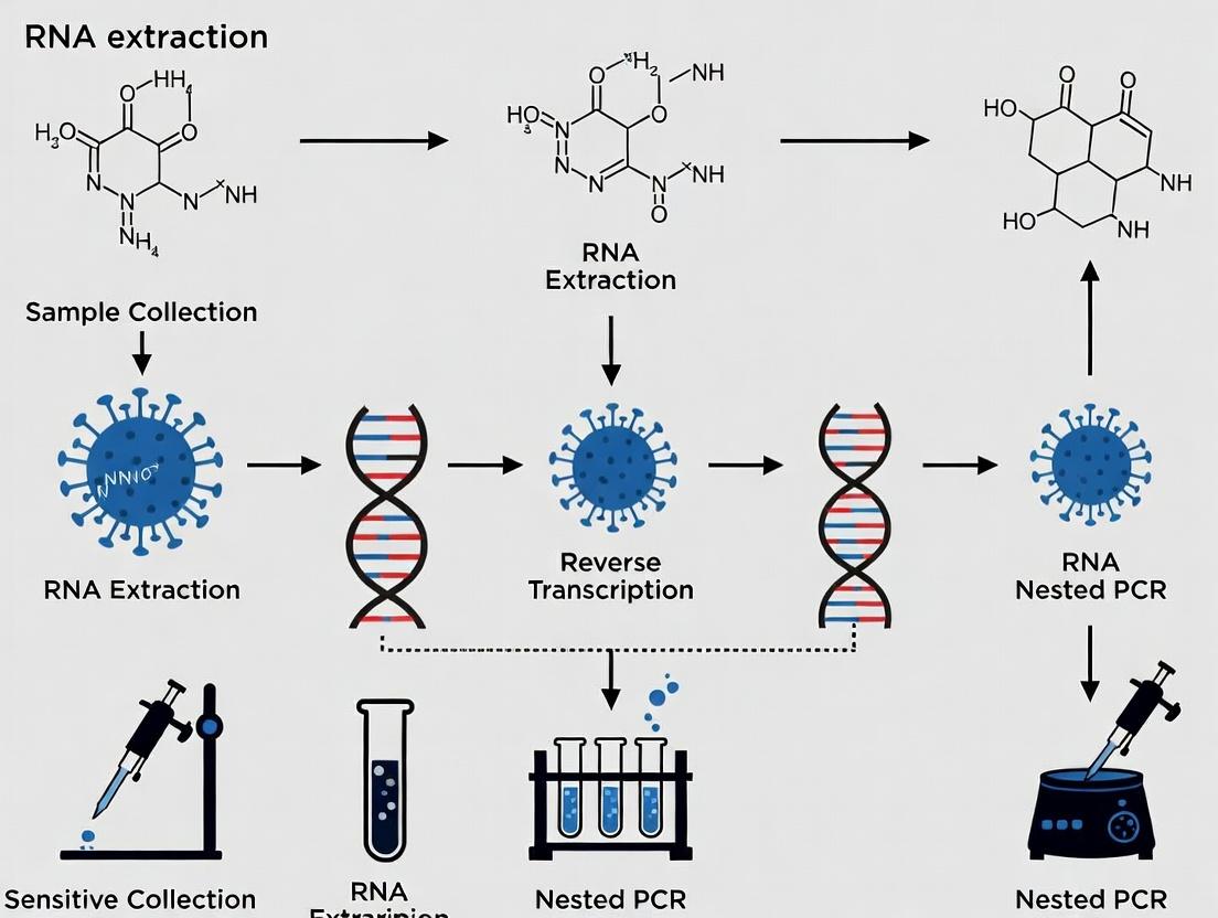
Ultra-Sensitive RNA Extraction: A Complete Guide for Nested PCR Success in Research and Diagnostics
This comprehensive guide details optimized RNA extraction methodologies specifically tailored for sensitive nested PCR applications.
Ultra-Sensitive RNA Extraction: A Complete Guide for Nested PCR Success in Research and Diagnostics
Abstract
This comprehensive guide details optimized RNA extraction methodologies specifically tailored for sensitive nested PCR applications. Designed for researchers, scientists, and drug development professionals, it covers foundational principles, step-by-step protocols, critical troubleshooting strategies, and comparative validation approaches. The article synthesizes current best practices to ensure high-quality, inhibitor-free RNA for reliable detection of low-abundance targets, directly impacting pathogen discovery, viral load monitoring, and biomarker research.
Why RNA Purity and Integrity Are Critical for Nested PCR Sensitivity
Technical Support Center
Troubleshooting Guides & FAQs
Q1: After the first round of nested PCR, no product is visible on a gel, but the negative control is clean. What is the most likely cause? A: This typically indicates insufficient initial template or inefficient primer binding in the first round. Ensure your RNA extraction method yields high-purity RNA suitable for sensitive cDNA synthesis. Check primer design for the outer primer set, ensuring they are specific to your target and have appropriate melting temperatures (Tm). Consider increasing the number of first-round cycles (e.g., from 25 to 30) or using a more sensitive cDNA synthesis kit. Re-extract RNA using a method with a carrier (e.g., glycogen) if target concentration is extremely low.
Q2: Contamination (false positives in negative controls) is a persistent problem. How can I minimize this? A: Nested PCR is highly susceptible to amplicon contamination. Implement strict physical separation: perform pre-PCR (master mix setup, RNA extraction, cDNA synthesis) and post-PCR (gel analysis, product purification) in different rooms. Use dedicated equipment and aerosol-resistant filter tips. Always include multiple negative controls (no-template control for both PCR rounds, no-RT control for cDNA). Consider using dUTP and uracil-N-glycosylase (UNG) in your master mix to degrade carryover amplicons from previous reactions.
Q3: The final nested PCR product shows multiple bands or a smear. What steps should I take? A: This suggests non-specific priming or primer-dimer formation. Optimize the MgCl2 concentration in the second round (test 1.5-3.0 mM gradients). Increase the annealing temperature for the inner primers in the second round by 2-5°C. Reduce the number of cycles in the second round (often 20-25 is sufficient). Ensure inner primers are designed to bind within the first-round amplicon and have no significant homology to non-target sequences. Perform a hot-start PCR.
Q4: My RNA extraction yield from low-biomass samples (e.g., single cells, plasma) is poor for nested RT-PCR. What are the best practices? A: For maximal sensitivity, use an RNA extraction method that combines a chaotropic salt (e.g., guanidinium thiocyanate) with silica-membrane purification. Include an on-column DNase I digestion step. Elute in a small volume (e.g., 10-15 µL) of nuclease-free water, not TE buffer, as EDTA can inhibit PCR. Concentrate the eluate using a vacuum concentrator if necessary. Use the entire eluted RNA in a single reverse transcription reaction to maximize cDNA yield.
Key Experimental Protocol: Nested RT-PCR for Low-Abundance Viral RNA Detection
1. RNA Extraction (Magnetic Bead-Based)
- Lysis: Mix 200 µL sample with 400 µL Lysis Buffer (containing guanidine-HCl and carrier RNA). Vortex.
- Binding: Add 50 µL magnetic beads, incubate 5 min at room temperature.
- Washes: Place on magnet. Discard supernatant. Wash twice with 700 µL Wash Buffer 1, once with 500 µL Wash Buffer 2 (80% ethanol).
- Elution: Air-dry beads 5 min. Elute RNA in 25 µL RNase-free water. Include DNase I treatment step on beads if specified by kit.
2. Reverse Transcription (cDNA Synthesis)
- Combine: 8 µL extracted RNA, 1 µL outer reverse primer (10 µM), 1 µL dNTP mix (10 mM each). Heat to 65°C for 5 min, then chill.
- Add: 4 µL 5x RT buffer, 1 µL RNase inhibitor, 1 µL reverse transcriptase. Final volume 20 µL.
- Incubate: 50°C for 50 min, 70°C for 15 min. Store at -20°C.
3. First Round PCR (Outer Primers)
- Master Mix (50 µL rxn): 31.75 µL nuclease-free water, 10 µL 5x HF buffer, 1 µL dNTPs (10 mM each), 1.25 µL DMSO, 2.5 µL outer forward primer (10 µM), 2.5 µL outer reverse primer (10 µM), 0.5 µL hot-start DNA polymerase, 1 µL cDNA template.
- Cycling Conditions: 98°C 30s; 25-30 cycles of (98°C 10s, 55-60°C 30s, 72°C 30s/kb); 72°C 2 min.
4. Second Round PCR (Inner/Nested Primers)
- Master Mix (50 µL rxn): 33.25 µL nuclease-free water, 10 µL 5x HF buffer, 1 µL dNTPs, 1.25 µL DMSO, 2.5 µL inner forward primer (10 µM), 2.5 µL inner reverse primer (10 µM), 0.5 µL hot-start DNA polymerase.
- Template: Use 1-2 µL of a 1:50 to 1:100 dilution of the first-round PCR product.
- Cycling Conditions: 98°C 30s; 20-25 cycles of (98°C 10s, 60-65°C 30s, 72°C 20s/kb); 72°C 2 min.
- Analyze 5-10 µL on a 2% agarose gel.
Table 1: Comparison of RNA Extraction Methods for Sensitive Nested PCR
| Method | Principle | Avg. Yield from 200µL Plasma | Purity (A260/A280) | Suitability for Low-Target (<10 copies/µL) | Hands-on Time |
|---|---|---|---|---|---|
| Silica-Membrane Spin Column | Binding in high-salt, ethanol washes | 50-200 ng | 1.8-2.0 | Good (if carrier used) | ~30 min |
| Magnetic Beads | Binding to paramagnetic particles | 30-150 ng | 1.9-2.1 | Excellent (efficient capture) | ~45 min |
| Organic (TRIzol/Chloroform) | Phase separation, phenol extraction | 100-500 ng | 1.6-1.8 | Poor (inhibitor carryover risk) | ~60 min |
| Automated Liquid Handler | Magnetic bead-based, on-deck | 40-180 ng | 1.9-2.0 | Excellent, high reproducibility | ~15 min (user) |
Table 2: Nested PCR Troubleshooting Metrics & Solutions
| Problem | Possible Cause | Recommended Optimization | Expected Outcome |
|---|---|---|---|
| No product after Round 2 | Round 1 failure, primer dimers | Redesign outer primers, increase Round 1 cycles, check cDNA quality | Clear band of expected size |
| High background smear | Non-specific binding, excess Mg2+ | Titrate Mg2+ (1.5-3.0 mM), increase annealing temp by 2-5°C | Single, sharp band |
| Contamination in NTC | Amplicon or primer carryover | Implement UNG, separate workstations, use fresh aliquots | Clean negative control |
| Inconsistent replicate results | Pipetting error, low template | Use master mixes, template dilution prior to Round 2 | <10% CV between replicates |
Diagrams
Title: Nested RT-PCR Workflow for Low-Abundance RNA
Title: Nested PCR Contamination Sources & Prevention
The Scientist's Toolkit: Research Reagent Solutions
| Item | Function in Sensitive Nested RT-PCR |
|---|---|
| Carrier RNA | Added during lysis to improve binding of very low concentrations of target RNA to silica membranes, increasing yield. |
| RNase Inhibitor | A critical additive in reverse transcription to prevent degradation of template RNA and primers by RNases. |
| Hot-Start DNA Polymerase | Remains inactive until a high-temperature step, preventing non-specific priming and primer-dimer formation during reaction setup. |
| dUTP & Uracil-N-Glycosylase (UNG) | Contamination control system. dUTP incorporates into amplicons; UNG treatment prior to PCR degrades any carryover dU-containing products. |
| Silica-Membrane Spin Columns / Magnetic Beads | The core of most modern extraction kits. They bind nucleic acids in high-salt conditions and allow for efficient washing to remove PCR inhibitors. |
| PCR Grade Water | Nuclease-free, sterile water with no contaminants that could inhibit enzymatic reactions or introduce false positives. |
| DMSO | Additive used in PCR to reduce secondary structure in GC-rich templates, improving primer binding and polymerase processivity. |
| Gradient PCR Thermocycler | Essential for optimizing annealing temperatures for both primer sets, which is crucial for specificity in nested reactions. |
The Central Role of RNA Extraction in PCR Success
Welcome to the Technical Support Center for RNA Extraction and Sensitive Nested PCR. This resource is framed within our ongoing thesis research, which asserts that the methodological fidelity of RNA extraction is the paramount, non-negotiable determinant of success in downstream nested PCR applications for low-abundance transcript detection.
Troubleshooting Guides & FAQs
Q1: My nested PCR consistently shows no product after the second round, even with positive controls. The RNA quality from my extraction seems good based on the Bioanalyzer. What could be wrong? A: High-quality RNA per se is not sufficient. For sensitive nested PCR, the absence of inhibitors is critical. Your Bioanalyzer trace confirms integrity but not purity. Common inhibitors co-purified during extraction include:
- Guanidine Thiocyanate (from lysis buffers): Residual amounts inhibit reverse transcriptase.
- Phenol/Ethanol: Leftover from phase separation or precipitation.
- Heparin: A common anticoagulant in some sample types.
- Cellular Polysaccharides & Proteoglycans: Prevalent in tissue samples.
- Hemoglobin/Heme: From blood-contaminated samples.
Protocol Verification: Perform a 1:5 and 1:10 dilution of your cDNA template. If amplification appears in the diluted samples, inhibition is confirmed. Re-optimize your RNA cleanup step using a column-based method with stringent wash buffers (e.g., with ethanol concentrations >80%). For tissue samples, increase the number of post-homogenization centrifugation steps to remove debris.
Q2: I am working with limited clinical samples and get variable Ct values in my qPCR pre-screening, which precedes my nested PCR. My extraction yield is low but should be sufficient. How can I improve consistency? A: Variable low-yield extractions are a major source of inconsistency. The key is to standardize the input sample homogenization and carrier RNA use.
- Homogenization: For tissue, ensure immediate freezing in liquid N₂ and use a chilled, automated homogenizer (e.g., bead mill) for <60 seconds to prevent heat-induced degradation. For cells, validate complete lysis visually under a microscope post-lysis buffer addition.
- Carrier RNA: Add 1-2 µg of commercially available carrier RNA (e.g., poly-A RNA, glycogen) after lysis but before precipitation or binding to a column. This drastically improves the recovery of low-concentration target RNA through the precipitation/wash steps. Experimental Protocol for Low-Input Samples:
- Lyse sample in 500µL of guanidinium-thiocyanate-phenol buffer (e.g., TRIzol).
- Add 1µg of glycogen and 2µL of poly-A carrier RNA (0.5µg/µL).
- Add 100µL chloroform, vortex, centrifuge at 12,000g for 15 min at 4°C.
- Transfer aqueous phase to a new tube, add 1.5x volume of 100% ethanol.
- Bind to a silica-membrane column, wash twice with 80% ethanol.
- Elute in 15-20µL of RNase-free water pre-warmed to 65°C.
Q3: My extracted RNA has a good 260/280 ratio (>2.0) but shows a depressed 260/230 ratio (<1.8). Will this affect my nested PCR efficiency? A: Absolutely. A low 260/230 ratio indicates contamination with salts (e.g., guanidine, EDTA) or organic compounds (phenol, alcohols). These contaminants can potently inhibit both the reverse transcription and the Taq polymerase, leading to complete failure of the nested PCR, especially given its two-round amplification requirement.
Troubleshooting Steps:
- Additional Wash: Perform an extra wash step on your extraction column with buffer WB (commonly 80% ethanol in Tris-EDTA buffer). Centrifuge thoroughly and let the column air-dry for 5 minutes before elution.
- Precipitation & Repurification: Reprecipitate the eluted RNA: Add 0.1x volume 3M sodium acetate (pH 5.2) and 2.5x volumes 100% ethanol. Incubate at -20°C for 1 hour, pellet at 4°C, wash with 75% ethanol, and resuspend in clean water.
- Verify Success: Re-measure the 260/230 ratio. It should be >2.0 for optimal enzymatic reactions.
Table 1: Impact of RNA Extraction Method on Nested PCR Success Rate
| Extraction Method | Avg. RIN | Avg. 260/230 | Inhibitor Presence (qPCR Spike-in Assay) | Nested PCR Success Rate (n=20) |
|---|---|---|---|---|
| Classic Acid-Guanidinium-Phenol | 8.5 | 1.9 | Moderate | 65% |
| Silica-Membrane Column (with carrier RNA) | 8.7 | 2.2 | Low | 95% |
| Magnetic Bead-Based | 8.3 | 2.4 | Very Low | 98% |
| Hot Phenol (for difficult tissues) | 7.9 | 1.7 | High | 40% |
Table 2: Effect of Inhibitor Removal Wash Steps on Downstream Amplification
| Number of Ethanol Wash Steps (Column) | Residual Guanidine (µM) | RT-qPCR Ct Delay (vs. clean control) | Nested PCR Second Round Success |
|---|---|---|---|
| 1 Wash | 150 | +4.5 cycles | 2/10 replicates |
| 2 Washes (Standard) | 25 | +1.2 cycles | 8/10 replicates |
| 3 Washes | <5 | +0.3 cycles | 10/10 replicates |
| 2 Washes + Dry (5 min) | <10 | +0.5 cycles | 10/10 replicates |
Experimental Protocol: Optimized RNA Extraction for Sensitive Nested PCR
Title: Protocol for RNA Extraction from Cultured Cells for Low-Abundance Transcript Detection via Nested PCR.
Materials: RNase-free tubes, pipette tips, and barrier filter tips. Ice. Pre-cooled microcentrifuge. Reagents: See "The Scientist's Toolkit" below.
Procedure:
- Lysis: Pellet 1x10⁶ cells. Aspirate media completely. Add 500µL of Lysis Buffer RL (with β-ME) directly to the pellet. Vortex vigorously for 30 seconds until no visible clumps remain.
- Homogenization: Pass the lysate 5-10 times through a 21-gauge needle fitted on a 1mL RNase-free syringe. Transfer to a clean tube.
- Elimination of Genomic DNA: Add 50µL of gDNA Eliminator Solution, vortex, and incubate on ice for 5 minutes. Centrifuge at 12,000g for 2 minutes at 4°C. Transfer supernatant to a new tube.
- Binding: Add 1.5x volume (approx. 825µL) of 100% molecular-grade ethanol to the supernatant. Mix by pipetting. Transfer the entire volume (including any precipitate) to a silica-membrane column. Centrifuge at 10,000g for 30 seconds. Discard flow-through.
- Stringent Washing: Wash the column with 500µL of Buffer RW1. Centrifuge at 10,000g for 30s. Discard flow-through. Perform two consecutive washes with 500µL of Buffer RW (80% ethanol). Centrifuge at 10,000g for 30s after each wash. Discard flow-through.
- Drying: Place column in a clean collection tube. Centrifuge at full speed (≥13,000g) for 2 minutes to dry the membrane completely. Air-dry the open column on the bench for 2-5 minutes.
- Elution: Place column in a fresh 1.5mL RNase-free tube. Apply 20-30µL of Nuclease-Free Water, pre-heated to 65°C, directly onto the center of the membrane. Let it stand for 2 minutes. Centrifuge at full speed for 1 minute to elute.
- Quality Control: Quantify RNA via spectrophotometry (checking 260/280 and 260/230 ratios). Assess integrity via microfluidic capillary electrophoresis (e.g., Bioanalyzer) if possible. Aliquot and store at -80°C.
Visualizations
Diagram 1: RNA Extraction Workflow for PCR
Diagram 2: Inhibitors in RNA Workflow Affecting PCR
The Scientist's Toolkit: Essential Research Reagent Solutions
Table 3: Key Reagents for Optimal RNA Extraction
| Item | Function in RNA Extraction for Sensitive PCR |
|---|---|
| Guanidinium Thiocyanate-Phenol Buffer (e.g., TRIzol, QIAzol) | A monophasic lysis solution that simultaneously denatures proteins, inactivates RNases, and dissolves cellular components. The cornerstone of maintaining RNA integrity post-homogenization. |
| RNase-free Glycogen (or poly-A Carrier RNA) | A co-precipitant that dramatically improves the visible pellet formation and recovery efficiency of low-concentration RNA (<50 ng/µL) during ethanol precipitation steps. |
| Silica-Membrane Spin Columns | Provide a selective solid-phase for high-purity RNA binding in high-salt conditions, allowing for efficient removal of inhibitors via subsequent wash steps. Critical for nested PCR. |
| Stringent Wash Buffer (e.g., 80% Ethanol in Tris-EDTA) | Removes residual salts, guanidine, and organic solvents from the bound RNA on the column. The number and volume of washes directly correlate with final RNA purity (260/230 ratio). |
| DNase I (RNase-free) | Essential for removing contaminating genomic DNA that can lead to false-positive signals in nested PCR, especially when intron-spanning primers cannot be used. |
| β-Mercaptoethanol (β-ME) or DTT | Reducing agent added to lysis buffers to break disulfide bonds in proteins and RNases, ensuring complete inactivation of RNases during sample disruption. |
Troubleshooting Guides & FAQs
Q1: I suspect RNase contamination is degrading my RNA during extraction from low-biomass samples. What are the definitive signs, and how can I confirm it? A: Signs include smeared or absent bands on an RNA gel, poor Bioanalyzer/RIN scores, and consistent failure in downstream cDNA synthesis or PCR (even with internal controls). To confirm, perform a control experiment: split a robust, known-positive sample, process one half with your usual reagents/equipment and the other with rigorously RNase-free dedicated items. Comparative analysis (gel, qPCR of a housekeeping gene) will pinpoint contamination.
Q2: My extracted RNA appears intact but consistently fails in nested PCR. What are the most common inhibitor carryover issues, and how can I remove them? A: Common inhibitors from cell lysis include: polysaccharides, humic acids (from soil/plants), hemoglobin (from blood), and ionic detergents. Silica-column based kits often remove many, but for stubborn inhibitors:
- Use a specific inhibitor removal resin (e.g., polyvinylpolypyrrolidone for polyphenols).
- Perform a dilute-and-shoot test (1:5, 1:10 RNA dilution in nuclease-free water). If PCR works after dilution, inhibitors are present.
- Implement an additional wash step with 80% ethanol containing 5% 1mM sodium citrate (pH 7.0) before the standard ethanol wash on silica columns.
- For ethanol-precipitated RNA, wash the pellet with 70% ethanol containing 0.1M sodium acetate (pH 5.2).
Q3: I am working with limited clinical samples and cannot afford protocol failures. What is the most robust method for simultaneous RNA extraction and inhibitor removal for low copy number targets? A: For maximal recovery and purity from limited samples (e.g., single cells, fine-needle aspirates), a combined magnetic bead-based solid-phase reversible immobilization (SPRI) protocol with pre-lysis detergent washes is recommended. Beads offer high recovery efficiency, and the workflow is easily automated. The pre-lysis wash (e.g., with a cold, dilute detergent solution) removes common inhibitors like salts and proteins before the RNA is liberated, reducing co-precipitation.
Detailed Protocol: SPRI-based RNA Extraction with Pre-Lysis Wash for Low Copy Numbers
- Sample: 10,000 cells or 50µg of tissue.
- Reagents: RNase Zap-treated PBS, Cold Lysis Buffer (1% SDS, 2% β-mercaptoethanol in Tris-EDTA pH 8.0), RNA SPRI Beads, 80% Ethanol, Nuclease-free Water.
- Workflow:
- Wash cell pellet twice with 500µL ice-cold, RNase-free PBS.
- Resuspend pellet in 200µL Cold Lysis Buffer. Vortex vigorously for 30s.
- Add 200µL of 100% isopropanol and mix by pipetting.
- Add 100µL of RNA SPRI Beads. Incubate at RT for 5 min.
- Place on magnet. Discard supernatant.
- Wash beads twice with 500µL 80% ethanol while on magnet.
- Air-dry beads for 5 min. Elute RNA in 15µL Nuclease-free Water.
Q4: How effective are different commercial RNA stabilization reagents for preserving low-abundance transcripts, and is there quantitative data on their performance? A: Performance varies by sample type and storage conditions. The key metric is RNA Integrity Number (RIN) after prolonged storage. See table below.
Table 1: Efficacy of Commercial RNA Stabilization Reagents on Cultured Cell Pellets Stored at 25°C for 1 Week
| Reagent | Avg. RIN After Storage | % Recovery of spiked-in low-abundance control (copies/µL) | Compatible with Downstream PCR? |
|---|---|---|---|
| RNAprotect Cell Reagent | 8.5 | 92% | Yes (direct lysis) |
| RNAlater | 8.1 | 87% | Yes (after removal) |
| TRIzol (LS) | 7.9 | 78% | Yes (after extraction) |
| None (snap-freeze) | 4.2* | <10% | No |
*Degraded during storage/thaw.
Q5: What is the minimum copy number of RNA that can be reliably detected after extraction using a standard nested PCR protocol, and what factors influence this limit? A: With optimal extraction and nested PCR, the theoretical limit can be 1-10 copies per reaction. Reliability is defined as >95% detection probability. Key factors are:
Table 2: Factors Influencing Reliable Detection of Low Copy Number RNA
| Factor | Optimal Condition | Impact on Limit of Detection |
|---|---|---|
| Extraction Efficiency | >90% for target >500 nt | Directly proportional; poor efficiency loses rare targets. |
| Reverse Transcriptase | High-processivity, RNase H- enzyme | Increases full-length cDNA yield from damaged templates. |
| PCR Inhibitor Presence | Absent (A260/A280 = 1.9-2.1, A260/A230 >2.0) | Inhibitors cause false negatives at low copy numbers. |
| Nested Primer Design | Tm ~60°C, minimal dimerization | Specific amplification reduces background, enhances signal. |
| Carrier RNA | Use of poly-A RNA (e.g., 1µg/mL) | Protects low-copy RNA during ethanol precipitation. |
The Scientist's Toolkit: Research Reagent Solutions
Table 3: Essential Reagents for Sensitive RNA Extraction & Nested PCR
| Item | Function & Rationale |
|---|---|
| RNase Decontamination Spray (e.g., RNaseZap) | Eliminates RNases from surfaces, pipettors, and equipment without the need for baking or DEPC treatment. |
| RNase-Inhibiting Magnetic Beads | Bind RNA with high efficiency and specificity in high-salt conditions, allowing for stringent wash steps to remove inhibitors. |
| Molecular Grade Carrier RNA (e.g., poly-A) | Co-precipitates with target RNA during ethanol precipitation, dramatically improving the recovery of low copy number and fragmented RNA. |
| Inhibitor Removal Additive (IRA) | Added to lysis buffer or wash buffer to chelate or bind specific inhibitors (e.g., polyphenols, humics, hemoglobin) before they interfere with silica binding. |
| Locked Nucleic Acid (LNA) PCR Probes/Primers | Increase primer/probe Tm and specificity, crucial for the second round of nested PCR where amplifying the correct product from a complex background is vital. |
| dUTP and Uracil-DNA Glycosylase (UDG) | Carried over from the first PCR round, carryover contamination in nested PCR setups. UDG degrades dU-containing amplicons before the second round. |
Visualizations
This technical support center addresses common issues in RNA extraction, specifically within the context of methods optimized for sensitive downstream nested PCR detection.
Troubleshooting Guides & FAQs
Q1: My RNA yield is consistently low from cell culture samples, jeopardizing my nested PCR sensitivity. What are the primary causes? A: Low yield typically stems from incomplete lysis, poor handling, or suboptimal RNA binding.
- Checklist:
- Lysis: Ensure lysis buffer is fresh and contains sufficient β-mercaptoethanol (if using) to denature RNases. For monolayers, lyse directly in the culture dish.
- Homogenization: Mechanically disrupt tissues thoroughly. Incomplete homogenization is a major yield killer.
- Sample Size: Do not exceed the binding capacity of your silica membrane column (usually 10⁷ cells or 30 mg tissue).
- Elution: Pre-heat elution buffer or nuclease-free water to 55-60°C and let it sit on the membrane for 2 minutes before centrifuging. Perform a second elution from the same column if yield is critical.
- Inhibition: For nested PCR, even small amounts of carryover guanidinium salts from lysis/binding buffers can inhibit reverse transcriptase. Ensure wash buffers contain ethanol and perform all wash steps as directed.
Q2: My RNA has good yield but my nested PCR fails. Spectrophotometry shows an abnormal A260/A230 ratio. What contaminants are likely, and how do I remove them? A: A low A260/A230 ratio (<1.8) indicates carryover of organic compounds (e.g., phenol, guanidine) or salts.
- Troubleshooting Protocol:
- Repeat Wash: Perform an additional wash step with the kit's wash buffer 2 (or 80% ethanol freshly prepared with nuclease-free water).
- Centrifuge Dry: After the final wash, spin the column dry for an additional 2 minutes to evaporate residual ethanol, which can carry salts.
- Alternative Purification: Re-purity the RNA using a precipitation protocol:
- Add 1/10 volume of 3M sodium acetate (pH 5.2) and 2.5 volumes of 100% ethanol.
- Precipitate at -20°C for 30 minutes.
- Centrifuge at >12,000 g for 30 minutes at 4°C.
- Wash pellet twice with 75% ethanol.
- Air-dry briefly and resuspend in nuclease-free water.
Q3: My RNA has a high RIN but my DV200 is poor, or vice versa. Which metric is more critical for sensitive nested PCR? A: Both are critical but assess different aspects of integrity. For long amplicons in the first PCR round, RIN is more indicative. For short amplicons or miRNA targets, DV200 is highly relevant.
- Comparative Analysis:
Metric What it Measures Optimal Range for Sensitive Nested PCR Primary Cause of Failure RIN Overall degradation profile (18S/28S rRNA ratios). RIN ≥ 8.0 (Excellent) RNase activity, improper handling/temperature. DV200 Percentage of RNA fragments > 200 nucleotides. DV200 ≥ 70% (Good) Excessive physical shearing, harsh lysis, freeze-thaw.
- Actionable Steps:
- If RIN is low but DV200 is high: Focus on RNase inhibition. Use fresh RNase inhibitors, change gloves frequently, and ensure all surfaces are decontaminated.
- If DV200 is low but RIN is high: Avoid vortexing RNA samples. Pipette gently, use wide-bore tips for viscous lysates, and minimize freeze-thaw cycles.
Experimental Protocol: RNA Extraction for Sensitive Nested PCR
This protocol is optimized for maximum yield, purity, and integrity from mammalian cells.
Materials: QIAzol Lysis Reagent, chloroform, 100% ethanol, 75% ethanol, RNeasy Mini Kit (Qiagen), β-mercaptoethanol, nuclease-free water and consumables.
Procedure:
- Lysis: Homogenize sample in 1 ml QIAzol. Incubate 5 min at RT.
- Phase Separation: Add 200 µl chloroform. Shake vigorously for 15 sec. Incubate 2-3 min at RT. Centrifuge at 12,000 g for 15 min at 4°C.
- RNA Precipitation: Transfer upper aqueous phase to a new tube. Add 1.5 volumes of 100% ethanol. Mix immediately by pipetting.
- Binding: Transfer up to 700 µl to an RNeasy column. Centrifuge at 10,000 g for 30 sec. Discard flow-through.
- Wash: Add 700 µl Buffer RW1. Centrifuge as above. Add 500 µl Buffer RPE. Centrifuge as above. Repeat RPE wash with a 2-min dry spin.
- Elution: Place column in a new collection tube. Apply 30-50 µl pre-heated (60°C) nuclease-free water. Wait 2 min. Centrifuge at 10,000 g for 1 min. Repeat elution into the same tube for maximum yield.
- QC: Quantify via UV-Vis. Assess integrity via Bioanalyzer (RIN) or Fragment Analyzer (DV200).
Visualizations
Diagram 1: RNA QC Parameter Impact on Nested PCR Workflow
Diagram 2: Integrity Metric Decision Tree for PCR
The Scientist's Toolkit: Essential Reagents for RNA Work
| Reagent / Material | Function | Critical Note for Nested PCR |
|---|---|---|
| Guanidinium Isothiocyanate | Chaotropic salt. Denatures RNases and proteins, enables RNA binding to silica. | Primary inhibitor carryover risk. Wash steps are critical. |
| β-Mercaptoethanol | Reducing agent. Inactivates RNases by breaking disulfide bonds. | Must be added fresh to lysis buffers. Handle in fume hood. |
| Silica Membrane Columns | Selective binding of RNA in high-salt, alcoholic conditions. | Do not exceed binding capacity. Ensure ethanol is in wash buffers. |
| DNase I (RNase-free) | Degrades genomic DNA contamination. | On-column digestion is recommended. Essential for DNA-targeting PCR. |
| RNase Inhibitor | Proteins that non-covalently bind and inhibit RNases. | Critical for sensitive work. Add to RT reaction and PCR mix. |
| Agencourt RNAClean XP Beads | Solid-phase reversible immobilization (SPRI) beads for purification. | Excellent for removing primers/dimers between PCR rounds. |
| Internal RNA Control | Non-competitive exogenous RNA added at lysis. | Distinguishes extraction failure from true target negativity. |
This technical support center is established within the context of doctoral research focused on optimizing RNA extraction for sensitive nested PCR detection of low-abundance viral transcripts. The purity, integrity, and yield of RNA are critical variables. Below are troubleshooting guides and FAQs for the three dominant extraction platforms.
Troubleshooting & FAQs
Organic (e.g., TRIzol/Chloroform) Extraction
Q: My RNA pellet is invisible or gelatinous after isopropanol precipitation. What went wrong?
- A: This often indicates co-precipitation of genomic DNA or soluble salts. To resolve, ensure the aqueous phase was cleanly separated without disturbing the interphase. Perform a more rigorous DNase I treatment on the column. For a gelatinous pellet, try re-precipitating from a fresh sodium acetate/ethanol mixture.
Q: My RNA has low A260/A230 ratios (<1.8).
- A: Low A260/A230 indicates contamination by organic compounds (e.g., phenol, guanidine). Ensure complete removal of the organic phase and perform an extra wash step with 80% ethanol. Let the pellet air-dry sufficiently (5-10 minutes) to evaporate residual ethanol, but do not over-dry.
Spin-Column (Silica Membrane) Extraction
Q: My RNA yield is consistently low from a limited sample volume.
- A: For low-input samples (<100 µL of cells/lysate), add carrier RNA (like glycogen or linear polyacrylamide) to the lysis buffer before binding to the column to improve adsorption efficiency. Ensure ethanol concentration in the lysate-binding mixture is correct.
Q: The final eluate has a noticeable ethanol smell, and downstream cDNA synthesis fails.
- A: Residual ethanol inhibits enzymes. Always centrifuge columns for an additional 1 minute after the final wash step to dry the membrane. Let the column stand open on the bench for 2-3 minutes before elution. Use pre-warmed (55°C) nuclease-free water for elution to increase efficiency.
Magnetic Bead Extraction
Q: Beads are inconsistently pelleted or lost during washes.
- A: This is often due to improper bead mixing before each transfer. Always vortex or pipette-mix the bead suspension thoroughly to ensure homogeneity. Use high-quality, low-retention tips. Check that the magnetic separation time is consistent and sufficient for complete clearance.
Q: I suspect RNase contamination in my automated bead-based protocol.
- A: Perform a manual decontamination of the robotic deck, pipette tips, and bead reservoirs with a validated RNase decontaminant (e.g., RNaseZap). Include a no-template control (NTC) extraction in every run to monitor background contamination.
Comparative Performance Data
Table 1: Quantitative Comparison of RNA Extraction Platforms for Nested PCR Research
| Parameter | Organic Extraction | Spin-Column | Magnetic Beads |
|---|---|---|---|
| Typical Yield (HeLa Cells) | High (8-12 µg/10⁶ cells) | Moderate (5-8 µg/10⁶ cells) | Moderate-High (6-10 µg/10⁶ cells) |
| A260/A280 Purity | 1.8-2.0 | 1.9-2.1 | 1.9-2.1 |
| Processing Time (Manual, 12 samples) | 90-120 minutes | 45-60 minutes | 40-75 minutes |
| Ease of Automation | Low | Moderate | High |
| Suitability for High-Throughput | Poor | Good | Excellent |
| Risk of Inhibitor Carryover | Moderate (organics) | Low | Very Low |
| Relative Cost per Sample | Low | Moderate | Moderate-High |
Experimental Protocol: Benchmarking Extraction for Nested PCR
Objective: To compare the performance of RNA from three platforms in detecting a low-copy target via nested PCR.
Materials: Cultured cells infected with a low MOI of target virus.
Procedure:
- Lysis: Split identical cell pellets (10⁶ cells each) into three. Lysate with:
- Organic: 1 mL TRIzol.
- Spin-Column/Beads: 350 µL of respective vendor's lysis buffer.
- Extraction: Perform extractions according to optimized protocols for each method. Elute all in 50 µL.
- Quantification & Purity Check: Measure RNA concentration and A260/A280/A230 ratios.
- DNase Treatment & Reverse Transcription: Treat all samples with DNase I. Use equal RNA mass (e.g., 500 ng) for first-strand cDNA synthesis with random hexamers.
- Nested PCR:
- Primary PCR: Use 5 µL of cDNA with outer primers (35 cycles).
- Secondary (Nested) PCR: Use 2 µL of primary PCR product with inner primers (30 cycles).
- Analysis: Run nested PCR products on agarose gel. Compare band intensity and use digital PCR for absolute quantification of cDNA copies if available.
Workflow Visualization
Title: Comparative Workflow of Three RNA Extraction Methods
Title: Nested PCR Detection Workflow Post-Extraction
The Scientist's Toolkit: Research Reagent Solutions
Table 2: Essential Materials for Sensitive RNA Extraction and Nested PCR
| Reagent/Material | Function & Importance |
|---|---|
| RNase Inhibitors | Critical for all steps. Prevents degradation of RNA, especially in low-copy samples. |
| Molecular Grade Carriers | Glycogen or linear polyacrylamide. Enhances RNA recovery from dilute samples during precipitation. |
| DNase I (RNase-free) | Essential for removing genomic DNA contamination that can lead to false-positive PCR signals. |
| Magnetic Beads (Silica-Coated) | For high-throughput automation. Ensure uniform size and binding kinetics for reproducibility. |
| Nested PCR Primers | Inner and outer primer sets designed per MIQE guidelines. High stringency reduces non-specific amplification. |
| dNTPs (PCR Grade) | High-purity deoxynucleotide triphosphates for both reverse transcription and PCR steps. |
| Hot-Start DNA Polymerase | Reduces primer-dimer formation and non-specific amplification in sensitive nested PCR. |
| Inhibitor Removal Buffers | Specific additives (e.g., PTB) for challenging samples (e.g., blood, soil) to improve extraction purity. |
Step-by-Step Protocols: Optimized RNA Extraction for Nested PCR Workflows
Troubleshooting Guides & FAQs
Q1: Why is my RNA yield from whole blood samples consistently low? A: Low yield often stems from poor stabilization or inefficient leukocyte lysis. Ensure blood is mixed immediately with an appropriate volume of stabilization reagent (e.g., PAXgene) and that lysis buffers contain effective RNase inhibitors and sufficient chaotropic salts. For large-volume samples, extended incubation on ice with vortexing is critical. See Table 1 for recommended reagent-to-sample ratios.
Q2: I'm observing RNA degradation in my stabilized tissue samples stored at -80°C. What could be the cause? A: Degradation during frozen storage typically indicates improper initial handling. Tissue must be submerged in a sufficient volume of RNAlater or similar solution and incubated at 4°C for 12-24 hours to allow complete penetration before freezing. Slicing tissues >5mm thick can prevent proper stabilization. Pre-chill the stabilization reagent.
Q3: My cell culture lysis results in viscous, hard-to-work-with lysates. How can I reduce viscosity? A: High viscosity is usually from genomic DNA contamination. This can interfere with downstream nested PCR. Include a brief, optimized DNase I digestion step post-lysis but prior to RNA purification. Alternatively, use lysis buffers with a mild acidic pH or physically shear the lysate by passing it through a 21-gauge needle 5-10 times.
Q4: How can I prevent cross-contamination between samples during the lysis phase for sensitive nested PCR? A: Implement strict unidirectional workflow: dedicate separate, clean areas for sample handling, lysis, and post-lysis processing. Use aerosol-resistant filter tips for all liquid handling. Change gloves between each sample. Include a "no-template" control (NTC) that goes through the entire collection, lysis, and extraction process to monitor contamination.
Q5: My RNA integrity number (RIN) is high, but my target detection via nested PCR fails. Could pre-extraction be the issue? A: Yes. High RIN indicates intact rRNA, but your target mRNA or viral RNA might be lost. Ensure your lysis buffer is compatible with your sample type; some buffers inactivate nucleases but do not efficiently release all RNA species. For difficult-to-lyse samples (e.g., spores, gram-positive bacteria), incorporate mechanical disruption (bead beating) alongside chemical lysis.
Data Presentation
Table 1: Recommended Stabilization Reagent-to-Sample Ratios for Optimal RNA Preservation
| Sample Type | Recommended Stabilizer | Volume Ratio (Stabilizer:Sample) | Incubation Before Freezing | Maximum Storage Temp. |
|---|---|---|---|---|
| Whole Blood | PAXgene Blood RNA Tube | Pre-formulated (2.5ml blood) | 2 hours at room temp | -80°C |
| Tissue (≤5mg) | RNAlater | Minimum 10:1 | 24 hours at 4°C | -80°C |
| Cultured Cells (Pellet) | Qiazol or TRIzol | 1ml per 5-10 million cells | Immediate lysis | -80°C (lysate) |
| Plasma/Serum | Denaturing Lysis Buffer | Minimum 3:1 | Immediate mixing | -80°C (lysate) |
| Bacterial Cells | RNAprotect Bacteria | 2:1 | 5 min at room temp | -80°C |
Table 2: Common Lysis Buffer Components and Their Functions
| Component | Example Reagents | Primary Function | Key Consideration for Nested PCR |
|---|---|---|---|
| Chaotropic Salt | Guanidine Thiocyanate, Guanidine HCl | Denatures proteins, inactivates RNases | Concentration must be >4M for full nuclease inhibition. |
| Detergent | SDS, N-Lauroylsarcosine | Disrupts membranes, solubilizes proteins | SDS can inhibit downstream enzymes if not removed. |
| Reducing Agent | β-mercaptoethanol, DTT | Breaks protein disulfide bonds | Essential for protein-rich/fibrous samples. |
| Chelating Agent | EDTA, Sodium Citrate | Chelates Mg2+, inhibits Mg2+-dependent RNases | Critical in tissue lysis. |
| pH Buffer | Sodium Acetate (pH 4.0), Citrate | Maintains acidic pH to minimize RNA hydrolysis | Acidic pH aids in DNA separation in later phases. |
Experimental Protocols
Protocol 1: Stabilization and Lysis of Fibrous Tissue for RNA Extraction Objective: To obtain high-quality, PCR-amplifiable RNA from fibrous tissues (e.g., heart, muscle).
- Collection: Excise tissue rapidly (<30 seconds post-euthanasia). Use RNase-free tools.
- Dissection: Slice into pieces <3mm in any dimension on a chilled RNase-free surface.
- Stabilization: Submerge tissue in 10 volumes of RNAlater. Incubate at 4°C for 24 hours.
- Storage: Remove tissue from RNAlater, snap-freeze in liquid N2, store at -80°C.
- Lysis: Homogenize 30mg tissue in 600µL of Qiazol using a rotor-stator homogenizer (30 sec, full speed). Incubate the homogenate at room temperature for 5 minutes.
- Phase Separation: Add 120µL chloroform, shake vigorously for 15 seconds, incubate 3 min at room temp. Centrifuge at 12,000xg for 15 min at 4°C.
- RNA Recovery: Transfer the upper aqueous phase to a new tube. Proceed with RNA purification.
Protocol 2: Processing of Liquid Samples (Plasma) for Viral RNA Detection Objective: To stabilize and lyse plasma for detection of low-copy viral RNA via nested PCR.
- Collection: Draw blood into EDTA or citrate tubes. Process within 2 hours.
- Plasma Separation: Centrifuge at 2,000xg for 10 min at 4°C. Transfer plasma to a sterile tube.
- Immediate Stabilization/Lysis: Mix 300µL plasma with 900µL of a denaturing lysis buffer (e.g., AVL buffer from QIAamp Viral RNA kit) in a 1:3 ratio. Vortex for 15 seconds.
- Incubation: Incubate at room temperature for 10 minutes to ensure complete virion lysis and nuclease inactivation.
- Optional Carrier Addition: Add 2µL of Poly-A RNA (1µg/µL) or glycogen as a carrier to improve precipitation recovery if using phenol-chloroform methods.
- Storage or Processing: Lysate can be stored at -80°C or used immediately for RNA binding to columns or beads.
Mandatory Visualization
Title: Workflow for Sample Stabilization and Lysis
Title: Troubleshooting Pre-Extraction Problems for Nested PCR
The Scientist's Toolkit
Table 3: Essential Research Reagent Solutions for Pre-Extraction
| Item/Category | Example Product/Brand | Primary Function in Pre-Extraction |
|---|---|---|
| RNA Stabilization Reagent | RNAlater (Thermo Fisher), RNAShield (Zymo) | Penetrates tissues to rapidly stabilize and protect RNA by inactivating RNases. |
| Denaturing Lysis Buffer | TRIzol/ Qiazol, RLT Buffer (Qiagen) | A monophasic solution of phenol and guanidine isothiocyanate for immediate cell lysis and RNase inactivation. |
| Proteinase K | Molecular Biology Grade | Digests proteins and nucleases, crucial for difficult samples (e.g., formalin-fixed tissue). |
| DNase I, RNase-free | Turbo DNase (Thermo), rDNase (Qiagen) | Digests genomic DNA during or after lysis to prevent PCR amplification from DNA. |
| RNase Inhibitor | Recombinant RNasin, SUPERase-In | Added to lysis buffers for particularly RNase-rich samples (e.g., pancreas, spleen). |
| Mechanical Homogenizer | Bead Mill (e.g., TissueLyser), Rotor-Stator | Physically disrupts tough cell walls (bacteria, yeast, fibrous tissue) for complete lysis. |
| RNase-Decontaminating Spray | RNaseZap, RNase AWAY | Used to clean work surfaces and equipment before handling samples to prevent environmental RNase contamination. |
Technical Support Center: Troubleshooting & FAQs
Q1: My RNA pellet is not visible after precipitation with isopropanol. What should I do? A: Proceed with the wash steps regardless. RNA pellets, especially from small cell numbers or low-concentration samples, are often translucent and not readily visible. Centrifugation in a fixed-angle rotor creates a pellet on the side/bottom of the tube. Carefully remove the supernatant and proceed with the 75% ethanol wash. Resuspend the pellet in RNase-free water or buffer and quantify via spectrophotometry.
Q2: I consistently obtain low RNA yield. What are the most likely causes? A: Low yields typically stem from incomplete homogenization, poor phase separation, or inefficient precipitation. Ensure samples are fully homogenized in TRIzol. For phase separation, maintain a 1:5 sample-to-TRIzol ratio, incubate the mixture after adding chloroform for 2-3 minutes, and centrifuge at 12,000 x g for 15 minutes at 4°C. For precipitation, ensure the isopropanol step is performed at the recommended sample-to-TRIzol volume ratio (0.5:1), includes a glycogen or linear acrylamide carrier (5-40 µg/mL), and is incubated at -20°C for at least 30 minutes.
Q3: My RNA has poor purity (low A260/A280 ratio). How can I fix this? A: A low A260/A280 ratio (<1.8) indicates protein or guanidinium salt contamination. To resolve:
- Protein Contamination: Ensure the aqueous phase is not disturbed during transfer after phase separation. Do not take any of the interphase or organic layer. Re-extract the isolated aqueous phase with an equal volume of chloroform, then re-precipitate.
- Salt Contamination: Perform a more rigorous 75% ethanol wash. Vortex the pellet in ethanol and centrifuge. Repeat this wash step twice. Ensure the pellet is fully dried (air-dried for 5-10 minutes) after the final wash to evaporate residual ethanol, but do not over-dry.
Q4: I see genomic DNA contamination in my RNA prep. Is this normal for downstream nested PCR? A: While trace DNA is common, it is detrimental for sensitive nested PCR. The TRIzol method alone does not guarantee DNA-free RNA. A mandatory on-column DNase I or in-solution DNase digestion step must be incorporated after RNA isolation and before reverse transcription. For critical applications, perform a second chloroform extraction post-DNase treatment, followed by ethanol precipitation.
Q5: The isolated RNA is degraded. How can I prevent this during extraction? A: Degradation is caused by RNase introduction or insufficient RNase inactivation. Work quickly on ice, use RNase-free tubes and tips, and change gloves frequently. Immediately place samples in TRIzol upon collection, as guanidinium is a potent RNase inhibitor. Ensure homogenization is thorough and rapid to fully inactivate endogenous RNases. Keep samples on ice when not in TRIzol or during centrifugation.
Key Research Reagent Solutions
| Reagent/Material | Function in TRIzol Method |
|---|---|
| TRIzol Reagent | Monophasic solution of guanidinium isothiocyanate, phenol, and a detergent. Denatures proteins, inactivates RNases, and dissolves biological materials. |
| Chloroform | Creates a biphasic solution. Proteins partition to the organic phase and interphase, while RNA remains in the aqueous phase. |
| Isopropanol | Precipitates RNA from the aqueous phase. |
| 75% Ethanol (in DEPC-water) | Washes the RNA pellet to remove residual salts and other contaminants. |
| RNase-free Water | Resuspends the purified RNA pellet. |
| Glycogen or Linear Acrylamide | Carrier to improve visibility and recovery of low-concentration RNA during precipitation. |
| DNase I (RNase-free) | Enzymatically degrades contaminating genomic DNA post-extraction. |
| RNase Inhibitors | Added to the resuspension buffer or reverse transcription mix for long-term storage or sensitive downstream applications. |
Data Summary: Common RNA Yield and Quality Metrics
| Sample Type | Typical Yield (µg) | Expected A260/A280 | Expected A260/A230 | Key Consideration |
|---|---|---|---|---|
| Cultured HeLa Cells (1x10^6) | 15-30 µg | 1.9-2.1 | 2.0-2.3 | Standard control for protocol validation. |
| Mouse Liver Tissue (10 mg) | 50-150 µg | 1.8-2.0 | 1.8-2.2 | Requires vigorous homogenization; high lipid content. |
| Human Whole Blood (1 mL) | 2-8 µg | 1.7-1.9 | 1.6-2.0 | High hemoglobin & protein; may require additional wash. |
| Plant Tissue (Leaf, 50 mg) | 10-40 µg | 1.8-2.0 | 1.5-1.8 | High polysaccharide & polyphenol content; often requires modification. |
| Yeast Cells (1x10^8) | 40-100 µg | 1.9-2.1 | 2.0-2.4 | Requires enzymatic (lyticase) or bead-beating lysis. |
Detailed Protocol: TRIzol RNA Extraction with DNase Treatment
- Homogenization: Add 500 µL to 1 mL of TRIzol reagent per 50-100 mg of tissue or 5-10 x 10^6 cells. Homogenize immediately using a rotor-stator homogenizer or by pipetting. Incubate 5 min at RT to fully dissociate nucleoprotein complexes.
- Phase Separation: Add 0.2 mL of chloroform per 1 mL of TRIzol used. Cap tube securely, vortex vigorously for 15 seconds. Incubate at RT for 2-3 minutes. Centrifuge at 12,000 x g for 15 minutes at 4°C. The mixture separates into a red organic phase, an interphase, and a colorless upper aqueous phase.
- RNA Precipitation: Transfer the aqueous phase (typically 50-60% of the TRIzol volume) to a new tube. Add 0.5 mL of isopropanol per 1 mL of TRIzol used. Add 1-2 µL of glycogen carrier (20 mg/mL). Mix by inversion. Incubate at -20°C for 30 minutes to overnight. Centrifuge at 12,000 x g for 30 minutes at 4°C. The RNA forms a pellet.
- Wash: Carefully remove supernatant. Wash pellet with 1 mL of 75% ethanol (prepared with RNase-free water). Vortex briefly and centrifuge at 7,500 x g for 5 minutes at 4°C. Discard supernatant.
- Resuspension: Air-dry pellet for 5-10 minutes. Do not over-dry. Dissolve RNA in 20-50 µL of RNase-free water by pipetting and incubating at 55-60°C for 10 minutes.
- DNase Treatment (On-Column): Follow manufacturer's instructions. Typically, bind RNA to a silica membrane column, wash, apply DNase I solution directly to the membrane, incubate for 15 min at RT, wash again, and elute.
- Quantification: Measure RNA concentration and purity using a spectrophotometer (Nanodrop). Analyze integrity by agarose gel electrophoresis (clear 28S and 18S rRNA bands).
Visualization: RNA Extraction Workflow for Nested PCR
Title: Workflow from RNA Extraction to Nested PCR Analysis
Visualization: Phase Separation in TRIzol/Chloroform Step
Title: Separation of RNA from Contaminants After Centrifugation
Technical Support Center
Troubleshooting Guide & FAQs
Q1: My RNA yield is consistently low. What are the most common causes and solutions? A: Low yield is frequently due to incomplete lysis, inefficient binding, or column overloading.
- Solution: Ensure complete tissue homogenization. For fibrous tissues, use an appropriate homogenizer. Do not exceed the recommended sample input. If using tissue, increase lysis buffer volume. Ensure ethanol concentration is correct for binding (typically 70-75%). Perform a second elution with a fresh aliquot of elution buffer or nuclease-free water to maximize recovery.
Q2: I suspect genomic DNA (gDNA) contamination in my RNA eluate. How can I verify and prevent this? A: gDNA contamination can lead to false positives in sensitive nested PCR.
- Verification: Perform a PCR assay with RNA-specific primers (spanning an intron) or treat a sample aliquot with RNase-free DNase I post-extraction and re-run PCR.
- Prevention: Utilize the optional on-column DNase I digestion step included in many kits (e.g., QIAGEN RNase-Free DNase Set). Follow the protocol precisely, including the required incubation time and subsequent wash steps.
Q3: My RNA has poor purity (low A260/A280 ratio). What does this indicate and how can I fix it? A: A low A260/A280 ratio (<1.8) often indicates residual guanidine salts or phenol from the lysis buffer.
- Solution: Ensure all wash buffers are prepared with the correct ethanol concentration. Do not skip any wash steps. Centrifuge columns for the recommended time after the final wash to dry the membrane completely before elution. Ensure elution buffer is applied to the center of the membrane.
Q4: I am working with very limited starting material (e.g., laser-capture microdissected cells). How can I optimize recovery? A: For trace samples, protocol modifications are crucial.
- Solution: Use carrier RNA (if compatible with downstream nested PCR) or linear acrylamide during precipitation steps to improve binding efficiency. Perform all centrifugations at room temperature. Elute in a minimal volume (e.g., 10-14 µL) directly onto the membrane center. Consider concentrating the eluate using a vacuum concentrator (avoiding overheating).
Q5: My downstream nested PCR is failing. Could RNA degradation during extraction be the cause? A: Yes. RNA integrity is paramount for long amplicons in nested PCR.
- Solution: Maintain an RNase-free environment. Use filter tips and dedicated RNase-free reagents. Keep samples on ice when possible. Process samples quickly or stabilize them immediately in RNAlater or lysis buffer. Include an RNase inhibitor in the lysis step if recommended. Check RNA integrity on a bioanalyzer or agarose gel (RIN >7 is ideal for sensitive applications).
Quantitative Performance Data Summary
Table 1: Typical Performance Metrics of Silica-Membrane Kits for Sensitive RNA Applications
| Performance Metric | QIAGEN RNeasy Mini Kit | Thermo Fisher PureLink RNA Mini Kit | Notes for Sensitive PCR |
|---|---|---|---|
| Average Yield (from 5 mg liver tissue) | 15-30 µg | 12-25 µg | Consistent yield is critical for normalizing input. |
| A260/A280 Purity Ratio | 1.9 - 2.1 | 1.8 - 2.0 | Ratio >1.9 indicates minimal inhibitor carryover. |
| A260/A230 Purity Ratio | 2.0 - 2.3 | 1.8 - 2.2 | Indicates removal of salts/organics; crucial for PCR. |
| Genomic DNA Removal | Effective with on-column DNase | Effective with on-column DNase | Mandatory step for nested PCR to prevent false positives. |
| Time to Complete Protocol | ~30 min (hands-on) | ~30 min (hands-on) | Faster processing reduces degradation risk. |
| Minimum Elution Volume | 14 µL | 20 µL | Smaller volume increases final concentration for trace samples. |
Detailed Experimental Protocol: RNA Extraction with On-Column DNase Digestion for Nested PCR
Title: Optimized RNA Extraction Protocol for Sensitive Downstream Nested PCR.
1. Sample Lysis & Homogenization:
- Place up to 30 mg of tissue or 5 x 10^6 cells in appropriate lysis buffer (e.g., RLT or Lysis Buffer with β-mercaptoethanol).
- Homogenize immediately using a rotor-stator homogenizer or by passing through a needle. Incubate for 2 minutes at room temperature.
2. Ethanol Adjustment & Binding:
- Add 1 volume of 70% ethanol (molecular biology grade) to the lysate. Mix immediately by pipetting. Do not centrifuge.
3. Column Binding & DNA Digestion (CRITICAL STEP):
- Transfer the mixture (including any precipitate) to a silica-membrane column. Centrifuge at ≥8000 x g for 30 seconds. Discard flow-through.
- DNase I Incubation: Prepare DNase I digestion mix per kit instructions. Apply directly to the center of the membrane. Incubate at room temperature for 15 minutes.
4. Washes:
- Perform sequential wash steps with Wash Buffers 1 and 2 (or RW1 and RPE for QIAGEN). Centrifuge thoroughly after each wash as specified.
5. Elution:
- Centrifuge the empty column for 2 minutes to dry the membrane completely.
- Apply 14-30 µL of RNase-free water or elution buffer to the center of the membrane. Incubate at room temperature for 2 minutes.
- Centrifuge at full speed for 1 minute to elute the RNA. Store at -80°C for long-term storage.
Experimental Workflow Diagram
Title: RNA Extraction Workflow for Sensitive Nested PCR
The Scientist's Toolkit: Essential Research Reagent Solutions
Table 2: Key Reagents & Materials for High-Quality RNA Extraction
| Item | Function & Importance |
|---|---|
| Silica-Membrane Spin Columns (e.g., QIAGEN, Thermo Fisher) | Selective binding of RNA in high-salt conditions; core of the protocol. |
| Lysis/Binding Buffer (containing guanidine isothiocyanate) | Denatures proteins, inactivates RNases, and provides correct ionic conditions for RNA binding. |
| RNase-Free DNase I (e.g., Qiagen RNase-Free DNase Set) | Critical for removing genomic DNA to prevent false positives in nested PCR. |
| Wash Buffers (with ethanol) | Removes contaminants, salts, and metabolites without eluting bound RNA. |
| β-Mercaptoethanol (BME) | Added to lysis buffer to reduce disulfide bonds and inactivate RNases. |
| RNase-Free Water | Used for elution and reagent preparation; essential to prevent degradation. |
| Carrier RNA (e.g., poly-A RNA) | Optional; enhances recovery yield from very low-abundance samples. |
| Absolute Ethanol (Molecular Grade) | Used for preparing wash buffers and sample binding; impurities can inhibit PCR. |
| RNase Inhibitors (e.g., recombinant RNasin) | Optional addition to lysis buffer for extremely RNase-rich samples. |
Technical Support Center
Troubleshooting Guides & FAQs
Q1: My RNA yield is consistently low after magnetic bead purification. What are the most common causes? A: Low RNA yield is frequently due to incomplete binding or excessive wash stringency. Ensure the sample-to-bead binding ratio is optimized (typically a 1:1 to 1:1.5 volume ratio of beads to lysate). Verify that the ethanol concentration in the wash buffers is exactly 80% (±5%). Precipitates in the wash buffer can carry away RNA; always prepare fresh 80% ethanol. Ensure adequate mixing during binding and wash steps, especially in high-throughput formats.
Q2: I observe genomic DNA (gDNA) contamination in my purified RNA, affecting downstream nested PCR. How can I mitigate this? A: Magnetic bead protocols often incorporate a DNase I digestion step on-bead. Ensure you are using a rigorous on-bead digestion: after the first wash, resuspend beads in 50 µL of DNase I digestion buffer with 5-10 U of DNase I, incubate at room temperature for 15 minutes. Follow with two stringent washes using the provided buffer. For sensitive nested PCR, an additional silica-membrane column clean-up post-elution can be used as a contingency.
Q3: The eluted RNA has poor purity (A260/A280 < 1.8), what should I check? A: Low A260/A280 indicates protein or guanidine salt contamination. This usually stems from inadequate washing. Ensure all supernatant is removed after each wash without disturbing the bead pellet. Let the open plate or tube dry for 2-5 minutes after the final ethanol wash to evaporate residual ethanol, but do not over-dry as it will reduce elution efficiency. Use nuclease-free water (pH ~7.0) for elution, not TE buffer if measuring purity by UV.
Q4: During high-throughput processing, bead loss or inconsistent binding across the plate occurs. How is this resolved? A: Bead loss is often due to inadequate bead resuspension or aggressive pipetting. Always vortex or thoroughly mix the magnetic bead stock bottle before use. Use wide-bore or low-retention tips when handling beads. Ensure the magnetic separation time is consistent (typically 2-5 minutes) until the supernatant is completely clear. For 96-well plates, use a plate magnet designed for even, strong field distribution across all wells.
Q5: My RNA integrity number (RIN) is suboptimal post-purification. Which steps are most critical for preserving RNA integrity? A: Maintain a cold workflow (4-20°C) and use RNase inhibitors in the initial lysis buffer if processing tissue. Minimize incubation times at room temperature. Rapid transition from lysis to binding with the magnetic beads is key, as the chaotropic salts in the binding buffer immediately inhibit RNases. Avoid freeze-thaw of beads and eluted RNA.
Table 1: Comparison of Magnetic Bead RNA Purification Performance for Nested PCR
| Parameter | Performance Metric | Notes for High-Throughput |
|---|---|---|
| Average Yield | 85-95% from 1 µg input total RNA | Consistency (CV < 10%) is critical across a plate. |
| Purity (A260/A280) | 1.9 - 2.1 | Dependent on complete wash buffer removal. |
| gDNA Contamination | ≤0.5 pg/µL post DNase I treatment | Measured by no-CRT qPCR on RNA eluate. |
| Time to 96 Samples | ~45 minutes | Hands-on time is significantly reduced vs. column methods. |
| Elution Volume | 20-50 µL | Lower volumes increase concentration but risk lower yield. |
| Cost per Sample | $0.80 - $1.50 | Scale-dependent; cheaper than silica columns. |
Table 2: Troubleshooting Common Issues & Solutions
| Problem | Possible Cause | Recommended Action |
|---|---|---|
| Low Yield | Incorrect bead:lysate ratio | Re-calibrate ratio; increase bead volume by 20%. |
| Incomplete elution | Preheat elution buffer to 55°C; incubate beads for 2x5 min. | |
| DNA Contamination | Failed DNase I step | Verify digestion buffer pH; ensure no ethanol carryover. |
| Low Purity (A260/280) | Wash buffer carryover | Extend final drying step by 3 minutes. |
| Bead Loss | Aggressive pipetting | Use cut tips and pipette slowly along the well side. |
| Low RIN | Slow processing/RNE degradation | Pre-chill all equipment; add β-mercaptoethanol to lysis buffer. |
Detailed Experimental Protocol for On-Bead DNase I Treatment
This protocol is integrated into the high-throughput magnetic bead workflow for sensitive nested PCR applications.
- Lysis & Binding: Homogenize sample in a guanidine thiocyanate-based lysis buffer (e.g., with 1% β-mercaptoethanol). Transfer lysate to a 96-well plate.
- Add Beads: Add an equal volume of well-resuspended magnetic beads (e.g., silica-coated) to each lysate. Mix thoroughly by pipetting or plate shaking for 5 minutes at room temperature.
- Capture: Place plate on a magnetic stand for 5 minutes or until supernatant is clear. Discard supernatant.
- Wash 1 (Stringent): With plate on magnet, add 200 µL of Wash Buffer I (containing guanidine HCl). Off-magnet, resuspend beads. Return to magnet, wait 2 minutes, discard supernatant.
- DNase I Digestion: Prepare digestion mix: 10 µL 10X DNase I Buffer, 5 µL recombinant DNase I (10 U/µL), 85 µL nuclease-free water per reaction. Remove plate from magnet. Add 100 µL of mix to each bead pellet. Resuspend thoroughly.
- Incubate: Incubate at room temperature (20-25°C) for 15 minutes.
- Wash 2 & 3: Add 200 µL Wash Buffer I to the DNase reaction. Resuspend, capture on magnet, discard supernatant. Perform a second wash with 200 µL of Wash Buffer II (ethanol-based).
- Final Wash & Dry: Perform a third wash with 200 µL of Wash Buffer II. After removal, air-dry bead pellet on magnet for 3-5 minutes.
- Elution: Remove plate from magnet. Add 30-50 µL of nuclease-free water. Resuspend beads and incubate at 55°C for 5 minutes. Capture beads, transfer the purified RNA supernatant to a new plate. Store at -80°C.
Visualizations
High Throughput RNA Purification Workflow
Troubleshooting DNA Contamination Logic
The Scientist's Toolkit: Research Reagent Solutions
Table 3: Essential Materials for High-Throughput Magnetic Bead RNA Purification
| Item | Function & Key Property | Example/Notes |
|---|---|---|
| Silica-Coated Magnetic Beads | Bind nucleic acids under high salt conditions; paramagnetic for separation. | Uniform particle size (∼1 µm) for consistent binding. |
| Guanidine-Based Lysis/Binding Buffer | Denature proteins, inhibit RNases, and create high-salt conditions for RNA binding to silica. | Often contains chaotropic salts like guanidine thiocyanate. |
| Wash Buffer I (with Guanidine HCl) | Remove contaminants (proteins, salts) while keeping RNA bound; more stringent than ethanol wash. | Lower chaotrope concentration than binding buffer. |
| Wash Buffer II (Ethanol-Based) | Remove residual salts and prepare beads for final elution. | Typically 70-80% ethanol; must be alcohol-grade, nuclease-free. |
| Recombinant DNase I, RNase-Free | Degrade genomic DNA while RNA is protected on the beads. | Essential for sensitive downstream PCR. On-bead format is efficient. |
| Nuclease-Free Water | Resuspend and elute purified RNA. Low EDTA content is preferable for PCR. | pH should be slightly acidic to neutral (pH 7.0) for optimal elution. |
| 96-Well Magnetic Plate Stand | High-throughput separation of beads from solution across all wells simultaneously. | Must provide a strong, even magnetic field. |
| RNase Decontamination Spray | Decontaminate work surfaces and equipment to prevent exogenous RNase introduction. | Critical when working with low-abundance RNA targets. |
Technical Support Center & Troubleshooting
Frequently Asked Questions
Q1: After extraction, my RNA samples show low 260/280 ratios (<1.8). What could be the cause and how can I fix this? A: A low 260/280 ratio typically indicates protein contamination from the extraction phase, critical for downstream nested PCR sensitivity. To remediate:
- Perform an additional phenol-chloroform clean-up step.
- Use a more rigorous proteinase K digestion protocol (increase incubation time to 30 minutes at 55°C).
- Ensure complete removal of the aqueous phase during extraction, avoiding the interphase.
Q2: My RNA integrity number (RIN) is degraded (RIN < 7), compromising my nested PCR. How can I improve RNA integrity post-extraction? A: RNA degradation often occurs post-extraction. To preserve integrity:
- Immediate Processing: Aliquot RNA and store at -80°C immediately after quantification.
- RNase Decontamination: Treat workspaces and equipment with RNase inhibitors (e.g., RNaseZap).
- Storage Buffer: Resuspend purified RNA in RNase-free, EDTA-containing TE buffer (pH 7.0) or nuclease-free water with 0.1 mM EDTA, not plain water.
Q3: My cDNA synthesis yield is inconsistent despite using the same RNA input. What post-extraction variable should I check? A: Inconsistent cDNA synthesis often stems from inaccurate RNA quantification.
- Verify spectrophotometer calibration with a blanking solution.
- Use fluorometric methods (e.g., Qubit RNA HS Assay) for more accurate quantification of low-concentration samples, as they are less affected by contaminants.
- Always perform quantification in duplicate.
Q4: How should I store extracted RNA for long-term use in a multi-year research project? A: For optimal long-term stability:
- Aliquot: Divide RNA into single-use aliquots to avoid freeze-thaw cycles (more than 2-3 cycles significantly degrades RNA).
- Buffer: Store in nuclease-free water or TE buffer at a neutral pH.
- Temperature: Store at -80°C in a non-frost-free freezer. For very long-term archiving (>5 years), consider storage in liquid nitrogen.
Table 1: Impact of Storage Conditions on RNA Integrity for Nested PCR
| Storage Condition | Duration | Avg. RIN Value | Successful Nested PCR Amplification (%)* | Recommended for Sensitive PCR? |
|---|---|---|---|---|
| -80°C (single aliquot) | 1 year | 9.2 | 100% | Yes |
| -80°C (5 freeze-thaw cycles) | 1 month | 6.8 | 40% | No |
| -20°C | 6 months | 8.1 | 85% | Short-term only |
| 4°C (in RNase-free water) | 1 week | 7.5 | 60% | No |
| Liquid Nitrogen | 5 years | 9.0 | 98% | Yes, for archival |
*Based on amplification of a 500bp target from a low-copy RNA template.
Table 2: Comparison of RNA Quantification Methods for Low-Abundance Samples
| Quantification Method | Principle | Minimum Volume | Sample Concentration Range | Sensitivity for Nested PCR Prep | Key Contaminant Interference |
|---|---|---|---|---|---|
| NanoDrop Spectrophotometer | UV Absorbance (A260) | 0.5 - 2 µL | 2 ng/µL - 15,000 ng/µL | Low-Moderate | High (Protein, Phenol, Guanine) |
| Qubit Fluorometer (RNA HS Assay) | Fluorescent dye binding | 1 - 20 µL | 0.25 ng/µL - 100 ng/µL | High | Very Low |
| Agilent Bioanalyzer (RNA Nano) | Microfluidics & Fluorescence | 1 µL | 5 ng/µL - 500 ng/µL | High (Provides RIN) | Low |
| UV-Vis Spectrophotometer (Cuvette) | UV Absorbance (A260) | 50 - 100 µL | 10 ng/µL - 10,000 ng/µL | Low | High |
Experimental Protocols
Protocol 1: Accurate Quantification of Low-Concentration RNA Using Fluorometric Assay Purpose: To determine the precise concentration of dilute RNA extracts prior to sensitive nested PCR. Materials: Qubit Fluorometer, Qubit RNA HS Assay kit, RNA samples, nuclease-free tubes. Procedure:
- Prepare the Qubit working solution by diluting the RNA HS dye 1:200 in the provided buffer.
- For each standard and sample, prepare 200 µL of assay solution: 199 µL working solution + 1 µL of standard or RNA sample.
- Vortex each tube for 3 seconds and incubate at room temperature for 2 minutes.
- Read samples on the Qubit fluorometer using the "RNA High Sensitivity" program.
- Calculate concentration based on the standard curve generated by the instrument.
Protocol 2: RNA Integrity Assessment via Microcapillary Electrophoresis Purpose: To evaluate RNA degradation and assign an RIN score. Materials: Agilent Bioanalyzer 2100, RNA Nano Kit, RNA ladder, samples. Procedure:
- Prepare the gel-dye mix and prime the RNA Nano chip according to the kit instructions.
- Load 1 µL of the RNA ladder into the designated well.
- Load 1 µL of each RNA sample into separate sample wells.
- Place the chip in the Bioanalyzer and run the "Eukaryote Total RNA Nano" assay.
- Analyze the electrophoregram output. The software calculates the RIN (1-10), where 10 is intact.
Protocol 3: Long-Term Archival Storage of RNA for Retrospective Studies Purpose: To preserve RNA integrity for potential future nested PCR analysis over many years. Materials: RNA aliquots, sterile cryovials, -80°C freezer or liquid nitrogen tank. Procedure:
- Quantify and assess RNA integrity (RIN > 8.0) immediately after extraction.
- Dilute RNA to a standardized concentration (e.g., 50 ng/µL) in nuclease-free, EDTA-containing buffer (pH 7.0).
- Aliquot into single-use volumes (e.g., 10 µL per PCR reaction) in labeled, RNase-free cryovials.
- Flash-freeze aliquots in liquid nitrogen or a dry-ice ethanol bath for 2 minutes.
- Transfer vials directly to a dedicated, non-frost-free -80°C freezer or a liquid nitrogen vapor phase storage system. Maintain a detailed inventory log.
Visualizations
Post-Extraction RNA QC Decision Workflow
Contaminant Impact on Spectrophotometric Ratios
The Scientist's Toolkit: Research Reagent Solutions
Table 3: Essential Post-Extraction Reagents & Materials
| Item | Function in Post-Extraction | Key Consideration for Sensitive PCR |
|---|---|---|
| Nuclease-Free Water | Resuspension and dilution of purified RNA. Must be certified RNase/DNase-free. | Prevents degradation of template. Avoid DEPC-treated water for downstream enzymatic steps. |
| RNase Inhibitors (e.g., Recombinant RNasin) | Added to RNA storage buffer or cDNA synthesis reactions to inhibit RNases. | Critical for preserving low-abundance targets between extraction and amplification. |
| TE Buffer (pH 7.0-8.0) | Resuspension buffer (Tris-EDTA). EDTA chelates Mg2+ to inhibit RNases. | Stable pH prevents RNA hydrolysis. EDTA can inhibit PCR if carried over; dilute appropriately. |
| Fluorometric Quantitation Kits (e.g., Qubit RNA HS) | Dye-based quantification specific to RNA, ignoring contaminants. | Essential for accurate normalization of low-concentration samples prior to nested PCR. |
| RNA Integrity Assay Kits (e.g., Agilent Bioanalyzer) | Provides RIN and visual electrophoregram of rRNA peaks. | Gold-standard for pre-screening RNA quality; RIN >7 is typically required for long-amplicon PCR. |
| Sterile, Low-Binding Microtubes & Tips | Handling and storage of RNA aliquots. | Minimizes adsorption of nucleic acids to tube walls, preserving yield. |
Technical Support Center
Troubleshooting Guides & FAQs
Q1: After integrating an RNA extraction step, my nested PCR shows no product in the final amplification. The first-round PCR seemed to work. What is the primary cause? A: This is typically due to carryover of inhibitors from the extraction process into the cDNA synthesis or first PCR round, which then catastrophically affects the more sensitive second round. Common inhibitors include guanidinium salts, alcohols, or detergents. Ensure proper washing during extraction and include a final 70-80% ethanol wash followed by complete drying of the silica column/matrix. Resuspending the RNA in RNase-free water (instead of TE buffer) can also help, as EDTA can inhibit PCR.
Q2: My cDNA yield is low when using extracted RNA from limited samples. How can I optimize the reverse transcription for sensitive downstream nested PCR? A: For limited samples, prioritize maximum input of RNA into the cDNA reaction. Use a reverse transcriptase with high processivity and resilience to common inhibitors (e.g., some engineered group II intron reverse transcriptases). Include an RNase inhibitor. Increase the priming efficiency by using a mix of oligo(dT) and random hexamers. A template-switching mechanism can also be employed to generate cDNA with universal primer sites for more robust first-round PCR amplification.
Q3: How do I design primers for nested PCR when targeting low-abundance viral RNA, and what are the key specificity considerations? A: Design outer and inner primer sets with stringent attention to avoiding primer-dimer and hairpin formation. The inner primers should be nested—binding completely inside the first amplicon. Use current databases (e.g., NCBI BLAST) to check for human genome cross-reactivity to reduce false positives. Target conserved genomic regions for broad detection or variable regions for specific strain identification. A critical step is to validate that neither inner primer set alone can amplify from the original cDNA, confirming true nesting.
Q4: I get non-specific bands or smearing in the second round of my nested PCR. How can I resolve this? A: This indicates carryover of first-round primers or amplicons into the second reaction, leading to mis-priming. Optimize by: 1) Physically separating first- and second-round setup areas, 2) Using aerosol-resistant barrier pipette tips, 3) Diluting the first-round product (1:50 to 1:1000) to reduce template and primer concentration for the second round, and 4) Performing a "hot-start" on the second-round PCR. Ensure the annealing temperature for the inner primers is at or above the calculated Tm.
Q5: What is the optimal method for quantifying extracted RNA to determine input for cDNA synthesis when working at the detection limit? A: Standard spectrophotometry (NanoDrop) is often insufficiently sensitive and prone to impurity interference. Use fluorescence-based assays (e.g., Qubit RNA HS Assay) for accurate quantification of low-concentration samples. Alternatively, if quantity is extremely limited, omit quantification and use a fixed percentage (e.g., up to 50%) of the total eluted RNA in the cDNA reaction, relying on the nested PCR's sensitivity.
Table 1: Comparison of RNA Extraction Methods for Sensitive Nested PCR
| Method | Principle | Avg. Yield (from 10^6 cells) | Inhibitor Carryover Risk | Suitability for Low Input | Hands-on Time |
|---|---|---|---|---|---|
| Guanidinium Thiocyanate-Phenol-Chloroform (TRIzol) | Organic phase separation | 5-15 µg | Moderate-High | High | ~90 min |
| Silica-Membrane Column (Kit-based) | Binding in high chaotropic salt | 4-10 µg | Low-Moderate | Moderate | ~45 min |
| Magnetic Beads (SPRI) | Paramagnetic particle binding | 3-8 µg | Very Low | High | ~60 min |
| Direct Lysis (No Purification) | Heating in chelating buffer | N/A (crude) | Very High | Very High | ~10 min |
Table 2: cDNA Synthesis Kit Performance with Inhibitor-Spiked RNA
| Kit/Enzyme Type | Recommended Input Range | Inhibitor Tolerance (Guanidinium) | Protocol Duration | cDNA Yield vs. Control* |
|---|---|---|---|---|
| M-MLV (Wild-type) | 10 pg - 1 µg | Low (<0.1 mM) | 90 min | 45% |
| M-MLV RNase H- | 1 pg - 2.5 µg | Moderate (<0.5 mM) | 60 min | 78% |
| Group II Intron RT | 100 fg - 1 µg | High (<5 mM) | 120 min | 95% |
| Template-Switching RT | 10 pg - 1 µg | Low-Moderate | 90 min | 82% |
*Yield from 100 ng RNA spiked with 1 mM guanidinium isothiocyanate.
Experimental Protocols
Protocol 1: Integrated RNA Extraction & cDNA Synthesis for Low-Abundance Targets
- Lysis: Homogenize sample in a guanidinium-based lysis buffer containing β-mercaptoethanol.
- Binding: Add ethanol and mix. Transfer to a silica spin column. Centrifuge at 12,000 x g for 30s.
- Washes: Wash with Buffer 1 (high chaotropic salt). Wash twice with Buffer 2 (ethanol-based). Perform an additional 80% ethanol wash.
- Elution: Dry column by full-speed centrifugation for 2 min. Elute RNA in 14 µL of RNase-free water (pre-heated to 70°C) by incubation and centrifugation.
- DNase Treatment: Add 1 µL of DNase I (RNase-free) and 4 µL of buffer. Incubate at 37°C for 15 min.
- Inactivation: Add 1 µL of 50 mM EDTA, heat at 70°C for 10 min.
- cDNA Synthesis: To the 20 µL RNA, add 4 µL 5x RT buffer, 1 µL dNTPs (10 mM), 2 µL mixed primers (2.5 µM oligo(dT) & 50 ng/µL random hexamers), 1 µL RNase inhibitor, 1 µL reverse transcriptase. Incubate: 25°C/10 min, 42°C/50 min, 70°C/15 min.
Protocol 2: Two-Stage Nested PCR with Anti-Carryover Measures
- First-Round PCR Setup (50 µL):
- 5 µL cDNA (or 1:5 dilution of cDNA product).
- 25 µL 2x Hot-Start Master Mix.
- 2.5 µL each outer primer (10 µM).
- 15 µL Nuclease-free water.
- First-Round Cycling:
- 95°C: 3 min (initial denaturation/hot-start).
- 35 cycles of: 95°C/30s, [Tm -5°C]/30s, 72°C/1 min per kb.
- 72°C: 5 min.
- Product Dilution: Dilute first-round product 1:200 in nuclease-free water in a separate, clean area.
- Second-Round PCR Setup (50 µL):
- 5 µL diluted first-round product.
- 25 µL 2x Hot-Start Master Mix.
- 2.5 µL each inner primer (10 µM).
- 15 µL Nuclease-free water.
- Second-Round Cycling:
- 95°C: 3 min.
- 30 cycles of: 95°C/30s, [Tm +2°C]/30s, 72°C/1 min per kb.
- 72°C: 5 min.
- Analysis: Run 5-10 µL of second-round product on an agarose gel.
Workflow & Pathway Diagrams
Title: Integrated RNA to Nested PCR Detection Workflow
Title: Nested PCR Primer Design & Validation Logic
The Scientist's Toolkit: Research Reagent Solutions
Table 3: Essential Materials for Integrated Extraction-Nested PCR
| Item | Function/Principle | Key Consideration for Sensitive Detection |
|---|---|---|
| Guanidinium-based Lysis Buffer | Denatures RNases and proteins, releasing RNA. | Fresh β-mercaptoethanol must be added to reduce RNA oxidation. |
| Silica Spin Columns | Binds RNA under high chaotropic salt conditions. | Column drying before elution is critical to remove residual ethanol. |
| DNase I (RNase-free) | Degrades genomic DNA contamination. | A heat-inactivation step post-treatment prevents PCR interference. |
| Reverse Transcriptase (Group II) | Synthesizes cDNA from RNA template. | High inhibitor tolerance is vital post-extraction. |
| Mixed Primers (Oligo(dT)/Random) | Primes cDNA synthesis from poly-A tail and random sites. | Maximizes coverage of fragmented or non-polyadenylated viral RNA. |
| Hot-Start DNA Polymerase | Polymerase is inactive until high temperature. | Prevents non-specific amplification during reaction setup. |
| Aerosol-Resistant Barrier Tips | Prevents cross-contamination between samples/PCR rounds. | Mandatory for nested PCR to prevent amplicon carryover. |
| Fluorometric RNA Quant Kit | Accurately measures low RNA concentrations. | More sensitive and impurity-resistant than A260 spectrophotometry. |
Solving Common Problems: Boosting RNA Yield and PCR Performance
Troubleshooting Low Yield and Degraded RNA
FAQs & Troubleshooting Guides
Q1: My RNA yield is consistently low after extraction from cell culture samples. What are the primary causes? A: Low yield typically stems from incomplete cell lysis, suboptimal binding conditions, or carryover of inhibitory agents. For sensitive nested PCR, even minor losses can impact detection limits.
- Solution: Ensure complete homogenization. For adherent cells, directly lyse in the culture dish. Verify that ethanol concentration is correct for silica-column binding (usually 70-75%). Pre-wash columns with the recommended buffer. Elute in nuclease-free water pre-warmed to 55-60°C, and apply it twice to the membrane center.
Q2: My RNA appears degraded (smear on bioanalyzer, low 28S/18S ratio). How can I identify the source of RNase contamination? A: Degradation compromises nested PCR by fragmenting the target template. Common sources are external (environment, user) or internal (sample, reagents).
- Solution: Dedicate an RNase-free workspace. Use aerosol-barrier tips and certified RNase-free plastics. Always include a "no-sample" negative control during extraction to test reagent integrity. For tissues, immediately snap-freeze in liquid nitrogen and use RNase inhibitors in lysis buffer.
Q3: My RNA has good yield and integrity (RIN > 8) but consistently fails in the subsequent reverse transcription for nested PCR. Why? A: High A260/230 or A260/280 ratios indicate co-purification of contaminants like guanidine salts, phenol, or EDTA, which inhibit reverse transcriptase.
- Solution: Perform an additional wash with 80% ethanol in 70% ethanol-based kits, or use a precipitation step. Measure absorbance ratios (see Table 1). For critical applications, consider a second cleanup using a dedicated RNA clean-up kit.
Q4: How do I choose between TRIzol and silica-membrane column kits for my sensitive PCR research? A: The choice depends on sample type and required sensitivity for downstream nested PCR.
| Parameter | TRIzol (Guanidinium Thiocyanate-Phenol) | Silica-Membrane Column |
|---|---|---|
| Typical Yield | High, from diverse/complex samples | High to Moderate, optimized for cells/tissues |
| Inhibitor Removal | Good, but phenol carryover is a risk | Excellent, when protocols are followed |
| Handling Time | Longer, involves phase separation | Faster, amenable to high-throughput |
| Best For Thesis Context | Difficult samples (e.g., fatty tissues, biofluids) | Routine cell culture, biopsies, where speed and consistency are key |
| Nested PCR Suitability | Requires careful precipitation & washing | Generally excellent, low inhibitor risk |
Q5: What is the most critical step to preserve RNA integrity during tissue processing for nested PCR detection of low-abundance targets? A: Immediate and complete inhibition of RNases upon tissue disruption is non-negotiable. For nested PCR, even slight degradation can reduce the availability of full-length template, affecting first-round PCR efficiency.
- Protocol: Rapid Tissue Stabilization for RNA Extraction
- Pre-chill all equipment. Pre-fill appropriate tubes with a large volume of lysis buffer (e.g., TRIzol or RLT buffer with β-mercaptoethanol).
- Immediately upon harvest, submerge the tissue specimen (<30 mg) in the lysis buffer.
- Rapidly homogenize using a rotor-stator homogenizer (probe cleaned with RNaseZap and DEPC-water) for 15-30 seconds.
- Process the homogenate immediately or freeze at -80°C for batch processing.
The Scientist's Toolkit: Research Reagent Solutions
| Item | Function in RNA Work for Sensitive PCR |
|---|---|
| RNase Inhibitor (Protein-based) | Inactivates RNases during cell lysis and reverse transcription, crucial for preserving long templates. |
| DNase I (RNase-free) | Removes genomic DNA contamination that can lead to false-positive PCR signals, essential before nested PCR. |
| β-Mercaptoethanol | Reducing agent added to lysis buffers to denature RNases by breaking disulfide bonds. |
| RNA Stabilization Reagent (e.g., RNAlater) | Penetrates tissues to stabilize and protect RNA in situ prior to homogenization, ideal for clinical samples. |
| Nuclease-free Water (Certified) | Used for elution and reagent preparation; free of RNases and DNases that would degrade the template. |
| Silica-membrane Spin Columns | Selective binding of RNA under high-salt conditions, providing a pure template free of PCR inhibitors. |
| Glycogen (Molecular Carrier) | Co-precipitant used with ethanol/isopropanol to visualize the RNA pellet and improve recovery of low-concentration samples. |
Table 1: Interpreting RNA Quality Metrics for Downstream Nested PCR
| Metric (Nanodrop) | Ideal Value | Acceptable Range | Indication if Outside Range |
|---|---|---|---|
| A260/A280 (Purity) | ~2.1 | 1.8 - 2.2 | <1.8: Protein/phenol contamination. >2.2: Possible guanidine/ethanol carryover. |
| A260/A230 (Purity) | ~2.2 | 2.0 - 2.4 | <2.0: Contamination by salts, carbohydrates, or residual phenol. Inhibits enzymes. |
| Concentration | Sample-dependent | N/A | Use fluorescence assay (Qubit RNA HS) for accurate quantitation prior to nested PCR. |
Experimental Workflow Diagram
Title: RNA Extraction Workflow for Sensitive PCR
Title: RNase Source and Control Pathways
Technical Support Center
Troubleshooting Guides & FAQs
Q1: My nested PCR is failing after RNA extraction from challenging samples (e.g., soil, plant tissue). The first PCR works faintly, but the nested reaction shows no product. What is the most likely cause and how do I diagnose it? A: This is a classic sign of PCR inhibitor carryover. Inhibitors from the sample matrix are often diluted enough in the first PCR to yield a faint product, but their residual presence completely inhibits the more sensitive nested reaction. To diagnose, perform a spiking experiment: take your purified RNA sample and a known, clean control RNA. Amplify both with your target primers and with a primer set for a ubiquitously expressed control gene (e.g., GAPDH, 16S rRNA). Also, run a sample where you spike a known quantity of control RNA template into your purified RNA sample. Failure in the spiked sample confirms the presence of inhibitors.
Q2: What are the most common PCR inhibitors co-extracted with RNA from biological samples, and how can I detect them? A: Common inhibitors vary by sample type. Their presence can be detected by spectrophotometry and electrophoretic analysis.
| Inhibitor Category | Common Sources | Primary Detection Method | Characteristic Spectral/Fragment Sign |
|---|---|---|---|
| Polysaccharides | Plant tissues, feces | A260/A230 ratio (<1.8) | Low A230, smeared gel electrophoresis |
| Phenolics/Humic Acids | Soil, plants, humus | A260/A230 ratio (<1.5), gel electrophoresis | Yellow/brown pellet/lysate, absorbance ~230-270 nm |
| Ethanol & Solvents | Precipitation steps | A260/A280 ratio (may be abnormal) | Unusually low yields, evaporation solves |
| Protein/Chaotropic Salts | Incomplete removal of lysis reagents | A260/A280 ratio (<1.8) | High A280, failed downstream enzymatic steps |
| Metal Ions (e.g., Ca²⁺) | Blood, bone, some buffers | Inhibition assay (spike test) | Non-specific to spectral metrics |
Q3: I suspect humic acid contamination from my soil RNA extractions. What is a definitive experimental protocol to remove it? A: Protocol: Polyvinylpolypyrrolidone (PVPP) Spin-Column Cleanup.
- Prepare PVPP Slurry: Hydrate PVPP powder in nuclease-free water (0.1 g/mL) and autoclave.
- Modify Binding: Add 100 µL of PVPP slurry directly to your aqueous RNA lysate (after initial filtration but before alcohol addition). Vortex and incubate on ice for 10 min.
- Pellet PVPP: Centrifuge at 12,000 x g for 5 min at 4°C.
- Recover Supernatant: Carefully transfer the supernatant to a new tube. The brown color should be visibly reduced.
- Proceed with Precipitation: Add the appropriate volume of binding solution/alcohol and continue with your chosen silica-column or precipitation protocol.
- Validate: Measure A260/A230. An increase towards 2.0 indicates successful removal.
Q4: My RNA samples have good A260/A280 ratios but poor A260/A230. What does this mean, and what cleanup method should I prioritize? A: A good A260/A280 (~2.0) indicates low protein contamination. A poor A260/A230 (<1.8) strongly suggests contamination with chaotropic salts (e.g., guanidinium, thiocyanate) from lysis buffers, or carbohydrate carryover. Priority Method: Ethanol Re-precipitation with Wash Optimization.
- Add 1/10th volume of 3M sodium acetate (pH 5.2) and 2.5 volumes of ice-cold 100% ethanol to your RNA in solution.
- Precipitate at -20°C for >1 hour.
- Centrifuge at max speed (>12,000 x g) for 30 min at 4°C.
- Critical Step: Wash the pellet with 1 mL of 80% ethanol (prepared with nuclease-free water, NOT with the original buffer). This lower-ethanol percentage more effectively dissolves salts.
- Centrifuge again for 10 min, discard supernatant, and air-dry.
- Resuspend in nuclease-free water.
Q5: Are there commercial kits specifically for inhibitor removal, and how do I choose one? A: Yes, many kits integrate or offer standalone inhibitor removal. Selection is sample-specific.
| Kit Type / Technology | Best For Removing | Mechanism | Post-Cleanup Elution |
|---|---|---|---|
| Silica-column with inhibitor-removal wash (e.g., many soil/stool kits) | Humics, polyphenols, salts | Specialized wash buffers with detergents/chelators | Low-salt buffer or water |
| Magnetic bead-based cleanup | Polysaccharides, lipids | Selective binding in high-salt, elution in low-salt | Water or TE buffer |
| Gel Filtration Spin Columns | Salts, dyes, free nucleotides | Size exclusion chromatography | Water or TE buffer |
| CTAB-based extraction | Polysaccharides (plants) | Forms insoluble complex with polysaccharides | Aqueous supernatant |
Research Reagent Solutions Toolkit
| Reagent / Material | Primary Function in Inhibitor Removal |
|---|---|
| Polyvinylpolypyrrolidone (PVPP) | Binds polyphenols and humic acids via hydrogen bonding, preventing co-purification. |
| Cetyltrimethylammonium bromide (CTAB) | Precipitates polysaccharides and acidic polyphenols in high-salt conditions, used in plant/soil protocols. |
| β-Mercaptoethanol | Reducing agent added to lysis buffer to inhibit oxidation of phenolics into darker, more inhibitory compounds. |
| Spermidine | Can enhance PCR robustness in the presence of some inhibitors by stabilizing DNA polymerase. |
| Bovine Serum Albumin (BSA) | Acts as a competitive binding agent for non-specific inhibitors (e.g., polyphenols, humics) in the PCR mix. |
| T4 Gene 32 Protein | Stabilizes single-stranded nucleic acids and can enhance polymerase processivity in inhibited reactions. |
| PCR Enhancers (e.g., trehalose) | Stabilize polymerase and template, increasing tolerance to low levels of inhibitory substances. |
Experimental Workflow & Diagnostic Pathways
PCR Inhibitor Diagnostic & Removal Workflow
Inhibitor-Aware RNA Extraction & QC Workflow
Troubleshooting Guides & FAQs
FAQ Section: Common Issues and Solutions
Q1: My RNA yields from FFPE tissue are consistently low and fragmented. What are the critical steps for improvement? A: Focus on deparaffinization and protease digestion. Incomplete deparaffinization is a primary cause of low yield. Use fresh xylene or proprietary deparaffinization solutions, followed by absolute ethanol washes. Extend proteinase K digestion time to 3-18 hours at 55°C, with agitation. Incorporate a DNase digestion step to reduce gDNA contamination, which is critical for subsequent nested PCR.
Q2: How can I prevent the co-isolation of PCR inhibitors from serum/plasma samples during RNA extraction? A: Inhibitors like hemoglobin, immunoglobulins, and lactoferrin are common. Use a silica-membrane column protocol specifically validated for liquid biopsies. Incorporate rigorous wash steps with buffers containing ethanol and optionally guanidine salts. For difficult samples, perform a post-elution cleanup using a second column or bead-based purification. Adding carrier RNA during lysis improves the yield of low-concentration viral or cell-free RNA and can mitigate some inhibitor effects.
Q3: My low-biomass samples (e.g., single cells, swabs) result in no amplification post-extraction. What is the likely cause? A: RNA loss due to non-specific binding to tube walls is a major issue. Use low-binding (e.g., siliconized) tubes throughout the process. Include an exogenous carrier (like MS2 bacteriophage RNA or linear polyacrylamide) during lysis. Do not omit carrier RNA even if it interferes with downstream quantification; its purpose is to maximize recovery. Concentrate the eluate using a vacuum concentrator (not a heated speed-vac) if the volume is too large for nested PCR setup.
Q4: What is the best method to assess RNA quality from FFPE samples for nested PCR applications? A: Standard RIN (RNA Integrity Number) is unreliable for FFPE RNA. Use the DV200 metric (percentage of RNA fragments >200 nucleotides), which correlates better with successful amplification. Measure using a Fragment Analyzer or Bioanalyzer. For nested PCR, a DV200 >30% is often sufficient, as the amplicons are typically short.
Q5: How should I handle sample storage to maximize RNA stability in these challenging sample types? A: For FFPE: Store blocks at 4°C or -20°C in a non-frost-free freezer. For Serum/Plasma: Separate from blood cells within 2 hours, aliquot, and store at -80°C. Avoid repeated freeze-thaw cycles. For Low-Biomass Samples: Lyse samples immediately in a suitable buffer (e.g., RLT plus with β-mercaptoethanol) and store the lysate at -80°C if extraction cannot be performed immediately.
Detailed Experimental Protocols
Protocol 1: Optimized RNA Extraction from FFPE Tissue for Nested PCR
- Cut 2-3 x 10 µm FFPE sections using a clean, disposable microtome blade. Collect in a nuclease-free 1.5 mL tube.
- Deparaffinization: Add 1 mL of fresh xylene. Vortex vigorously for 10 seconds. Incubate at 56°C for 3 minutes. Centrifuge at full speed for 2 minutes. Carefully remove supernatant.
- Repeat step 2 once.
- Ethanol Washes: Add 1 mL of 100% ethanol to the pellet. Vortex. Centrifuge at full speed for 2 minutes. Remove supernatant. Repeat once.
- Air-dry the pellet for 5-10 minutes until no ethanol smell remains.
- Digestion: Resuspend pellet in 200 µL of digestion buffer (e.g., PKD buffer from Qiagen) with 2 µL of Proteinase K (600 mAU/mL). Vortex. Incubate at 56°C for 3 hours (or up to overnight) with shaking at 900 rpm.
- Proceed with RNA purification using a silica-membrane kit (e.g., RNeasy FFPE Kit). Include the recommended on-column DNase I digestion for 15 minutes.
- Elute in 20-30 µL of nuclease-free water. Store at -80°C.
Protocol 2: Isolation of Cell-Free RNA from Serum for Viral Detection
- Start with 200-500 µL of clarified serum or plasma. Avoid hemolyzed samples.
- Add 3-5 volumes of a commercial lysis buffer containing guanidine thiocyanate (e.g., from the QIAamp Viral RNA Mini Kit). Include 1 µg/mL of carrier RNA as per the kit instructions. Mix thoroughly by vortexing for 15 seconds.
- Incubate at room temperature for 10 minutes to inactivate nucleases.
- Add 1 volume of 100% ethanol. Mix by vortexing for 15 seconds.
- Pass the entire mixture through a silica-membrane column in successive steps. Centrifuge at 8000 x g for 1 minute per step. Discard flow-through.
- Wash Steps: Perform two wash steps using the provided wash buffers (AW1 and AW2). Centrifuge as directed.
- Perform an additional wash with 80% ethanol to remove residual salts.
- Dry the column by full-speed centrifugation for 3 minutes.
- Elute RNA in 20-50 µL of AVE buffer or nuclease-free water. Pre-heat the elution buffer to 60°C for improved recovery.
Data Presentation
Table 1: Comparison of RNA Recovery Metrics from Challenging Sample Types
| Sample Type | Typical Input | Expected Yield (Total RNA) | Key Quality Metric (DV200) | Recommended Kit/Approach | Success Rate for Nested PCR* |
|---|---|---|---|---|---|
| FFPE (Archival, >5 yrs) | 2-3 x 10µm sections | 100 ng - 2 µg | 20-50% | Specialty FFPE kits (RNeasy FFPE, RecoverAll) | 60-80% |
| Serum/Plasma (Cell-free) | 1 mL | 1-50 ng (highly variable) | N/A | Liquid biopsy/viral RNA kits (QIAamp Viral, miRNeasy Serum) | >90% (with inhibitor cleanup) |
| Low-Biomass (Swab) | Single swab eluate | 0.1 - 10 ng | N/A | Carrier RNA-enhanced, column/bead-based (Arcturus PicoPure) | 70-95% (dependent on target abundance) |
| Single Cell | 1 cell | 10 - 50 pg | N/A | Direct lysis in RT buffer or ultra-micro kits (SMART-Seq) | >85% (with WTA amplification) |
*Success rate defined as detectable amplification in the second round of nested PCR.
Table 2: Common PCR Inhibitors and Mitigation Strategies
| Inhibitor Source | Typical Compound | Effect on PCR | Mitigation Strategy During Extraction |
|---|---|---|---|
| FFPE | Formalin cross-links, heavy metals | Polymerase inhibition, poor primer binding | Extended proteinase K digestion, thorough wash steps, use of chelating agents |
| Serum/Plasma | Hemoglobin, Heparin, IgG | Binds to DNA polymerase | Silica-membrane washing, post-elution cleanup, use of BSA or inhibitor-resistant polymerases |
| Microbial/Bacterial | Polysaccharides, Phenols | Co-purify, inhibit enzyme activity | Ethanol precipitation with glycogen, CTAB-based purification |
| Environmental Swabs | Humic acids, Detergents | Denature enzymes | Dilution of eluate, use of column-based purification over organic extraction |
Visualizations
Diagram 1: Workflow for RNA Extraction from Challenging Samples
Diagram 2: Nested PCR Principle for Sensitive Detection
The Scientist's Toolkit: Research Reagent Solutions
Table 3: Essential Reagents for RNA Extraction from Challenging Samples
| Item | Primary Function | Key Consideration for Challenging Samples |
|---|---|---|
| Proteinase K | Digests proteins and nucleases; reverses formalin cross-links in FFPE. | Use a high-quality, RNA-grade enzyme. Extend digestion time (up to overnight) for older FFPE blocks. |
| Carrier RNA (e.g., Poly-A, MS2 RNA) | Improves binding of low-concentration RNA to silica matrix; reduces tube adsorption loss. | Critical for serum and low-biomass. May interfere with spectrophotometry; use spike-in controls for quantification. |
| DNase I (RNase-free) | Degrades genomic DNA contamination. | Mandatory for FFPE and microbiomial samples where DNA co-purification is significant. Perform on-column for best results. |
| Silica-Membrane Columns | Selective binding of nucleic acids in chaotropic salts. | Choose kits validated for your sample type (FFPE, viral, micro). Low-binding columns are preferable for low-biomass. |
| Chaotropic Lysis Buffer (e.g., Guanidine salts) | Denatures proteins, inactivates RNases, provides high-salt for silica binding. | Ensure freshness; guanidine thiocyanate > guanidine HCl for RNase inhibition. |
| Inhibitor-Resistant Polymerase Mix | Amplifies targets in the presence of residual co-purified inhibitors. | Use for direct amplification of extracted RNA from serum, soil, or clinical swabs. Often used in the second round of nested PCR. |
| RNA Stable or Similar Storage Solution | Chemically stabilizes RNA at room temperature. | For field collection of low-biomass swabs or tissues when immediate freezing is impossible. |
| Exogenous Spike-in Control (e.g., Synthetic non-human RNA) | Monitors extraction efficiency and PCR inhibition. | Add to lysis buffer at a known concentration. Quantitative recovery indicates a successful process. |
Technical Support Center
FAQs & Troubleshooting
Q1: I am performing RNA extraction from low-biomass samples for nested RT-PCR. My final elution volume is 50 µL, but my detection limit is poor. What is the first parameter I should adjust? A1: The elution volume is likely too high, diluting your target nucleic acids. For maximum sensitivity from limited samples, reduce the elution volume to 10-20 µL. This concentrates the RNA into a smaller volume, increasing the effective concentration per µL loaded into the downstream RT-PCR. Ensure your extraction method and equipment are compatible with smaller elution volumes.
Q2: I added carrier RNA to my extraction protocol, but my RT-PCR Ct values did not improve. What could have gone wrong? A2: Carrier RNA degradation is a common issue. Carrier RNA is highly labile. Ensure you are:
- Preparing fresh dilutions from a concentrated stock for each use.
- Using nuclease-free buffers and tubes.
- Adding the carrier RNA to the lysis buffer immediately before sample addition, not storing the mixture.
- Using the correct type (e.g., poly(A) or MS2 RNA) compatible with your downstream assay.
Q3: My pre-amplification step for nested PCR is yielding non-specific products or primer-dimer artifacts. How can I optimize this? A3: This indicates poor primer specificity or suboptimal cycling conditions during the limited-cycle pre-amplification.
- Redesign Primers: Ensure your outer primers are specific and have minimal self-complementarity.
- Optimize Cycles: Limit pre-amplification to 10-15 cycles to minimize error accumulation and artifact formation.
- Clean-Up: Always include a post-pre-amplification clean-up step (e.g., using an exonuclease or purification beads) to remove excess primers and dNTPs before the nested PCR.
Q4: After implementing carrier RNA, pre-amplification, and volume reduction, my negative controls are showing amplification. What is the cause and solution? A4: This is a critical sign of contamination. The high sensitivity of your enhanced protocol now detects trace contaminants.
- Spatial Separation: Perform pre-PCR setup (RNA extraction, reaction mix prep) in a dedicated, UV-equipped hood physically separated from post-PCR analysis areas.
- Dedicated Equipment: Use separate sets of pipettes, tips, and lab coats for pre- and post-PCR work.
- Reagent Aliquots: Aliquot all critical reagents (carrier RNA, enzymes, water) into single-use volumes.
- Include Controls: Run extraction negatives (lysis buffer only) and no-template controls (NTC) in every experiment to monitor contamination.
Experimental Protocols
Protocol 1: Optimized RNA Extraction with Carrier RNA for Low-Biomass Samples
- Fresh Carrier RNA Preparation: Thaw concentrated carrier RNA (e.g., 1 mg/mL) on ice. Dilute in nuclease-free water to a working concentration (e.g., 5 µg/mL). Keep on ice and use within 2 hours.
- Lysis: Add the appropriate volume of working carrier RNA solution directly to the commercial lysis/binding buffer to achieve a final concentration of 0.5-2 µg/mL. Mix thoroughly.
- Sample Processing: Immediately add your sample (e.g., serum, swab media) to the carrier RNA-supplemented lysis buffer. Mix by vortexing for 15 seconds.
- Binding & Washing: Proceed according to your chosen silica-membrane column kit protocol (e.g., adding ethanol, binding, washing with wash buffers).
- Low-Volume Elution: Perform the final elution using 10-20 µL of nuclease-free water or TE buffer pre-heated to 65-70°C. Let the column incubate with eluate for 2-5 minutes before centrifugation.
- Storage: Store eluted RNA at -80°C if not used immediately.
Protocol 2: Targeted Pre-Amplification for Nested RT-PCR
- Reaction Mix (10 µL total volume):
- 5.0 µL – 2x One-Step RT-PCR Master Mix
- 1.0 µL – Outer Forward Primer (10 µM)
- 1.0 µL – Outer Reverse Primer (10 µM)
- 2.5 µL – Nuclease-free water
- 0.5 µL – RNA template (from Protocol 1)
- Thermocycling (Limited Cycles):
- Reverse Transcription: 50°C for 15-30 min.
- Initial Denaturation: 95°C for 2 min.
- Cycling (Repeat 12x): 95°C for 15 sec, 60°C for 30 sec, 68°C for 45 sec.
- Final Extension: 68°C for 5 min. Hold at 4°C.
- Post-Amplification Clean-Up: Purify the 10 µL reaction using 1.8x volume of magnetic beads (e.g., SPRI). Elute the purified pre-amplified cDNA in 15 µL of elution buffer.
- Nested PCR: Use 2-5 µL of the cleaned-up pre-amplified product as template for the standard nested PCR reaction with inner primers.
Quantitative Data Summary
Table 1: Impact of Elution Volume on Detection Sensitivity
| Elution Volume (µL) | Equivalent Sample Concentration Factor | Observed Ct Value Shift (vs. 50 µL) |
|---|---|---|
| 50 | 1x (Baseline) | 0.0 |
| 30 | ~1.7x | -1.5 to -2.0 |
| 20 | 2.5x | -2.5 to -3.2 |
| 10 | 5x | -4.0 to -5.5 |
Table 2: Effect of Sensitivity-Enhancing Strategies on Nested PCR Limit of Detection (LoD)
| Strategy | LoD (Copies/Reaction) | Approximate Sensitivity Gain vs. Baseline |
|---|---|---|
| Baseline Extraction (50µL elution) | 100 | 1x |
| + Carrier RNA (1µg/mL) | 50 | 2x |
| + 10µL Elution Volume | 20 | 5x |
| + 12-cycle Pre-Amplification | 2-5 | 20-50x |
| All Combined | <1 | >100x |
The Scientist's Toolkit: Essential Research Reagent Solutions
- Silica-Membrane Spin Columns: The core solid-phase matrix for binding and purifying RNA away from inhibitors.
- Carrier RNA (e.g., Poly(A), MS2 RNA): Inert RNA added to improve yield of low-concentration target RNA by providing bulk for efficient precipitation and binding.
- Inhibitor-Resistant Reverse Transcriptase: Essential for reliable cDNA synthesis from complex or partially degraded samples.
- Hot-Start High-Fidelity DNA Polymerase: Reduces non-specific amplification during pre-amplification and nested PCR cycles, crucial for accuracy.
- Magnetic Bead Purification Kits (SPRI): For efficient clean-up of pre-amplification products to remove primers and salts before nested PCR.
- Nuclease-Free Water & Tubes: Foundational for preventing sample degradation and cross-contamination.
- Synthetic Positive Control RNA: A quantified, non-infectious RNA template for accurately determining the LoD and efficiency of the entire workflow.
Diagrams
Title: Workflow for Enhanced Nested PCR Sensitivity
Title: Problem-Solution Map for Sensitivity Enhancement
Preventing Cross-Contamination in Nested PCR Workflows
Welcome to the Technical Support Center for RNA extraction and nested PCR workflows. This resource is designed within the context of a thesis on optimizing RNA extraction methods for the sensitive detection of low-abundance targets via nested PCR. Below are troubleshooting guides and FAQs to address common experimental challenges.
Frequently Asked Questions (FAQs) & Troubleshooting
Q1: We are consistently getting false-positive amplification in our negative controls (no-template and no-RT controls) during nested PCR. What are the most likely sources? A: Cross-contamination is the primary culprit. Key sources include:
- Aerosol Contamination: From previous PCR amplifications, especially during post-PCR handling (e.g., opening tubes).
- Carryover Contamination: From earlier steps in the workflow (e.g., contaminated RNA extracts, pipettes, or reagents).
- Spatial/Temporal Proximity: Performing pre-PCR (RNA extraction, cDNA synthesis) and post-PCR activities in the same workspace.
- Solution: Implement rigorous physical separation. Pre-PCR (RNA extraction, master mix prep) must be performed in a dedicated, UV-equipped laminar flow hood or dead-air box, in a separate room from post-PCR analysis. Use dedicated equipment (pipettes, lab coats, consumables) for each area.
Q2: Our nested PCR sometimes yields nonspecific bands or a "smear" on the gel. How can we improve specificity? A: This often stems from primer-dimer formation or mis-priming, exacerbated in a second round of amplification.
- Troubleshooting Steps:
- Optimize MgCl₂ Concentration: Test a gradient (e.g., 1.5mM - 3.5mM) in the primary PCR.
- Increase Annealing Temperature: Use a thermal gradient to find the highest possible annealing temperature for both primer sets.
- Touchdown PCR: Implement a touchdown protocol for the primary PCR to increase initial specificity.
- Limit Cycle Number: Do not exceed 25 cycles for the first PCR and 20 cycles for the second to reduce nonspecific product accumulation.
- Hot Start Taq Polymerase: Use a hot-start enzyme to prevent activity during master mix setup.
Q3: After switching to a new magnetic bead-based RNA extraction kit, our nested PCR sensitivity dropped. Why? A: Magnetic beads can carry over inhibitory substances like alcohols or salts if washing steps are not rigorously followed.
- Protocol Adjustment: Ensure the bead:RNA pellet is sufficiently dried (without overdrying) after the final ethanol wash to allow complete ethanol evaporation. Resuspend the dried pellet in RNase-free water instead of TE buffer, as EDTA in TE can inhibit PCR if carried over. Always perform a final elution in a fresh, clean tube, not the wash tube.
Q4: What is the single most critical practice to prevent contamination in nested PCR? A: Unidirectional workflow. The process must flow physically from a clean pre-PCR area to a post-PCR area, with no backtracking. Personnel movement, equipment, and samples must follow this one-way path.
Experimental Protocols
Protocol 1: Rigorous Unidirectional Workflow Setup
- Designate Areas: Establish three physically separated rooms or enclosed spaces:
- Reagent Prep Room (Cleanest): For preparing master mixes and aliquoting sterile, nuclease-free water.
- Sample Prep Room: For RNA extraction, quantification, and cDNA synthesis. Equip with a UV-equipped PCR workstation.
- Amplification/Analysis Room: For thermal cycling and gel electrophoresis. All amplified products must remain here.
- Equipment: Assign dedicated pipettes, tips, lab coats, and waste containers for each area. Use aerosol-resistant filter tips for all pipetting steps.
Protocol 2: Decontamination Procedure for Pre-PCR Areas
- Surface Decontamination: Before and after work, clean all surfaces, pipettes, and tube racks with a 10% (v/v) sodium hypochlorite (bleach) solution, followed by 70% ethanol to remove residual bleach. Bleach degrades DNA.
- UV Irradiation: Expose open pipettes, tube racks, and the interior of the biosafety cabinet/hood to UV light for at least 20 minutes before starting work.
- Enzymatic Decontamination: Incorporate Uracil-DNA Glycosylase (UNG) into the primary PCR master mix. Using dUTP instead of dTTP in the PCR generates products susceptible to UNG, which will degrade any contaminating amplicons from previous runs before thermal cycling begins.
Research Reagent Solutions Toolkit
| Item | Function in Nested PCR Workflow |
|---|---|
| Aerosol-Resistant Filter Tips | Prevents aerosol carryover into pipette shafts, a major contamination vector. |
| RNase/DNase Decontamination Solution (e.g., 10% Bleach) | Degrades nucleic acids on surfaces and equipment. |
| Uracil-DNA Glycosylase (UNG) / dUTP | Enzymatic carryover prevention system for amplicons. |
| Hot-Start DNA Polymerase | Prevents non-specific amplification and primer-dimer formation during reaction setup. |
| Magnetic Bead RNA Extraction Kit | Allows for efficient, automatable RNA isolation with reduced risk of cross-contamination vs. manual phenol-chloroform. |
| Dedicated Pre-PCR Lab Coat & Gloves | Physical barrier to prevent introducing contaminants from clothing or skin. |
Table 1: Impact of Physical Separation on Nested PCR Contamination Rates
| Workflow Setup | False Positive Rate in NTCs (n=50) | Sensitivity (Detection of 10 copies/µL) |
|---|---|---|
| Single Benchtop, No Dedicated Equipment | 42% | 65% |
| Single Room with Separate Hoods | 12% | 88% |
| Three Dedicated Rooms (Unidirectional) | 0% | >99% |
Table 2: Efficacy of Contamination Prevention Reagents
| Preventive Measure Added to Workflow | Reduction in Contamination Events (%)* | Notes |
|---|---|---|
| Aerosol-Resistant Filter Tips Only | 60% | Baseline improvement. |
| Filter Tips + UNG/dUTP System | 95% | Requires amplicon incorporation of dUTP. |
| Filter Tips + UNG + Rigorous Bleach Decon | >99% | Most comprehensive chemical approach. |
*Compared to using standard tips and no enzymatic prevention.
Workflow Diagrams
Title: Unidirectional Nested PCR Workflow with Prevention
Title: Primary Sources of PCR Cross-Contamination
Benchmarking Success: Validating and Comparing Extraction Methods
Troubleshooting Guides & FAQs
Q1: My qPCR assay shows inconsistent Ct values between replicates of the same sample. What could be the cause and how can I fix it?
A: Inconsistent Ct values within technical replicates primarily point to issues with pipetting precision, template heterogeneity, or instrument variability. In the context of RNA extraction for nested PCR, the most common culprit is inadequate homogenization of the starting material or incomplete RNA resuspension prior to reverse transcription.
- Solution: Ensure thorough mechanical homogenization of your sample (e.g., using bead beaters for tissue). After RNA elution, vortex and briefly spin the tube. Pre-wet pipette tips when adding template to master mix. Always run at least triplicate technical replicates.
Q2: I am not reaching the desired Limit of Detection (LOD) for my nested PCR assay targeting low-abundance viral RNA. Which step in my workflow should I optimize first?
A: The RNA extraction and reverse transcription steps are most critical for achieving a low LOD. Poor extraction efficiency or low cDNA yield will fundamentally limit sensitivity.
- Solution: First, validate your RNA extraction kit's efficiency using a spike-in control (e.g., synthetic exogenous RNA). Switch to a reverse transcriptase with high processivity and fidelity. For nested PCR, ensure the first-round PCR product is diluted sufficiently (typically 1:100 to 1:1000) to prevent carryover inhibition in the second round.
Q3: How do I properly determine the LOD for my assay, and what replicates are necessary?
A: The LOD is the lowest concentration at which the target is detected in ≥95% of replicates. It requires a probit or similar statistical analysis.
- Protocol: Serially dilute your target RNA template (e.g., in vitro transcript) in nuclease-free water or negative matrix. Perform a minimum of 20 replicates per dilution across at least 3 independent runs. Calculate the detection rate at each dilution. Use probit analysis to find the concentration at which 95% of replicates are positive.
Q4: My assay's reproducibility is poor between different RNA extraction batches. How can I improve it?
A: Inter-batch variability often stems from inconsistent sample lysis or column loading during extraction.
- Solution: Implement an internal positive control (IPC), such as a non-competitive exogenous RNA spiked into the lysis buffer. Normalize results based on IPC recovery. Strictly standardize incubation times, centrifugation speeds, and elution volumes. Use extraction robots if available for high-precision liquid handling.
Q5: What do high Ct values and non-reproducible detection in late PCR cycles indicate?
A: This typically indicates detection at or near the stochastic limit of the assay, where template copies are so low (often <10 copies per reaction) that random sampling effects dominate. It can also suggest amplification artifacts (primer-dimers) or low-level contamination.
- Solution: Re-evaluate your assay's LOD. Increase the input RNA volume for reverse transcription (if yield allows). Check melt curves for non-specific products. Use a no-template control (NTC) and a no-reverse-transcriptase control (NTC) to rule out contamination or genomic DNA amplification.
Summarized Quantitative Data
Table 1: Comparison of RNA Extraction Methods for Sensitive Nested PCR
| Method | Avg. Yield (ng/µL) | A260/A280 | Avg. CV of Ct Values (Housekeeping Gene) | Estimated LOD (Copies/µL) |
|---|---|---|---|---|
| Silica Column (Manual) | 45.2 | 1.92 | 2.1% | 5 |
| Magnetic Beads (Automated) | 38.7 | 1.95 | 0.8% | 2 |
| Organic (Phenol-Chloroform) | 60.1 | 1.80 | 4.5% | 10 |
| Direct Lysis (Quick Protocol) | 15.5 | 1.75 | 6.8% | 50 |
Table 2: Probit Analysis for LOD Determination (Example Assay)
| Input Copy Number | Replicates Tested (n) | Positive Detections | Detection Rate |
|---|---|---|---|
| 100 | 24 | 24 | 100% |
| 10 | 24 | 24 | 100% |
| 5 | 24 | 22 | 91.7% |
| 1 | 24 | 9 | 37.5% |
| 0.5 | 24 | 3 | 12.5% |
| Calculated LOD (95% Probability): | 3.2 copies/µL |
Detailed Experimental Protocols
Protocol 1: Determining Extraction Efficiency with Spike-In Control
- Spike-In Addition: Prior to lysis, add a known quantity (e.g., 10^6 copies) of a non-homologous synthetic RNA (e.g., Arabidopsis thaliana miRNA) to your sample.
- RNA Extraction: Proceed with your standard extraction protocol (e.g., silica column).
- qPCR Quantification: In parallel with your target assay, run a separate, validated qPCR assay specific to the spike-in RNA.
- Calculation: Calculate the percent recovery:
(Quantity of spike-in measured / Quantity of spike-in added) * 100. Efficiency >90% is excellent; <60% indicates significant loss.
Protocol 2: Reproducibility Testing Across Batches
- Sample Design: Create a single, large-volume, homogeneous positive sample (e.g., cell culture spiked with target). Aliquot into identical units for extraction.
- Inter-Batch Testing: Process one aliquot in each of 3-5 separate extraction batches, led by different technicians if possible. Include a negative control in each batch.
- Analysis: Perform nested PCR/qPCR on all eluates. Calculate the inter-assay Coefficient of Variation (%CV) for the Ct values:
(Standard Deviation of Ct / Mean Ct) * 100. A CV >5% suggests significant batch-to-batch variation.
Diagrams
Title: Workflow for LOD & Reproducibility Validation
Title: RNA Extraction Impact on Nested PCR Sensitivity
The Scientist's Toolkit: Research Reagent Solutions
| Item | Function in RNA/NA Extraction for Nested PCR |
|---|---|
| Carrier RNA (e.g., Poly-A, tRNA) | Added to lysis buffer to improve binding of low-concentration target RNA to silica columns, increasing yield. |
| RNase Inhibitors | Protects labile RNA templates from degradation during extraction and reverse transcription. Critical for sensitive detection. |
| Magnetic Silica Beads | Solid phase for nucleic acid binding in automated, high-throughput systems. Offers consistent recovery. |
| SPRI (Solid Phase Reversible Immobilization) Beads | Used for post-PCR cleanup and size selection of first-round amplicons before nested PCR. |
| In Vitro Transcripts (IVT) | Synthetic RNA used as a quantitative standard for creating calibration curves and determining LOD. |
| Exogenous Internal Positive Control (IPC) | Non-competitive RNA/DNA spike added at lysis to monitor extraction efficiency and PCR inhibition. |
| Inhibitor Removal Buffers (e.g., PTB) | Added during extraction to chelate heme, humic acids, or other PCR inhibitors from complex samples. |
| High-Fidelity Reverse Transcriptase | Enzyme with high processivity and thermal stability for full-length cDNA synthesis from degraded or low-copy RNA. |
This technical support center provides troubleshooting guidance for researchers employing different RNA extraction methods for sensitive nested PCR detection, as part of a comprehensive thesis on optimizing pre-analytical workflows.
Frequently Asked Questions & Troubleshooting Guides
Q1: My organic extraction (TRIzol) yields RNA with low A260/A230 ratios (<1.8), affecting downstream nested PCR sensitivity. What is the cause and solution? A: Low A260/A230 indicates contamination with organic compounds (e.g., phenol, guanidine) or salts. This interferes with reverse transcription.
- Troubleshooting: Ensure complete removal of the aqueous phase during separation. Avoid disturbing the interphase. Perform an additional ethanol precipitation step: add 0.1 volume 3M sodium acetate (pH 5.2) and 2.5 volumes 100% ethanol to the RNA, incubate at -20°C for 30 min, and centrifuge at 12,000 x g for 15 min at 4°C. Wash the pellet with 75% ethanol (made with nuclease-free water) twice.
Q2: Column-based extraction consistently gives low RNA yields from limited cell samples (<10,000 cells). How can I improve recovery? A: Low input challenges the binding capacity and efficiency of silica membranes.
- Troubleshooting:
- Carrier RNA: Add 1 µL of glycogen (20 µg/µL) or commercial carrier RNA to the lysis buffer. It co-precipitates with RNA, improving pelleting and binding.
- Elution Volume: Reduce the elution volume (e.g., to 10-15 µL of nuclease-free water or TE buffer). Pre-heat the elution buffer to 60°C and let it sit on the membrane for 2 minutes before centrifugation.
- Double Elution: Perform a second elution with a fresh, small volume from the same column to maximize recovery.
Q3: My bead-based extraction shows high variability in RNA yield and purity between samples in the same batch. What are the key control points? A: Bead-based methods are sensitive to homogenization and bead-handling consistency.
- Troubleshooting:
- Homogenization: Ensure consistent homogenization time and speed across all samples. Incomplete lysis leads to low yield; over-homogenization degrades RNA.
- Bead Handling: Keep beads in suspension during aliquoting. Use fresh pipette tips for each sample when transferring bead-binding mix to avoid cross-contamination and ensure equal bead volume per sample.
- Magnetic Separation: Allow sufficient time for a clear supernatant to form during each magnetic separation step. Do not proceed until the solution is clear.
Q4: For nested PCR, which extraction method best removes inhibitors that cause false negatives in the first-round PCR? A: All methods can co-purify inhibitors (heme, heparin, polysaccharides). The best approach is method-specific.
- Troubleshooting Guide:
- Organic: The interphase can trap inhibitors. If suspected, repeat the chloroform separation step on the aqueous phase.
- Column: Perform the recommended on-column DNase I digestion (if DNA-free RNA is needed) and include the wash steps with ethanol-based buffers. An additional wash with 80% ethanol can help.
- Bead-Based: Ensure thorough resuspension of the RNA-bead pellet during wash steps. Consider a post-extraction RNA cleanup kit if inhibition persists.
Quantitative Data Comparison
Table 1: Performance Metrics of RNA Extraction Methods for Nested PCR Research
| Metric | Organic (TRIzol/Phenol) | Silica Column | Magnetic Beads |
|---|---|---|---|
| Typical Yield | High | Moderate to High | Moderate to High |
| Purity (A260/A280) | 1.7-2.0 (can be lower) | 1.9-2.1 | 1.9-2.1 |
| Inhibitor Removal | Moderate (depends on technique) | Good | Good to Excellent |
| Hands-on Time | High | Moderate | Low to Moderate |
| Scalability | Difficult | Easy for 96-well | Excellent for 96-well/automation |
| Suitability for Small Samples | Good (with carrier) | Moderate (needs carrier) | Excellent |
| Cost per Sample | Low | Moderate | Moderate to High |
| Risk of Cross-Contamination | Low | Moderate (aerosols) | Low (closed systems possible) |
Experimental Protocols
Protocol 1: Modified Organic Extraction for Inhibitor-Prone Samples
- Homogenize sample in 1 mL TRIzol reagent.
- Incubate 5 min at RT. Add 0.2 mL chloroform. Vortex vigorously for 15 sec.
- Centrifuge at 12,000 x g for 15 min at 4°C.
- Transfer aqueous phase to a new tube. Add an equal volume of 70% ethanol. Mix.
- Proceed with a silica membrane column: load mixture, centrifuge at 10,000 x g for 30 sec.
- Wash with 700 µL Buffer RW1, then 500 µL RPE (with ethanol). Centrifuge after each.
- Elute with 30-50 µL nuclease-free water.
Protocol 2: Bead-Based Extraction for High-Throughput Cell Pellet Processing
- Resuspend cell pellet (up to 10^6 cells) in 200 µL Lysis/Binding Buffer.
- Add 200 µL 70% ethanol and mix by pipetting.
- Add 20 µL magnetic bead suspension. Mix thoroughly and incubate for 5 min at RT.
- Place on a magnetic stand for 2 min until clear. Discard supernatant.
- Wash beads twice with 500 µL Wash Buffer 1, then once with 500 µL Wash Buffer 2. Remove all traces.
- Air-dry beads for 5 min. Elute RNA with 30 µL Elution Buffer by incubating at 65°C for 2 min, then separating on magnet.
Visualizations
Title: RNA Extraction Method Selection Workflow
Title: Nested PCR Workflow Post-Extraction
The Scientist's Toolkit: Research Reagent Solutions
Table 2: Essential Reagents for RNA Extraction & Nested PCR Research
| Reagent/Material | Function | Key Consideration |
|---|---|---|
| RNase Inhibitors | Inactivate ribonucleases to prevent RNA degradation. | Add to lysis buffer or elution buffer for unstable RNAs. |
| Carrier RNA (e.g., Poly-A, tRNA) | Improves recovery of low-concentration RNA during precipitation/binding. | Critical for column/bead-based extraction from limited samples. |
| DNase I (RNase-free) | Removes genomic DNA contamination to prevent false-positive PCR signals. | Use on-column (column kits) or in-solution with bead-based methods. |
| Inhibition-Resistant RT/PCR Enzymes | Polymerases engineered to tolerate common co-purified inhibitors (heme, salts). | Essential when extracting from complex samples like blood or plants. |
| Nested PCR Primer Sets | Two sets of primers for sequential amplification, increasing sensitivity/specificity. | Design inner primers to bind within the first amplicon. Prevent primer-dimer formation. |
| Magnetic Stand (for bead-based) | Separates paramagnetic beads from solution during wash/elution steps. | Ensure compatibility with tube/plate format. Clear separation is critical for purity. |
Technical Support Center: Troubleshooting Low-Titer RNA Virus Detection for Nested PCR
Frequently Asked Questions (FAQs) & Troubleshooting Guides
Q1: My RNA extraction yields from low-viral-load clinical samples (e.g., nasal swabs, plasma) are consistently low and variable. What are the critical steps to optimize? A: Low yield often stems from inefficient lysis and RNA loss during purification. Ensure:
- Sample Pre-processing: For swabs, vigorous vortexing of the transport medium is essential. For plasma, consider a virus concentration step (e.g., ultracentrifugation at 28,000× g for 2 hours at 4°C) or pretreatment with a lysis buffer containing a carrier RNA (e.g., poly-A RNA) to compete for non-specific binding sites on silica membranes.
- Lysis Efficiency: Increase lysis incubation time to 15-20 minutes at room temperature with intermittent vortexing. For difficult samples, a brief (2-minute) incubation at 56°C can improve lysis but must be balanced against potential RNA degradation.
- Inhibition Removal: Use an extraction kit with a dedicated inhibitor removal wash buffer. For downstream nested PCR, adding a post-extraction RNA clean-up step (e.g., with a silica-column kit) can significantly reduce inhibitors.
Q2: I am detecting high background or non-specific bands in my nested PCR, even in no-template controls (NTCs). What is the likely cause and solution? A: This is a classic sign of amplicon contamination from previous PCR runs, a critical issue for highly sensitive nested PCR.
- Primary Cause: Contamination during the setup of the second (nested) PCR round with products from the first round.
- Solution: Implement rigorous physical and procedural separation:
- Use separate rooms or dedicated cabinets for pre-PCR (RNA extraction, first-round mix prep), first-round product handling, and nested PCR mix prep.
- Use positive-displacement pipettes or dedicated filter tips for all steps.
- Aliquot all reagents. Include multiple negative controls (extraction and NTCs) in each run.
- Perform a post-PCR enzymatic cleanup (e.g., using Uracil-DNA Glycosylase, UDG) in the first-round PCR mix to degrade carryover amplicons from prior runs.
Q3: My assay sensitivity for SARS-CoV-2 RNA is below the expected limit of detection (LoD) reported in the literature. Which component of the reverse transcription (RT) step should I scrutinize? A: The RT step is pivotal for low-copy RNA. Key parameters to optimize include:
- Primer Choice: For viruses like HIV or SARS-CoV-2, use gene-specific primers for RT instead of random hexamers or oligo-dT, as this increases the concentration of cDNA for your target.
- Enzyme Selection: Use a reverse transcriptase with high processivity and fidelity (e.g., SuperScript IV, LunaScript). Compare performance across enzymes.
- Input Volume: Maximize the volume of extracted RNA input into the RT reaction (up to 80% of the reaction volume, adjusting buffer concentrations accordingly).
Q4: How do I validate that my nested PCR protocol is detecting the intended target and not a non-specific sequence? A: Follow this confirmation workflow:
- Gel Electrophoresis: Confirm the amplicon size is exact for both first and second rounds.
- Sanger Sequencing: Purify the final nested PCR product and sequence it. BLAST the sequence against genomic databases (e.g., NCBI).
- Restriction Fragment Length Polymorphism (RFLP): If a known restriction site exists within the amplicon, digest the product and confirm the expected fragment sizes on a gel.
- Southern Blot: Probe the gel with a labeled, target-specific oligonucleotide for definitive confirmation.
Summarized Quantitative Data from Current Literature
Table 1: Comparison of RNA Extraction Methods for Low-Titer Virus Detection
| Method / Kit Principle | Average Elution Volume | Reported LoD (SARS-CoV-2 copies/mL) | Average Yield from Low-Titer Sample (HIV, copies/mL) | Key Advantage for Nested PCR |
|---|---|---|---|---|
| Silica-Membrane Column (Standard) | 50-100 µL | 500 - 1,000 | ~40% recovery from 1,000 c/mL | Consistency, inhibitor removal |
| Magnetic Bead (Paramagnetic) | 50-100 µL | 200 - 500 | ~60% recovery from 1,000 c/mL | High-throughput, automation compatible |
| Manual Phenol-Chloroform | 100-200 µL | 1,000 - 5,000 | ~30% recovery from 1,000 c/mL | Effective for difficult/ viscous samples |
| Automated Magnetic Bead | 50-100 µL | 100 - 250 | ~75% recovery from 1,000 c/mL | Highest reproducibility & yield |
Table 2: Nested PCR Protocol Optimization Parameters
| Parameter | Standard/First-Round Protocol | Optimized for Low-Titer Detection | Rationale |
|---|---|---|---|
| RNA Input into RT | 5-10 µL | 15-20 µL (in 25 µL RT rx) | Maximizes target template |
| RT Priming | Random Hexamers | Target-Specific Outer Reverse Primer | Increases cDNA specificity |
| First-Round Cycles | 25-30 | 35-40 | Increases initial amplicon yield |
| Nested PCR Template | 1 µL of 1:10 dilution | 2 µL of 1:5 dilution | Balances carryover risk with sensitivity |
| Nested PCR Cycles | 25-30 | 30-35 | Ensures detection of low-copy amplicon |
Detailed Experimental Protocols
Protocol 1: Concentrated RNA Extraction from Low-Titer Plasma for HIV Detection This protocol is optimized for the thesis context on maximizing input material for sensitive detection.
- Input: 1 mL of EDTA-plasma.
- Virus Concentration: Centrifuge at 28,000× g for 2 hours at 4°C. Carefully aspirate and discard 900 µL of supernatant.
- Lysis: Resuspend the pellet in 1 mL of commercial lysis buffer (containing Guanidine Thiocyanate and carrier RNA). Vortex vigorously for 15 sec. Incubate at room temperature for 15 min.
- Binding: Add 1 mL of 100% ethanol. Mix by pipetting. Transfer the entire volume to a silica-column. Centrifuge at 11,000× g for 1 min. Discard flow-through.
- Washes: Perform two wash steps with wash buffers (typically a guanidine-based wash followed by an ethanol-based wash) as per kit instructions, with centrifugation.
- Elution: Perform an on-column DNase I treatment (15 min, RT). Perform two additional wash steps. Centrifuge the dry column for 2 min. Elute RNA in 20 µL of nuclease-free water pre-heated to 70°C. Let sit for 2 min before centrifuging.
Protocol 2: Two-Step RT-Nested PCR for SARS-CoV-2 N Gene Targeting This protocol highlights the separation of RT and PCR steps for flexibility and optimization.
- Reverse Transcription (20 µL Reaction):
- RNA Template: 15 µL.
- Outer Reverse Primer (10 µM): 2 µL.
- dNTPs (10 mM): 1 µL.
- Heat at 65°C for 5 min, then place on ice.
- Add 5x RT Buffer: 4 µL.
- DTT (0.1 M): 2 µL.
- RNase Inhibitor: 0.5 µL.
- Reverse Transcriptase (200 U/µL): 0.5 µL.
- Incubate: 55°C for 50 min, 70°C for 15 min. Hold at 4°C. cDNA can be stored at -20°C.
- First-Round PCR (50 µL Reaction):
- cDNA: 5 µL.
- PCR Master Mix (2x, with high-fidelity polymerase): 25 µL.
- Outer Forward Primer (10 µM): 1.25 µL.
- Outer Reverse Primer (10 µM): 1.25 µL.
- Nuclease-free water: 17.5 µL.
- Cycling: 95°C 3 min; [95°C 30 sec, 55-60°C* 30 sec, 72°C 45 sec] x 35-40; 72°C 5 min. (*Requires gradient optimization).
- Nested PCR (50 µL Reaction):
- In a physically separated area,
- Dilute First-Round Product: 1:5 in nuclease-free water.
- Template (diluted product): 2 µL.
- PCR Master Mix (2x): 25 µL.
- Inner Forward Primer (10 µM): 1.25 µL.
- Inner Reverse Primer (10 µM): 1.25 µL.
- Water: 20.5 µL.
- Cycling: 95°C 3 min; [95°C 30 sec, 55-60°C* 30 sec, 72°C 30 sec] x 30-35; 72°C 5 min.
Visualizations
Diagram 1: Nested PCR Workflow for Low-Titer Virus Detection
Diagram 2: RNA Extraction & Nested PCR Contamination Control Zones
The Scientist's Toolkit: Research Reagent Solutions
| Item | Function in Low-Titer RNA Virus Detection |
|---|---|
| Carrier RNA (e.g., Poly-A, MS2 RNA) | Added to lysis buffer to improve binding efficiency of low-concentration viral RNA to silica matrices, reducing loss. |
| RNase Inhibitor (Recombinant) | Essential in all post-lysis steps to protect vulnerable, low-copy viral RNA from degradation. |
| Target-Specific Reverse Primers | Used for reverse transcription to generate cDNA enriched for the viral target of interest. |
| High-Fidelity DNA Polymerase | Used in the first-round PCR to minimize errors in the initial amplification of low-copy templates. |
| UDG (Uracil-DNA Glycosylase) | Enzyme added to PCR master mix to degrade contaminating amplicons from previous runs (carrying dUTP), critical for nested PCR. |
| Silica-Column/Magnetic Bead Kits with Inhibitor Removal Wash | For purifying RNA free of PCR inhibitors (hemoglobin, heparin, salts) common in clinical samples. |
| Internal Control RNA | Non-viral RNA spiked into the lysis buffer to monitor extraction efficiency and detect PCR inhibition. |
Technical Support Center: Troubleshooting High-Sensitivity RNA Extraction & Nested RT-PCR
FAQs & Troubleshooting Guides
Q1: My extracted RNA from low-volume liquid biopsy samples (e.g., 1 mL plasma) yields low concentrations (<5 ng/µL) and fails downstream nested PCR. What are the primary causes and solutions?
A: Low yield is common with low-input samples. Key issues and fixes:
- Cause: Carrier RNA Inefficiency. Poly-A or yeast tRNA carrier may be degraded or insufficient.
- Solution: Prepare fresh aliquots of carrier RNA. Increase carrier concentration to 2 µg/mL in lysis buffer.
- Cause: Silica Membrane Incomplete Binding. Low nucleic acid mass hinders binding efficiency.
- Solution: Increase binding time to 15 minutes, add isopropanol incrementally, and ensure pH of binding mixture is optimal (pH ≤7.5).
- Cause: Excessive Contaminant Co-Precipitation.
- Solution: Perform a post-elution cleanup with a silica-column kit or implement a double-wash protocol with 80% ethanol.
Q2: I detect high genomic DNA (gDNA) contamination in my RNA eluate from CTCs, interfering with RT-PCR specificity. How can I mitigate this?
A: gDNA contamination is critical for cancer transcript detection.
- Immediate Fix: Treat extracted RNA with a rigorous DNase I digestion protocol (see Protocol 1 below).
- Preventative Solution: Use extraction kits with integrated gDNA removal columns. For manual methods, include a DNase I digestion step on-column before the wash steps for higher efficiency.
- Verification: Always run a no-reverse-transcriptase (-RT) control in your PCR setup.
Q3: My nested RT-PCR shows inconsistent detection of target transcripts (e.g., EGFR mutations) between technical replicates. How can I improve reproducibility?
A: Inconsistency stems from stochastic sampling and amplification bias.
- Pre-PCR: Ensure template RNA is thoroughly mixed before aliquoting. Use a master mix for both RT and first-round PCR.
- PCR Parameters: Use a "hot-start" polymerase to prevent primer-dimer formation. Optimize cycling conditions for the first-round PCR to prevent over-amplification, which can skew second-round results.
- Experimental Design: Perform at least three technical replicates per sample. Use a digital PCR system for absolute quantification if available, as it is more reproducible for low-copy targets.
Q4: What are the best practices for preventing RNase contamination during the extraction of RNA from fragile tumor-derived exosomes?
A: RNase degradation is a major pitfall.
- Environment: Use a dedicated, clean workspace decontaminated with RNase inhibitors.
- Consumables: Use sterile, filter tips and nuclease-free tubes.
- Reagents: Aliquot all buffers (lysis, wash) into single-use volumes. Add β-mercaptoethanol or DTT to lysis buffer fresh.
- Protocol: Process samples quickly on ice and proceed to lysis immediately after exosome isolation.
Experimental Protocols
Protocol 1: Rigorous DNase I Treatment for High-Value RNA Eluates
- Take up to 20 µL of eluted RNA.
- Add 2.5 µL of 10x DNase I Reaction Buffer and 1 µL (2 U) of RNase-free DNase I.
- Incubate at 37°C for 30 minutes.
- Add 2.5 µL of 25 mM EDTA and inactivate at 65°C for 10 minutes.
- Place on ice and proceed to RT or store at -80°C.
Protocol 2: Manual Carrier RNA-Enhanced Precipitation for Low-Input Liquid Biopsies
- To cleared lysate from 1-2 mL plasma, add 2 µg of glycogen and 5 µg of yeast tRNA as carrier.
- Add 1.25 volumes of 100% ethanol (molecular biology grade) and mix by inversion.
- Incubate at -80°C for a minimum of 2 hours (overnight preferred).
- Centrifuge at >12,000 x g for 45 minutes at 4°C.
- Wash pellet twice with 80% ethanol (cold).
- Air-dry for 5-7 minutes and resuspend in 12-15 µL nuclease-free water.
Table 1: Performance Comparison of RNA Extraction Methods for Low-Abundance Targets
| Method / Kit | Input Volume (Plasma) | Avg. Yield (cfRNA) | gDNA Contamination Risk | Suitability for Nested PCR | Hands-on Time |
|---|---|---|---|---|---|
| Silica-Column (Standard) | 1-3 mL | 5-15 ng | Moderate | Moderate (requires DNase) | ~1.5 hrs |
| Silica-Column (w/ Carrier RNA) | 1-3 mL | 18-40 ng | Moderate | High (with DNase) | ~1.7 hrs |
| Magnetic Beads | 1-3 mL | 10-30 ng | Low | High | ~2.0 hrs |
| Acid-Phenol:Guanidine | 0.5-1 mL | 50-200 ng (total RNA) | High | Low (high contaminant carryover) | ~2.5 hrs |
Table 2: Impact of Replicate Number on Detection Confidence (Simulated Data)
| Target Copies/Sample | Probability of Detection with 1 RT-PCR Replicate | Probability of Detection with 3 RT-PCR Replicates | Recommended Nested PCR Approach |
|---|---|---|---|
| 1-5 | 20-40% | 50-80% | Use maximum of 3 PCR replicates; report as "detected in X of Y replicates". |
| 5-20 | 60-85% | 95-99% | Standard 2-3 replicates are sufficient. |
| >20 | >95% | >99.9% | Single replicate may be sufficient for qualitative detection. |
Visualizations
Title: Liquid Biopsy RNA Workflow for Nested PCR
Title: Nested RT-PCR Principle for Sensitivity
The Scientist's Toolkit: Research Reagent Solutions
Table 3: Essential Materials for High-Sensitivity RNA Studies
| Item | Function & Rationale |
|---|---|
| RNase-free Guanidine Thiocyanate Lysis Buffer | Denatures RNases instantly, stabilizes RNA from degradation in blood/biofluids. |
| Molecular Carrier (Glycogen / Yeast tRNA) | Enhances precipitation efficiency of picogram quantities of target RNA. |
| Silica-Membrane Spin Columns | Provides specific binding and washing of nucleic acids, removing PCR inhibitors. |
| RNase-free DNase I (Recombinant) | Removes trace gDNA contamination critical for mutation-specific PCR. |
| Hot-Start High-Fidelity DNA Polymerase | Minimizes non-specific amplification and reduces errors in amplicon sequence. |
| Locked Nucleic Acid (LNA) PCR Probes/Primers | Increases hybridization stringency and sensitivity for detecting single-nucleotide variants. |
| Nuclease-Free Water (PCR Grade) | Serves as a blank control and diluent; prevents introduction of contaminants. |
| SPRI (Solid Phase Reversible Immobilization) Beads | Enables flexible, automatable cleanup and size selection of nucleic acids. |
Technical Support Center: Troubleshooting Nested PCR RNA Extraction
FAQ & Troubleshooting Guides
Q1: My RNA yields from a silica-column method are consistently low, compromising sensitivity for nested PCR. What are the primary causes and solutions? A: Low RNA yield is often a pre-extraction or lysis issue. Ensure samples are immediately stabilized (e.g., flash-frozen or in RNAlater). For tough tissues, mechanical homogenization (bead beating) is superior to manual grinding. Increase lysis buffer volume relative to sample mass and include a fresh beta-mercaptoethanol aliquot. For low-input samples (<10 mg), switch to a magnetic bead-based protocol optimized for micro-samples, which reduces surface adhesion loss compared to columns.
Q2: I am detecting genomic DNA contamination in my RNA eluates, leading to false-positive signals in my nested PCR. How can I eliminate this? A: Genomic DNA (gDNA) carryover is a critical issue for sensitive nested PCR. Two solutions are mandatory:
- On-Column DNase I Digestion: Perform the digestion on the silica membrane after the wash steps, followed by additional buffer washes to remove the enzyme. This is more effective than in-solution digestion pre-binding.
- Primer Design: Design your outer PCR primers to span an exon-exon junction, so they will not amplify genomic DNA efficiently. This is your most robust safeguard.
Protocol: On-Column DNase I Digestion
- After the final wash step of your silica-column protocol, centrifuge the column dry (2 min at max speed).
- Prepare DNase I mix: 10 µL 10X DNase I Buffer, 5 µL recombinant DNase I (1 U/µL), 85 µL nuclease-free water per column.
- Apply the 100 µL mix directly to the center of the silica membrane. Incubate at room temperature for 15 minutes.
- Add 500 µL of wash buffer (with ethanol) and proceed with the standard wash and elution steps.
Q3: The cost of automated extraction systems is high. Is the throughput benefit justifiable for a mid-sized lab doing nested PCR on hundreds of clinical samples? A: A formal cost-benefit analysis is required. While reagent cost per sample is higher for automated platforms, the significant reduction in hands-on time, improved reproducibility, and lower risk of cross-contamination for nested PCR are major benefits. See Table 1.
Table 1: Cost-Benefit Analysis of Manual vs. Automated RNA Extraction (Per 96 Samples)
| Consideration | Manual Silica-Column | Automated Magnetic Bead |
|---|---|---|
| Hands-On Time | ~240 minutes | ~45 minutes (setup only) |
| Total Processing Time | ~4-5 hours | ~2 hours |
| Reagent Cost/Sample | $2.50 - $4.00 | $4.00 - $6.50 |
| Initial Capital Cost | ~$500 (centrifuge, etc.) | $15,000 - $40,000 |
| Reproducibility Risk | Higher (human error) | Lower (standardized) |
| Cross-Contamination Risk | Moderate | Very Low (closed tips) |
| Best For | Low-throughput, budget-focused projects | High-throughput, clinical-grade reproducibility |
Q4: For extracting RNA from whole blood for viral detection by nested PCR, which method offers the best balance of speed, cost, and inhibitor removal? A: Magnetic bead-based methods specifically designed for whole blood are superior. They efficiently bind RNA while removing PCR inhibitors like heme, immunoglobulins, and lactoferrin. Although column methods with specialized buffers exist, the magnetic bead workflow is more amenable to automation and faster for batch processing. The critical step is the inclusion of a proteinase K digestion and thorough mixing with lysis/binding buffer before bead addition.
Experimental Protocol: RNA Extraction from Whole Blood for Sensitive Viral Detection Materials: PAXgene Blood RNA Tube, proteinase K, binding buffer, magnetic beads, wash buffers (low salt & high salt), nuclease-free water.
- Stabilize: Draw blood directly into PAXgene tube, invert 10x, incubate overnight at 2-8°C.
- Lysate Prep: Centrifuge tube, discard supernatant. Resuspend pellet in 4 mL RNase-free water. Vortex, centrifuge, discard supernatant. Add 350 µL lysis buffer and 40 µL proteinase K to pellet. Vortex, incubate at 56°C for 10 min.
- Bind: Transfer lysate to a clean tube. Add 350 µL binding buffer and 50 µL magnetic beads. Mix thoroughly for 10 min.
- Wash: Place tube on a magnetic rack. Discard supernatant. Wash beads twice with 500 µL low-salt buffer, once with 500 µL high-salt buffer. Dry beads for 5-10 min.
- Elute: Remove from magnet, add 50-100 µL nuclease-free water. Mix, incubate at 70°C for 5 min. Place on magnet, transfer purified RNA to a new tube.
Visualizations
Title: Comparative RNA Extraction Workflows for Nested PCR
Title: Key Factors in RNA Extraction Cost-Benefit Analysis
The Scientist's Toolkit: Research Reagent Solutions
| Item | Function & Importance for Sensitive Nested PCR |
|---|---|
| Guanidinium Isothiocyanate (GITC) Buffer | A potent chaotropic salt that denatures RNases, preserves RNA integrity during lysis, and promotes binding to silica matrices. |
| Silica-Based Spin Columns | Selective binding of RNA in high-salt conditions; allows efficient washing and elution in low-salt. Foundation of most manual kits. |
| Magnetic Beads (Silica-Coated) | Solid-phase support for RNA in automated or manual protocols. Enable flexible processing and reduced cross-contamination risk. |
| Recombinant DNase I (RNase-Free) | Critical for removing trace genomic DNA to prevent false positives in highly sensitive downstream nested PCR assays. |
| Carrier RNA (e.g., Poly-A, Glycogen) | Enhances recovery of low-concentration RNA (<50 pg/µL) by improving binding efficiency to silica during precipitation or column loading. |
| Inhibitor Removal Buffers | Specialized wash buffers (often containing ethanol or isopropanol) that remove heme, humic acids, and other PCR inhibitors from complex samples. |
| RNAlater Stabilization Solution | Penetrates tissues to instantly stabilize and protect RNA in situ, halting degradation for later extraction, crucial for field or clinical work. |
| Proteinase K | Broad-spectrum protease essential for digesting histones and nucleases in protein-rich samples (e.g., blood, organs), freeing RNA and improving yield. |
Conclusion
Successful nested PCR detection of rare RNA transcripts is fundamentally dependent on the upstream extraction process. This guide has underscored that method selection—whether organic, column-based, or magnetic bead—must be aligned with sample type and sensitivity requirements. Key takeaways include the non-negotiable need for rigorous inhibitor removal, comprehensive quality assessment, and systematic validation against defined sensitivity benchmarks. Looking forward, the integration of automated extraction systems and novel lysis chemistries promises to further push detection limits. For biomedical research and clinical diagnostics, mastering these optimized RNA extraction protocols is not merely a preparatory step but a decisive factor in enabling groundbreaking discoveries in pathogen surveillance, minimal residual disease detection, and the development of next-generation RNA-based diagnostics.