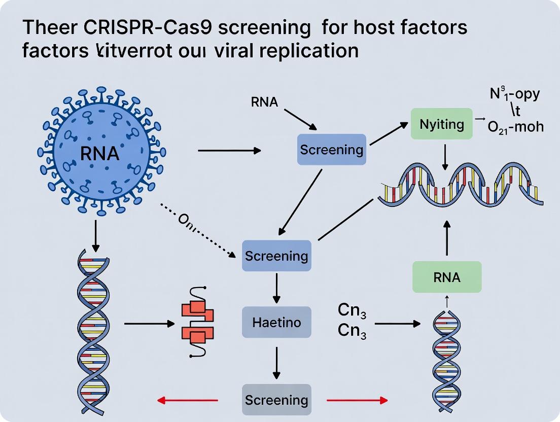
Unlocking Viral Secrets: A Comprehensive Guide to CRISPR-Cas9 Screening for Host Dependency Factors in Viral Replication
This article provides a comprehensive guide for researchers and drug development professionals on employing CRISPR-Cas9 screening to identify host factors essential for viral replication.
Unlocking Viral Secrets: A Comprehensive Guide to CRISPR-Cas9 Screening for Host Dependency Factors in Viral Replication
Abstract
This article provides a comprehensive guide for researchers and drug development professionals on employing CRISPR-Cas9 screening to identify host factors essential for viral replication. It explores the foundational principles of host-pathogen interactions, details state-of-the-art methodological workflows—from library design to hit validation—and addresses common troubleshooting and optimization challenges. Furthermore, it compares CRISPR screening with other genetic and pharmacological methods, validating its power and limitations. The synthesis offers a roadmap for translating screening hits into novel broad-spectrum antiviral targets and therapeutic strategies, bridging fundamental discovery with clinical application.
The Host as a Battlefield: Foundational Principles of Host-Virus Interactions and CRISPR Screening Strategy
Why Target the Host? The Rationale for Identifying Host Dependency and Restriction Factors.
Within viral replication research, the primary thesis is that viral pathogens are obligate intracellular parasites requiring host cell machinery for their lifecycle. Directly targeting viral components with antivirals faces challenges due to high mutation rates and the limited number of viral enzymes. Therefore, a complementary strategy focuses on identifying Host Dependency Factors (HDFs)—cellular proteins essential for viral replication—and Host Restriction Factors (HRFs)—cellular proteins that inhibit viral replication. CRISPR-Cas9 knockout screening provides a powerful, unbiased method to systematically identify these factors on a genome-wide scale, offering novel targets for broad-spectrum and resistance-resistant therapeutic strategies.
Key Concepts and Quantitative Data
Table 1: Advantages of Targeting Host vs. Viral Factors
| Aspect | Targeting Viral Factors | Targeting Host Factors (HDFs/HRFs) |
|---|---|---|
| Genetic Barrier to Resistance | Low (high viral mutation rate) | High (host genome is stable) |
| Spectrum of Activity | Narrow (often virus-specific) | Potentially Broad (exploiting common pathways) |
| Number of Potential Targets | Limited (few viral proteins) | Vast (entire host proteome) |
| Therapeutic Toxicity Risk | Low (absent in host) | Higher (potential on-target toxicity) |
| Validation Complexity | Straightforward (direct mechanism) | Complex (requires understanding of host biology) |
Table 2: Example Host Factors Identified via CRISPR-Cas9 Screens Data compiled from recent literature (2022-2024).
| Virus | Identified Host Dependency Factor (HDF) | Function in Viral Lifecycle | Potential Therapeutic Approach |
|---|---|---|---|
| SARS-CoV-2 | TMEM41B | Lipid membrane remodeling for replication organelle formation | Small-molecule inhibition |
| Influenza A | NXT1 | Nuclear export of viral mRNA | Repurposing of exportin inhibitors |
| HIV-1 | LEDGF/p75 | Integration of viral DNA into host genome | Peptide blockers (e.g., LEDGINs) |
| Virus | Identified Host Restriction Factor (HRF) | Antiviral Mechanism | Viral Countermeasure |
| HIV-1 | SAMHD1 | Depletes dNTP pool, limiting reverse transcription | Viral Protein Vpx degrades SAMHD1 |
| Influenza A | IFITM3 | Traps virus in endosomes, blocking fusion | Partially escaped by some strains |
| Herpesviruses | Tetherin (BST-2) | Retains virions on cell surface, inhibiting release | Viral ubiquitin ligase degradation |
Experimental Protocols
Protocol 1: Genome-wide CRISPR-Cas9 Knockout Screen for Host Factors in Viral Replication
Objective: To identify host genes whose loss of function alters viral infectivity or replication.
Materials:
- Cell Line: Permissive cell line (e.g., A549, Huh-7, THP-1) stably expressing Cas9 (Cas9-expressing lentivirus generated).
- CRISPR Library: Brunello (human) or Brie (mouse) genome-scale knockout lentiviral library.
- Virus: Reporter virus (e.g., GFP-expressing) or wild-type virus with quantifiable readout (plaque assay, qPCR).
- Reagents: Polybrene (8 µg/mL), Puromycin (for selection), PEG-it virus precipitation solution, TRIzol, NGS library prep kit.
Procedure: A. Library Amplification & Titering (1 week):
- Transform the plasmid library into Endura electrocompetent cells. Plate on large LB-ampicillin plates. Pool colonies and maxiprep DNA.
- Produce lentivirus in HEK293T cells by co-transfecting library plasmid with packaging psPAX2 and envelope pMD2.G plasmids using PEI.
- Harvest supernatant, concentrate with PEG-it, and titer on target cells.
B. Screen Execution (4 weeks):
- Infect & Select: Transduce Cas9-expressing target cells at an MOI of ~0.3 to ensure single guide RNA (sgRNA) integration. Use sufficient cell numbers to maintain >500x library representation. Select with puromycin (2 µg/mL) for 7 days.
- Split & Infect: Split selected cells into two arms: Virus-Infected and Mock-Infected Control. Infect the treatment arm with the target virus at an MOI that yields ~30-50% infection (to maintain selective pressure).
- Harvest Genomic DNA: At 5-7 days post-infection (or after clear phenotypic shift), harvest genomic DNA from both populations (minimum 50 million cells each) using a blood & cell culture DNA maxi kit.
C. Sequencing & Analysis (2 weeks):
- Amplify sgRNA inserts: Perform a two-step PCR on gDNA to add Illumina adapters and sample barcodes.
- Next-Generation Sequencing (NGS): Pool PCR products and sequence on an Illumina platform to >100x coverage of the original library.
- Bioinformatics: Align reads to the reference library. Use MAGeCK or PinAPL-Py to compare sgRNA abundance between infected and control groups. Significant enrichment or depletion of sgRNAs targeting a gene identifies potential HRFs or HDFs, respectively.
Protocol 2: Validation of Candidate Host Factors
Objective: To confirm the role of a top-hit gene from the primary screen.
A. CRISPR-Cas9 Knockout Validation:
- Design 3-4 new sgRNAs targeting the candidate gene using the Broad Institute GPP portal.
- Clone into a lentiviral sgRNA vector (e.g., lentiGuide-Puro).
- Transduce Cas9-expressing cells, select with puromycin, and confirm knockout via western blot or T7E1 assay.
- Challenge validated knockout pools with the virus and measure replication (e.g., by viral titer or reporter signal) vs. non-targeting sgRNA control.
B. Complementation/Rescue Assay:
- Clone a cDNA of the candidate gene, with silent mutations in the sgRNA target site to confer resistance, into an expression vector.
- Transiently or stably express this construct in the validated knockout cell line.
- Infect and assess if viral replication is restored to wild-type levels, confirming on-target effect.
Visualizations
CRISPR Screening & Validation Workflow
Host Factor Roles and Therapeutic Strategies
The Scientist's Toolkit: Key Research Reagents
Table 3: Essential Reagents for CRISPR-based Host Factor Screening
| Reagent / Material | Provider Examples | Function in Protocol |
|---|---|---|
| Cas9-Expressing Cell Line | Synthego, ATCC, generated in-house | Provides the CRISPR effector enzyme stably for screening. |
| Genome-wide sgRNA Library (e.g., Brunello) | Addgene, Dharmacon | Targets ~19,000 human genes with 4 sgRNAs/gene for pooled screening. |
| Lentiviral Packaging Mix (psPAX2, pMD2.G) | Addgene | Essential plasmids for producing transducible lentiviral particles of the sgRNA library. |
| Polybrene (Hexadimethrine bromide) | Sigma-Aldrich | Increases transduction efficiency by neutralizing charge repulsion. |
| Puromycin Dihydrochloride | Thermo Fisher | Selects for cells that have successfully integrated the sgRNA vector. |
| PEG-it Virus Precipitation Solution | System Biosciences | Concentrates lentiviral supernatants for higher titer infections. |
| Next-Generation Sequencing Kit (Illumina) | Illumina, New England Biolabs | For preparing and sequencing the amplified sgRNA inserts from genomic DNA. |
| Bioinformatics Software (MAGeCK) | Open Source | Statistical tool for identifying significantly enriched/depleted genes from screen NGS data. |
| Validation sgRNAs (synthego) | Synthego, IDT | Chemically synthesized, high-fidelity sgRNAs for independent knockout validation. |
Identifying host factors essential for viral replication is a cornerstone for developing novel antiviral therapies. CRISPR-Cas9 screening has revolutionized this search by enabling systematic, genome-wide interrogation of gene function. This primer details the three core screening modalities—knockout, activation, and interference—within the framework of discovering host-dependency and host-restriction factors for viruses.
Core Modalities: Mechanisms and Applications
CRISPR-Knockout (KO): Utilizes Streptococcus pyogenes Cas9 (SpCas9) nuclease and a single guide RNA (sgRNA) to create double-strand breaks (DSBs) in the target gene. Error-prone repair via non-homologous end joining (NHEJ) leads to insertion/deletion (indel) mutations, resulting in frameshifts and premature stop codons. Ideal for identifying host factors that viruses exploit (dependency factors).
CRISPR Activation (CRISPRa): Employs a catalytically dead Cas9 (dCas9) fused to transcriptional activation domains (e.g., VP64, p65, Rta). The dCas9-activator complex is guided to the promoter or enhancer region of a target gene, recruiting RNA polymerase II to upregulate transcription. Powerful for identifying host restriction factors whose overexpression inhibits viral replication.
CRISPR Interference (CRISPRi): Uses dCas9 fused to transcriptional repressive domains (e.g., KRAB, SID4x). The dCas9-repressor complex binds near the transcription start site, blocking RNA polymerase binding or elongation to downregulate gene expression. Offers a reversible, titratable alternative to knockout for studying essential host genes.
Quantitative Comparison of Screening Modalities
Table 1: Comparison of CRISPR Screening Modalities for Host-Pathogen Research
| Feature | CRISPR-KO | CRISPRa | CRISPRi |
|---|---|---|---|
| Cas9 Form | Nuclease (SpCas9) | dead Cas9 (dCas9) | dead Cas9 (dCas9) |
| Primary Effect | Permanent gene disruption | Transcriptional activation | Transcriptional repression |
| Best For Identifying | Host dependency factors | Host restriction factors | Essential host factors |
| Typical Fold-Change | Gene depletion (>5-fold) | Gene enrichment (2-10 fold) | Gene depletion (2-5 fold) |
| Key Advantage | Complete loss-of-function | Gain-of-function | Reversible, tunable knock-down |
| Common Library Size | 3-5 sgRNAs/gene | 5-10 sgRNAs/gene | 3-5 sgRNAs/gene |
| Primary Analysis | Depletion of sgRNAs post-infection | Enrichment of sgRNAs post-infection | Depletion of sgRNAs post-infection |
Experimental Protocols
Protocol 1: Genome-wide CRISPR-KO Screen for HIV-1 Host Dependency Factors
A. Library Lentiviral Production
- Transfection: Co-transfect 293T cells with the Brunello genome-wide KO library plasmid (Addgene #73179), psPAX2 packaging plasmid, and pMD2.G VSV-G envelope plasmid using PEI-Max reagent.
- Harvest: Collect lentiviral supernatant at 48h and 72h post-transfection. Pool, filter (0.45 µm), and concentrate via ultracentrifugation.
- Titer: Determine functional titer on HeLa cells via puromycin selection.
B. Screen Execution in Target Cells
- Transduction: Transduce proliferating CEM T-cells (MOI~0.3) with the lentiviral library to ensure ~200-500x coverage of each sgRNA. Include non-targeting control sgRNAs.
- Selection: Treat cells with puromycin (2 µg/mL) for 7 days to select successfully transduced cells.
- Challenge & Selection: Infect pooled cells with HIV-1 (NL4-3 strain, MOI=0.5) or maintain mock-infected controls. Culture for 14-21 days, allowing multiple viral replication cycles.
- Harvest Genomic DNA: Collect >50 million cells from both infected and control pools at endpoint. Extract gDNA using a Maxi Prep kit.
C. Next-Generation Sequencing (NGS) & Analysis
- Amplify sgRNA Loci: Perform two-step PCR on gDNA to add Illumina adaptors and sample barcodes.
- Sequencing: Pool amplicons and sequence on an Illumina NextSeq (75bp single-end).
- Bioinformatics: Align reads to the sgRNA library reference. Use MAGeCK or BAGEL2 algorithms to compare sgRNA abundance between infected and control conditions, identifying significantly depleted genes (FDR < 0.05).
Protocol 2: Targeted CRISPRa Screen for SARS-CoV-2 Restriction Factors
A. Library Design & Production
- Library: Use the Calabrese genome-scale CRISPRa SAM library (targeting ~20,000 promoters) or a custom sub-library of interferon-stimulated genes (ISGs).
- Virus Production: As in Protocol 1A, using the dCas9-VPR activation complex and the sgRNA library.
B. Screen in Lung Epithelial Cells
- Stable Line Generation: Generate A549 cells stably expressing dCas9-VPR via lentiviral transduction and blasticidin selection.
- Transduction & Selection: Transduce the stable line with the sgRNA library (MOI~0.3). Select with puromycin for 7 days.
- Challenge: Infect pooled cells with SARS-CoV-2 (WA1 strain) at a low MOI (0.1) to allow multi-cycle replication. Maintain a mock-infected control. Harvest genomic DNA 72-96 hours post-infection.
C. Analysis: Process as in Protocol 1C, but identify sgRNAs/genes that are enriched in the surviving cell population post-infection, indicating their activation conferred a protective effect.
Visualizing Screening Workflows & Mechanisms
Table 2: Key Research Reagent Solutions for CRISPR Screening
| Item | Function & Application in Viral Screens | Example/Supplier |
|---|---|---|
| Genome-wide sgRNA Libraries | Pre-designed pooled libraries for KO, activation, or interference screens. Essential for unbiased discovery. | Brunello KO (Addgene), SAM CRISPRa (Addgene), Dolcetto CRISPRi (Addgene) |
| Lentiviral Packaging Plasmids | For producing replication-incompetent lentiviral particles to deliver Cas9/dCas9 and sgRNAs. | psPAX2 (packaging), pMD2.G (VSV-G envelope) |
| dCas9 Effector Plasmids | Express dead Cas9 fused to activator (VPR) or repressor (KRAB) domains for CRISPRa/i. | pHAGE dCas9-KRAB (Addgene #50919), lenti-dCas9-VPR (Addgene #63798) |
| Cas9-Nuclease Cell Line | Stable cell lines expressing SpCas9, streamlining knockout screens by requiring only sgRNA delivery. | HEK293T Cas9 (ATCC), A549 Cas9 (commercial) |
| NGS Library Prep Kits | For amplifying and barcoding integrated sgRNAs from genomic DNA of pooled screens. | NEBNext Ultra II Q5 (NEB) |
| Bioinformatics Software | Algorithms to identify significantly enriched or depleted genes from NGS read counts. | MAGeCK, BAGEL2, CRISPhieRmix |
| Viral Titer Assay Kits | To accurately quantify infectious virus used for challenge (e.g., TCID50, plaque assays). | QuickTiter Lentivirus Titer Kit (Cell Biolabs) |
Within CRISPR-Cas9 screening for host factors in viral replication, the precise definition of the screened phenotype is paramount. This protocol details the design and execution of three critical, distinct screen types: Survival, Fitness, and Viral Entry/Replication. Each identifies host factors but interrogates different biological questions and requires tailored experimental setups.
Core Phenotype Definitions and Applications
Table 1: Comparative Overview of Screen Types
| Screen Type | Primary Phenotype | Biological Question | Typical Assay Readout | Key Identified Factors |
|---|---|---|---|---|
| Survival Screen | Cell viability post-infection. | Which host genes are required for cell survival during viral infection? | Genomic DNA abundance (NGS) at Tfinal vs. T0. | Anti-apoptotic factors, essential genes in infected state. |
| Fitness Screen | Proliferative capacity post-infection. | Which host genes confer a growth advantage/disadvantage during infection? | gRNA abundance over multiple cell divisions (NGS). | Immune modulators, metabolic regulators, proviral factors. |
| Viral Entry/Replication Screen | Direct measurement of viral infection. | Which host genes are essential for viral entry, replication, or spread? | FACS (e.g., viral GFP), luminescence, plaque assay. | Viral receptors, endocytic machinery, transcription factors. |
Detailed Experimental Protocols
Protocol 3.1: Pooled CRISPR-Cas9 Survival Screen
Objective: Identify host genes essential for survival during a lytic viral infection. Key Reagents: Brunello or similar genome-wide gRNA library, Polybrene, Puromycin, Viral Stock (e.g., HSV-1, Influenza A). Workflow:
- Cell Preparation: Generate a stably expressing Cas9 cell line (e.g., A549-Cas9). Validate Cas9 activity.
- Library Transduction: At a low MOI (~0.3) to ensure single gRNA integration, transduce cells with the pooled gRNA library. Use polybrene (8 µg/ml) to enhance transduction. Culture for 24h.
- Selection: Treat with puromycin (e.g., 2 µg/ml for A549) for 5-7 days to select transduced cells.
- Population Sampling (T0): Harvest 5x10^6 cells as the T0 reference. Extract genomic DNA (gDNA).
- Infection & Selection: Infect the remaining population with the target virus at a high MOI (e.g., MOI=5 for lytic virus). Critical: Include an uninfected control population.
- Phenotype Application: Allow infection to proceed until ~80-90% cytopathic effect (CPE) is observed in the infected control (e.g., 72-96h). The surviving cell population is harvested (Tfinal).
- gDNA Extraction & NGS: Extract gDNA from T0 and Tfinal samples. Amplify integrated gRNA sequences via PCR and submit for NGS.
- Analysis: Enrichment/depletion of gRNAs is calculated (e.g., MAGeCK, BAGEL2). Genes with depleted gRNAs in the infected vs. uninfected sample are hits.
Protocol 3.2: Pooled CRISPR-Cas9 Fitness Screen
Objective: Identify genes that alter cellular proliferation dynamics during persistent or non-lytic infection. Key Reagents: Brunello gRNA library, Blasticidin (for Cas9 selection), Persistent Virus (e.g., HCV replicon, SARS-CoV-2 non-lytic strain). Workflow:
- Library Transduction & Selection: Follow steps 1-3 from Protocol 3.1.
- Baseline (T0): Harvest reference cells.
- Infection & Passaging: Infect the experimental arm. Maintain both infected and uninfected populations in log-phase growth for ≥14 days (allowing ~10-12 population doublings). Passage cells regularly to avoid confluence.
- Serial Sampling: Harvest cells at multiple time points (e.g., T7, T14 days). This allows modeling of fitness effects over time.
- gDNA Extraction, NGS & Analysis: Extract gDNA from all time points. Analyze using tools like MAGeCK-MLE or PinAPL-Py to model gRNA abundance trajectories. Hits show progressive enrichment or depletion over time specifically in the infected condition.
Protocol 3.3: FACS-Based Viral Entry/Replication Screen
Objective: Isolate cells with defective viral entry or replication using a reporter virus. Key Reagents: Custom CRISPR sub-library (e.g., targeting membrane proteins, kinases), Reporter Virus (e.g., VSV-G pseudotyped GFP, Influenza A NS1-GFP). Workflow:
- Sub-library Transduction: Transduce Cas9-expressing cells with the focused sub-library. Select with puromycin.
- Infection with Reporter Virus: Infect the pooled, gene-edited population with the reporter virus at a low MOI (e.g., MOI=0.5-1) to ensure clear resolution of GFP+ (infected) vs. GFP- (non-infected) cells.
- FACS Sorting: At 24-48h post-infection, sort the population into GFP-Low/Negative (putative knockout cells resistant to infection) and GFP-High (control, susceptible) bins. Collect ≥1x10^6 cells per bin.
- gDNA Recovery & NGS: Extract gDNA from sorted populations and the pre-sort input. Amplify and sequence gRNA inserts.
- Analysis: Calculate significant enrichment of gRNAs in the GFP-Low population versus input or GFP-High control (MAGeCK). Top hits are candidate host dependency factors.
Diagrams
Diagram 1: Phenotype Screening Decision Logic
Diagram 2: Core Workflow for Pooled Survival/Fitness Screens
Diagram 3: FACS-Based Viral Entry Screen Workflow
The Scientist's Toolkit
Table 2: Essential Research Reagent Solutions
| Reagent / Material | Function / Purpose | Example Product/Note |
|---|---|---|
| Genome-Wide gRNA Library | Targets all human genes for loss-of-function screening. | Broad Institute Brunello library (4 gRNAs/gene, ~77k guides). Optimized for reduced off-target effects. |
| Focused Sub-Library | Targets specific gene families (e.g., kinases, membrane proteins) for deeper coverage. | Custom designed using tools like CHOPCHOP or purchased from vendors (e.g., Sigma Mission TRC). |
| Lentiviral Packaging Mix | Produces VSV-G pseudotyped lentivirus for efficient gRNA delivery. | 2nd/3rd generation systems (psPAX2, pMD2.G). Essential for biosafety. |
| Reporter Virus | Expresses a fluorescent (GFP) or luminescent (Luciferase) protein for infection readout. | VSV-G pseudotyped ΔG-GFP reporters; recombinant Influenza A expressing NS1-GFP. |
| Cas9-Expressing Cell Line | Provides constitutive Cas9 expression for CRISPR knockout. | Commercially available (e.g., A549-Cas9, HEK293T-Cas9) or generated via lentiviral transduction + blasticidin selection. |
| Next-Generation Sequencer | Quantifies gRNA abundance from pooled genomic DNA. | Illumina NextSeq 500/550 for medium throughput. Guide counts dictate required sequencing depth. |
| FACS Sorter | Physically isolates cells based on infection reporter signal (GFP fluorescence). | Must be capable of sterile sorting for cell culture recovery (e.g., BD FACSAria, Sony SH800). |
| Bioinformatics Pipeline | Statistically identifies significantly enriched/depleted genes from NGS data. | MAGeCK (Model-based Analysis of Genome-wide CRISPR-Cas9 Knockout) is the current standard. BAGEL2 for essential gene analysis. |
Within the context of a CRISPR-Cas9 screening thesis for identifying host dependency and restriction factors, the selection of appropriate viral pathogens and their permissive cell lines is paramount. This application note provides current guidelines and protocols for establishing these critical model systems, ensuring biologically relevant and high-throughput compatible readouts for functional genomics screens.
Pathogen & Cell Line Selection Criteria
The selection must balance viral biology, cell line permissiveness, assay feasibility, and relevance to human disease. Key considerations include biosafety level (BSL), availability of reverse genetics systems, and compatibility with high-content imaging or survival-based screens.
Table 1: Model Viral Pathogens for Host Factor Screening
| Pathogen | Primary Receptor(s) | Relevant Disease Models | Common Permissive Cell Lines for Screening | Typical Readout for CRISPR Screen | Biosafety Level |
|---|---|---|---|---|---|
| HIV-1 | CD4, CCR5/CXCR4 | AIDS | T-cell lines (e.g., Jurkat, CEM), HeLa-derived (e.g., TZM-bl), Primary CD4+ T-cells | Viral p24 ELISA, Luciferase reporter, Cell survival (if using cytopathic strain) | BSL-2/BSL-3* |
| Influenza A | Sialic acid | Seasonal/Pandemic Flu | MDCK, A549, Calu-3, Primary HAE | TCID50, Plaque assay, GFP reporter virus | BSL-2 |
| SARS-CoV-2 | ACE2 | COVID-19 | Vero E6, Caco-2, Calu-3, A549-ACE2, Primary HAE | Plaque assay, qRT-PCR (viral RNA), CPE-based survival | BSL-3 |
| Zika Virus | AXL, others | Congenital Zika Syndrome | Vero, C6/36 (mosquito), Huh-7, Neural Progenitor Cells | Plaque assay, Immunofluorescence, Cell viability | BSL-2 |
*BSL-3 for replication-competent infectious clones.
Table 2: Selected Cell Line Properties & Screening Suitability
| Cell Line | Origin | Key Applications | Advantages for Screening | Limitations |
|---|---|---|---|---|
| Vero E6 | African Green Monkey Kidney | SARS-CoV-2, Zika, other arboviruses | High viral yield, low interferon response | Non-human, limited physiological relevance |
| A549-ACE2 | Human Lung Carcinoma (engineered) | SARS-CoV-2 | Human, expresses ACE2 receptor, adaptable to HTS | Transformed cell line |
| Calu-3 | Human Airway Epithelium | SARS-CoV-2, Influenza | Polarized, relevant entry pathway, better mimic of respiratory tract | Slower growth, more challenging for HTS |
| Jurkat | Human T-cell Leukemia | HIV-1 | Suspension, relevant for T-cell tropic viruses, easy FACS analysis | Non-primary, transformed |
| Huh-7 | Human Hepatocellular Carcinoma | Zika, HCV, Dengue | Highly permissive for flaviviruses, easy to culture | Cancer cell line with altered pathways |
| Primary HAE | Human Airway Epithelium | SARS-CoV-2, Influenza, RSV | Gold standard for physiological relevance (polarized, mucus, cilia) | Costly, donor variability, low-throughput |
Core Protocols for Viral Infection in CRISPR Screening Workflows
Protocol 2.1: Lentiviral Delivery of CRISPR Library and Challenge with Influenza A Virus (IAV)
Objective: Generate knockout cells for screening host factors required for IAV replication.
Materials:
- Cas9-expressing A549 cell line (stable).
- Focused or genome-wide sgRNA lentiviral library.
- Influenza A/PR/8/34 (H1N1) or GFP-expressing reporter virus (e.g., PR8-GFP).
- MDCK cells for virus titration.
- Transduction enhancers (e.g., Polybrene).
- Cell culture media (DMEM, FBS, Pen/Strep).
- Lysis buffer for RNA extraction (e.g., TRIzol).
Procedure:
- Library Transduction: Seed Cas9-expressing A549 cells in 10-cm dishes at 60% confluency. Transduce with the sgRNA lentiviral library at an MOI of ~0.3 to ensure single sgRNA integration. Use 8 µg/mL polybrene. Spinoculate at 1000 × g for 1 hour at 32°C.
- Selection: 48 hours post-transduction, add puromycin (2 µg/mL) for 5-7 days to select for successfully transduced cells. Maintain library representation by keeping a minimum of 500 cells per sgRNA.
- Infection Challenge: Split cells and seed for infection. Wash cells with PBS and inoculate with IAV at an MOI of 0.5 in infection medium (DMEM, 0.3% BSA, TPCK-trypsin 1 µg/mL). Incubate 1 hour at 37°C, 5% CO₂.
- Readout Collection: For a replication/survival screen, incubate for 72-96 hours until significant cytopathic effect (CPE) is evident in control cells. Harvest genomic DNA from surviving cells (DNeasy Kit). For an early-entry screen, harvest RNA (TRIzol) at 8-12 hpi for qRT-PCR-based quantification of viral load.
- NGS & Analysis: Amplify integrated sgRNA sequences via PCR from gDNA using indexed primers. Sequence on an Illumina platform. Compare sgRNA abundance in infected vs. non-infected control populations using specialized algorithms (e.g., MAGeCK).
Protocol 2.2: SARS-CoV-2 Infection of CRISPR-Modified Calu-3 Cells
Objective: Identify host factors restricting SARS-CoV-2 replication in a physiologically relevant cell line.
Materials (BSL-3):
- Calu-3 cells with stable Cas9 expression.
- SARS-CoV-2 isolate (e.g., WA1/2020 or Omicron variant).
- Vero E6 cells for titration.
- qRT-PCR reagents (primers targeting SARS-CoV-2 N gene).
- Plaque assay materials (Avicel overlay, neutral red stain).
Procedure:
- Knockout Pool Generation: Perform steps as in Protocol 2.1 to generate a Calu-3 Cas9 knockout pool with your selected sgRNA library. Note: Calu-3 cells may require optimization of transduction conditions.
- Polarized Infection: Seed Transwell inserts with the knockout pool and culture until fully polarized (TER > 1000 Ω·cm²). Infect from the apical side with SARS-CoV-2 at an MOI of 1 in a minimal volume for 2 hours.
- Dual Readout Harvest: At 48 hpi:
- Apical Wash: Collect apical supernatant for viral titer determination by plaque assay on Vero E6 cells.
- Cell Harvest: Lyse cells for gDNA extraction (sgRNA abundance analysis) and parallel RNA extraction (viral RNA quantification via qRT-PCR).
- Data Integration: Correlate depletion/enrichment of specific sgRNAs (from gDNA NGS) with reduced or increased viral RNA yield (from qRT-PCR) to pinpoint high-confidence host factors.
Visualizing Screening Workflows & Viral Entry Pathways
Title: CRISPR Screen for HIV Host Factors
Title: SARS-CoV-2 Host Cell Entry Pathways
The Scientist's Toolkit: Essential Research Reagents
Table 3: Key Reagent Solutions for Viral CRISPR Screens
| Reagent/Category | Example Product/Description | Function in Viral Screening |
|---|---|---|
| CRISPR Library | Brunello, GeCKO, or focused antiviral libraries (e.g., Dharmacon) | Delivers sgRNAs to generate genome-wide or targeted knockouts. |
| Cas9 Cell Line | Lentiviral Cas9 (e.g., lentiCas9-Blast) or stable cell lines (A549-Cas9) | Provides the endonuclease for sgRNA-directed gene knockout. |
| Viral Titer Kit | QuickTiter Kit (Cell Biolabs) or plaque assay reagents | Quantifies infectious virus particles pre- and post-infection. |
| qRT-PCR Assay | TaqMan assays for viral RNA (e.g., CDC 2019-nCoV kit) | Precisely quantifies intracellular viral load as a screen readout. |
| Cell Viability Assay | CellTiter-Glo (Promega) or PrestoBlue | Measures virus-induced cytopathic effect (CPE) and cell survival. |
| NGS Library Prep Kit | NEBNext Ultra II DNA Library Prep Kit | Prepares sgRNA amplicons for next-generation sequencing. |
| Infection Enhancer | Polybrene (for lentivirus) or DEAE-Dextran (for some viruses) | Increases viral transduction/infection efficiency. |
| Biosafety Materials | BSL-2/3 Cabinets, Inactivation reagents (e.g., TRIzol, bleach) | Ensures safe handling of pathogenic viruses. |
Application Notes
CRISPR-Cas9 genome-wide knockout screening has become a pivotal tool for identifying host factors essential for viral replication. Hits from these screens—genes whose disruption impairs or enhances viral infection—require functional validation and pathway analysis. This process moves a screening "hit" toward a testable "hypothesis" regarding the biological pathway involved. Current research consistently implicates several core host pathways across diverse viral families.
Endosomal Trafficking and Membrane Fusion
Many viruses, including influenza, SARS-CoV-2, and Ebola, utilize endocytic pathways for cellular entry. Hits often cluster around genes regulating clathrin-mediated endocytosis (CLTC, AP2), endosomal maturation (Rab GTPases, ESCRT complex), and endosomal acidification (ATP6V0D1, ATP6V1A). Acidification triggers conformational changes in viral fusion proteins, enabling capsid release into the cytosol.
Table 1: Common Host Factors in Endosomal Entry Pathways
| Gene | Pathway/Complex | Viral Model(s) | Perturbation Phenotype (Avg. % Infection Reduction) | Key Functional Validation |
|---|---|---|---|---|
| CLTC | Clathrin-Mediated Endocytosis | Influenza A, VSV, SARS-CoV-2 | 70-85% | siRNA rescue, dominant-negative mutant |
| RAB5A | Early Endosome Formation | Ebola, HIV-1, Adenovirus | 60-80% | Constitutively active/dominant-negative mutants |
| ATP6V0D1 | V-ATPase (Endosomal Acidification) | Influenza A, Dengue, VSV | 75-90% | Bafilomycin A1 treatment control, pH reporter assays |
| NPC1 | Cholesterol Transport / Late Endosome | Ebola, Marburg | >95% | Cholesterol depletion/rescue experiments |
Endoplasmic Reticulum (ER) & Golgi Trafficking
Following replication, viruses like Hepatitis C, Dengue, and Coronaviruses hijack the ER and secretory pathway for protein processing, assembly, and egress. CRISPR hits frequently involve the ER-associated degradation (ERAD) pathway, oligosaccharyltransferase complex, and COPI/COPII vesicle coats.
Table 2: Host Factors in ER/Golgi-Dependent Viral Replication
| Gene | Pathway/Complex | Viral Model(s) | Perturbation Phenotype | Key Functional Validation |
|---|---|---|---|---|
| SEC61A1 | ER Translocation/ERAD | Dengue, HCV, SARS-CoV-2 | Replication reduced by 80-90% | Proximity ligation assay (PLA) with viral proteins |
| STT3A | Oligosaccharyltransferase Complex | HCV, Dengue, Zika | Infectivity reduced by 65-75% | Glycosylation status blot of viral glycoproteins |
| COPB2 | COPI Vesicle Coat | Coronavirus, Picornavirus | Viral titer reduced 2-3 log10 | Immunofluorescence for viral protein colocalization |
| UBE2J1 | ERAD Ubiquitin Conjugation | Influenza A, HIV-1 | Viral protein accumulation reduced by 70% | Cycloheximide chase assay for viral protein stability |
Innate Immune Sensing and Interferon Signaling
CRISPR screens selecting for enhanced viral replication often identify negative regulators of interferon (IFN) response (e.g., TRIM, SOCS families). Conversely, screens for resistance factors reveal essential pattern recognition receptors (RIG-I/MDA5, cGAS) and interferon-stimulated genes (ISGs).
Table 3: Immune Pathway Host Factors in Viral Replication
| Gene | Pathway/Function | Viral Model(s) | CRISPR Screen Phenotype (Fold-Change) | Validation Assay |
|---|---|---|---|---|
| MAVS | RLR Signaling Adaptor | VSV, SeV, HCV | Knockout increases replication 10-100x | IFN-β luciferase reporter assay |
| cGAS | Cytosolic DNA Sensor | HSV-1, Vaccinia, HIV-1 | Knockout increases replication 5-50x | STING phosphorylation blot |
| IFITM3 | Restriction Factor (Endosomal) | Influenza A, Dengue, SARS-CoV-2 | Knockout increases infection 3-10x | Viral entry pseudotyped particle assay |
| TRIM25 | Positive Regulator of RIG-I | Influenza A, SARS-CoV-2 | Knockout increases replication 20-50x | Co-Immunoprecipitation with viral RNA/RIG-I |
Nuclear Import & Transcriptional Regulation
DNA viruses (e.g., HSV, CMV) and retroviruses require nuclear entry and manipulation of host transcription. Common hits include components of the nuclear pore complex (NUPs), importins (KPNA, KPNB1), and transcriptional co-activators (EP300, MED complex).
Experimental Protocols
Protocol 1: CRISPR-Cas9 Knockout Screen for Host Viral Factors
Objective: Identify host genes essential for viral replication using a genome-wide knockout library. Materials: GeCKOv2 or Brunello CRISPR knockout library, HEK293T or A549 cells, Lentiviral packaging plasmids, Polybrene, Puromycin, Viral stock (e.g., GFP-reporter virus), FACS sorter, NGS reagents. Procedure:
- Library Lentivirus Production: Co-transfect HEK293T cells with the CRISPR library plasmid and packaging plasmids (psPAX2, pMD2.G). Harvest supernatant at 48h and 72h.
- Cell Transduction: Infect target cells (MOI ~0.3) with library lentivirus in the presence of 8 µg/mL Polybrene. Select with puromycin (2 µg/mL) for 7 days to generate the mutant pool.
- Viral Challenge: Split the mutant pool and infect with the target virus at a low MOI (e.g., 0.1-0.5) to ensure infection is dependent on host factors. Maintain an uninfected control.
- Sorting & Recovery: At 48-72h post-infection, use FACS to collect the bottom 10-20% (infection-negative) and top 10% (infection-positive) populations based on reporter signal or viral antigen staining.
- NGS Sample Prep: Isolate genomic DNA from sorted populations and the uninfected control. Amplify the integrated sgRNA sequences by PCR using primers adding Illumina adaptors.
- Sequencing & Analysis: Perform high-throughput sequencing. Compare sgRNA abundance between infected (low) and control populations using MAGeCK or similar algorithms to identify significantly depleted hits.
Protocol 2: Hit Validation via Individual sgRNA Knockout and Viral Titer Reduction Assay
Objective: Validate candidate host genes from primary screen. Materials: Individual sgRNA constructs (lentiviral), target cells, polyclonal selection reagents, viral stock, plaque assay or TCID50 reagents. Procedure:
- Generate Knockout Cell Lines: Transduce target cells with lentivirus carrying individual sgRNAs targeting the candidate gene. Include a non-targeting control (NTC) sgRNA. Select with puromycin.
- Confirm Knockout: After 7 days, harvest a portion of cells for western blot or T7E1 assay to confirm protein knockout or editing efficiency.
- Infect & Harvest: Infect knockout and NTC cells in triplicate with the target virus at a defined MOI (e.g., 0.01). Collect supernatant at various time points (e.g., 24, 48, 72h).
- Quantify Viral Output: Titrate harvested supernatant on permissive wild-type cells using a plaque assay or TCID50 method.
- Analyze: Calculate the log10 reduction in viral titer compared to NTC cells. A significant reduction (≥1 log10) confirms the host factor's role.
Protocol 3: Pathway Reconstitution via Complementation Assay
Objective: Rule out off-target effects and confirm pathway-specific function. Materials: cDNA construct of the target gene (wild-type and/or functional mutant), plasmid with sgRNA-resistant "safe harbor" site or silent mutations, transfection reagent. Procedure:
- Design Resistant cDNA: Create a cDNA of the target gene with silent mutations in the PAM/protospacer region targeted by the validation sgRNA.
- Generate Stable Complemented Line: Transfect the knockout cell line (from Protocol 2) with the resistant cDNA plasmid. Select with appropriate antibiotic (e.g., blasticidin).
- Challenge with Virus: Infect the parental, knockout, and complemented cell lines in parallel.
- Assay Function: Measure infection (e.g., by flow cytometry for reporter virus) or viral titer. Restoration of viral replication in the complemented line confirms the phenotype is due to loss of the specific gene.
Visualizations
Title: From CRISPR Screen Hit to Pathway Hypothesis Workflow
Title: Common Host Pathways in Viral Entry & Trafficking
Title: Innate Immune Sensing Pathways Targeted in Viral Screens
The Scientist's Toolkit: Research Reagent Solutions
Table 4: Essential Reagents for CRISPR Viral Host Factor Screens
| Reagent / Material | Function / Application | Example Product/Catalog |
|---|---|---|
| Genome-wide CRISPR Knockout Library | Provides pooled sgRNAs targeting all human genes for loss-of-function screening. | Brunello Human CRISPR Knockout Library (Addgene #73179) |
| Lentiviral Packaging Plasmids | Required for production of sgRNA library lentivirus. | psPAX2 (Addgene #12260), pMD2.G (Addgene #12259) |
| Polybrene (Hexadimethrine Bromide) | Cationic polymer that enhances viral transduction efficiency. | Sigma-Aldrich, TR-1003 |
| Puromycin Dihydrochloride | Antibiotic for selecting cells successfully transduced with the sgRNA library. | Thermo Fisher, A1113803 |
| Fluorescent Reporter Virus | Virus engineered to express GFP, mCherry, etc., enabling FACS-based readout of infection. | e.g., GFP-expressing Influenza A (PR8 strain) |
| FACS Sorter | Instrument for isolating cell populations based on infection (fluorescence) phenotype. | BD FACS Aria, Beckman Coulter MoFlo |
| NGS Library Prep Kit | For amplifying and preparing sgRNA sequences from genomic DNA for deep sequencing. | Illumina Nextera XT, NEBNext Ultra II |
| MAGeCK Software | Computational tool for analyzing CRISPR screen NGS data to identify enriched/depleted sgRNAs. | https://sourceforge.net/p/mageck |
| Validated sgRNA & cDNA Clones | For individual gene knockout and rescue experiments. | Horizon Discovery, Sigma MISSION shRNA; Addgene for cDNAs |
| Plaque Assay Materials | For quantifying infectious viral titer (agarose, cell stain like crystal violet). | Standard molecular biology supplies |
From Library to Leads: A Step-by-Step Workflow for Executing a Viral CRISPR-Cas9 Screen
Within a thesis investigating host factors in viral replication using CRISPR-Cas9 screening, the selection of an appropriate gRNA library is a foundational decision that dictates the scope, cost, and interpretability of results. This guide compares two primary strategies: genome-wide screens and focused library screens. The choice is framed by the research question: Is the goal to discover entirely novel host-pathogen interaction networks (favoring genome-wide), or to validate and deconvolute factors within a biologically or therapeutically relevant subset (favoring focused)?
Library Type Comparison: Application Notes
Genome-Wide Libraries (e.g., Brunello, Brie)
- Scope: Target all ~19,000-20,000 protein-coding genes in the human genome.
- Primary Application in Viral Research: Unbiased discovery of novel host factors involved in viral entry, replication, assembly, and egress. Essential for pathway analysis and building comprehensive interaction maps.
- Key Considerations: High cost, significant sequencing depth required, complex data analysis, and higher hit validation burden. Requires robust cell numbers and infection models.
Focused Libraries (e.g., Druggable Genome, Membrane Proteome, Custom Immune Gene sets)
- Scope: Target a defined subset of genes (e.g., ~5,000 druggable genes, ~2,500 membrane proteins).
- Primary Application in Viral Research: Hypothesis-driven investigation of specific gene families. The "Druggable Genome" library is particularly relevant for identifying immediate therapeutic targets. Membrane protein libraries are ideal for screening viral entry receptors and co-factors.
- Key Considerations: Lower cost, higher gRNA density per gene increases statistical power, streamlined analysis, and higher relevance for translational drug development.
Quantitative Comparison Table
| Parameter | Genome-Wide Library | Focused Library (e.g., Druggable Genome) |
|---|---|---|
| Number of Genes | ~19,000 | ~5,000 |
| gRNAs per Gene | 4-6 | 6-10 |
| Total gRNAs | ~75,000-100,000 | ~30,000-50,000 |
| Typical Cell Requirement | 200-500 million | 50-150 million |
| Sequencing Depth (reads) | 50-100 million | 15-30 million |
| Primary Data Analysis Complexity | High | Moderate |
| Hit Validation Workload | High | Focused |
| Therapeutic Target Yield | Indirect | Direct |
Experimental Protocols
Protocol 1: Genome-Wide CRISPR Screen for Viral Entry Factors
Objective: Identify host genes essential for viral entry using a whole-genome knockout screen.
- Library Amplification & Lentivirus Production: Amplify the Brunello library in E. coli and purify plasmid DNA. Produce lentivirus in HEK293T cells via transfection with packaging plasmids.
- Cell Transduction & Selection: Transduce the target cell line (e.g., A549 for respiratory viruses) at a low MOI (0.3-0.4) to ensure single integration. Select transduced cells with puromycin (2 µg/mL) for 7 days.
- Screen Execution: Split cells into infection and control arms. Infect the treatment arm with virus at a predetermined MOI that yields ~30-50% infection/cell death. Maintain the control arm in parallel.
- Harvest & Genomic DNA Extraction: Harvest cells 7-14 days post-infection (or after sufficient selection pressure). Extract gDNA using a large-scale kit (e.g., Qiagen Maxi Prep).
- gRNA Amplification & Sequencing: Amplify integrated gRNA sequences via two-step PCR, adding Illumina adaptors and sample barcodes. Pool and sequence on an Illumina NextSeq platform (minimum 50M reads).
- Bioinformatic Analysis: Align reads to the library reference. Use MAGeCK or similar tool to compare gRNA abundance between infection and control arms, identifying significantly depleted/enriched genes.
Protocol 2: Focused Druggable Genome Screen for Antiviral Compounds
Objective: Identify druggable host dependencies for viral replication.
- Library Selection: Procure a druggable genome library (e.g., the Asimov library targeting ~5,000 genes).
- Viral Challenge Model Optimization: Establish a replication-competent reporter virus or a quantifiable assay (e.g., plaque assay, qPCR) in the desired cell type. Determine a viral challenge that gives a robust, measurable signal window.
- Screen Conduct: Follow Protocol 1 steps 2-6, but scale down cell numbers proportionally to library size. Include a non-targeting gRNA control arm for normalization.
- Hit Triaging: Prioritize hits based on statistical significance (FDR < 5%), known drug availability, and pathway enrichment. Cross-reference with known viral interactomes.
Visualizations
CRISPR Screen for Host Factors in Viral Replication
Library Selection Decision Tree for Viral Research
The Scientist's Toolkit: Key Research Reagent Solutions
| Item | Function in CRISPR Screen for Viral Replication |
|---|---|
| CRISPR Knockout Library (e.g., Brunello) | Pooled lentiviral library of gRNAs targeting the human genome. Enables genome-scale loss-of-function screening. |
| Lentiviral Packaging Plasmids (psPAX2, pMD2.G) | Second- and third-generation packaging plasmids required for the production of replication-incompetent lentiviral particles. |
| Polybrene (Hexadimethrine Bromide) | A cationic polymer that enhances viral transduction efficiency by neutralizing charge repulsion between virus and cell membrane. |
| Puromycin Dihydrochloride | Selection antibiotic for cells stably expressing the Cas9 and gRNA constructs from lentiviral vectors with a puromycin resistance gene. |
| QuickExtract DNA Solution | Enables rapid, PCR-ready gDNA extraction from a large number of screening samples prior to NGS library prep. |
| MAGeCK (Model-based Analysis of Genome-wide CRISPR-Cas9 Knockout) | A robust computational tool for identifying positively and negatively selected gRNAs/genes from CRISPR screen data. |
| Reporter Virus (e.g., GFP-expressing) | A recombinant virus expressing a fluorescent or luminescent protein, enabling high-throughput quantification of infection via FACS or plate readers. |
| Cell Viability Assay (e.g., CellTiter-Glo) | Luminescent assay to measure ATP levels as a proxy for cell number/viability, critical for cytotoxicity counterscreens. |
Within the broader thesis investigating host factors essential for viral replication using genome-wide CRISPR-Cas9 knockout screening, this protocol details the core experimental pipeline. The objective is to generate a library of genetically perturbed cells, challenge them with a virus of interest, and identify host genes whose loss confers resistance or enhanced susceptibility to infection. This systematic approach enables the discovery of novel therapeutic targets for antiviral drug development.
Experimental Workflow & Timeline
Diagram 1: CRISPR-viral screening workflow
Detailed Protocols
Lentiviral Library Production & Delivery
Objective: Generate high-titer lentiviral particles encoding the CRISPR-Cas9 sgRNA library and transduce target cells to achieve low MOI and high coverage.
Protocol:
- Day -3: Seed HEK293T cells (5 x 10^6) in a 15 cm dish in DMEM + 10% FBS, no antibiotics.
- Day -2: Transfect using polyethylenimine (PEI):
- CRISPR sgRNA Library Plasmid (e.g., Brunello, 20 µg)
- psPAX2 packaging plasmid (15 µg)
- pMD2.G envelope plasmid (6 µg)
- Opti-MEM to 1.5 mL total.
- Mix with 120 µL PEI (1 mg/mL), incubate 20 min, add dropwise to cells.
- Day 0: 48h post-transfection, collect supernatant, filter through 0.45 µm PES filter. Concentrate via ultracentrifugation (70,000 x g, 2h at 4°C). Resuscentrate pellet in cold PBS, aliquot, and store at -80°C.
- Titer Determination: Transduce HEK293 cells with serial dilutions of vector in presence of 8 µg/mL polybrane. 72h later, select with puromycin (2 µg/mL) for 7 days. Count surviving colonies. Calculate titer (TU/mL) = (colonies counted * dilution factor) / volume (mL).
Cell Selection & Library Generation
Objective: Create a stable Cas9-expressing cell line and transduce with the sgRNA library at low MOI to ensure single-integration events.
Protocol:
- Stable Cas9 Cell Line: Lentivirally transduce your target cell line (e.g., A549, Huh7) with a Cas9-PuroR construct. Select with puromycin (dose determined by kill curve, typically 1-5 µg/mL) for 7 days. Validate Cas9 activity via SURVEYOR or T7E1 assay.
- Library Transduction: Seed 5 x 10^6 Cas9 cells per replicate in 10 cm dishes. The next day, transduce with the lentiviral sgRNA library at an MOI of ~0.3 to ensure >95% of cells receive ≤1 sgRNA. Include 8 µg/mL polybrane. Spinoculate (1000 x g, 90 min, 32°C).
- Selection: 24h post-transduction, replace medium with fresh medium containing puromycin. Select for 5-7 days until all cells in a non-transduced control dish are dead. Maintain cells at a minimum coverage of 500 cells per sgRNA (e.g., for a 75k sgRNA library, maintain >3.75 x 10^7 cells).
Viral Challenge & Sample Collection
Objective: Infect the pooled sgRNA library with the target virus and collect samples at defined time points to track sgRNA abundance changes.
Protocol:
- Day -1: Seed 2 x 10^7 library cells (per condition) to achieve 70% confluence at infection.
- Day 0 (T0 Baseline): Harvest 2 x 10^7 cells by trypsinization. Pellet, wash with PBS. Aliquot one pellet for genomic DNA (gDNA) extraction. Freeze pellet at -80°C. This serves as the pre-infection reference.
- Viral Infection: Infect remaining cells with virus at a predetermined MOI (e.g., 0.5 for influenza A/WSN/33) in infection medium (serum-free). Adsorb for 1h at 37°C. Replace with full growth medium.
- Post-Infection Sampling: Harvest cells at multiple time points (e.g., 72h post-infection for lytic viruses, or when significant cytopathic effect is observed in wild-type controls). Collect 2 x 10^7 cells per time point. Include an uninfected "cell pool" control harvested in parallel. Freeze pellets at -80°C.
Table 1: Core Protocol Timeline Summary
| Phase | Key Activity | Duration | Critical Parameters |
|---|---|---|---|
| Library Prep | Lentivirus Production & Titration | 10 days | Titer >1e8 TU/mL; Functional validation |
| Cell Prep | Cas9 Cell Line Generation & Selection | 14-21 days | 100% Cas9+; Puromycin kill curve completed |
| Library Generation | sgRNA Library Transduction & Selection | 10 days | MOI = 0.3; Coverage >500x; Puro selection complete |
| Viral Challenge | Infection & Time-Series Sampling | 1-7 days (virus-dependent) | Optimized MOI; Clear CPE in control; Viable cell count |
| Downstream | gDNA Extraction, NGS Library Prep, Sequencing | 10-14 days | >5 µg gDNA per sample; >500x read coverage per sgRNA |
Table 2: Example Quantitative Parameters for Influenza A Virus Screen (A549 Cells)
| Parameter | Value/Range | Rationale |
|---|---|---|
| Library | Brunello (4 sgRNAs/gene, 76,441 sgRNAs total) | Genome-wide, high-confidence |
| Library Coverage | 500 cells/sgRNA | Minimizes stochastic dropout |
| Transduction MOI | 0.2 - 0.4 | Ensures single integration |
| Puromycin Dose | 2 µg/mL (A549-Cas9) | Determined by 7-day kill curve |
| Infection MOI | 0.5 - 1.0 | Achieves ~70% infection without rapid total cell death |
| Sample Collection Post-Infection | T0, T72h, T120h | Captures early and late host factor effects |
| Cells per gDNA Prep | 2 x 10^7 | Yields ~50-60 µg gDNA, sufficient for PCR |
| Sequencing Depth | >300 reads/sgRNA for T0 | Ensures statistical power for dropout/enrichment analysis |
Diagram 2: Screening logic and outcomes
The Scientist's Toolkit: Essential Research Reagents
Table 3: Key Reagent Solutions for CRISPR-Viral Screening
| Reagent / Material | Supplier Examples | Function in Protocol |
|---|---|---|
| Genome-wide CRISPR Knockout Library (e.g., Brunello) | Addgene, Sigma-Aldrich | Provides pooled sgRNAs targeting all human genes for loss-of-function screening. |
| Lentiviral Packaging Plasmids (psPAX2, pMD2.G) | Addgene | Second- and third-generation system components for producing replication-incompetent lentivirus. |
| Polyethylenimine (PEI), Linear, MW 25,000 | Polysciences, Sigma-Aldrich | High-efficiency, low-cost cationic polymer for transfection of HEK293T cells. |
| Polybrene (Hexadimethrine Bromide) | Sigma-Aldrich, Millipore | Enhances lentiviral transduction efficiency by reducing charge repulsion. |
| Puromycin Dihydrochloride | Thermo Fisher, Sigma-Aldrich | Selectable antibiotic for cells expressing resistance gene from lentiviral constructs. |
| PCR Kit for NGS Library Prep (2-step) | Takara, NEB | Amplifies sgRNA cassettes from genomic DNA for next-generation sequencing. |
| NucleoSpin Blood XL Kit | Macherey-Nagel | Efficient large-scale gDNA extraction from cell pellets (>50 µg yield). |
| Quick-Cas9 Activity Kit (T7E1) | NEB, IDT | Validates Cas9 nuclease activity in generated stable cell lines. |
| Cell Counting Kit-8 (CCK-8) or MTS | Dojindo, Abcam | Assesses cell viability and cytopathic effect (CPE) post-viral infection. |
This application note details protocols for the deconvolution of pooled CRISPR-Cas9 screening data within a research thesis focused on identifying host factors essential for viral replication. The identification of these factors is a critical step in understanding viral life cycles and developing novel host-directed antiviral therapeutics. The workflow hinges on high-quality genomic DNA (gDNA) preparation from screening cells, optimal NGS sequencing depth, and precise bioinformatic alignment of reads to deconvolute guide RNA (gRNA) identities and quantify their abundance.
Application Notes
The Role of NGS in CRISPR Screen Deconvolution
In a pooled CRISPR-Cas9 knockout screen, a library of lentivirally delivered gRNAs is transduced into cells at a low multiplicity of infection (MOI) to ensure one gRNA per cell. Following selection and a challenge (e.g., viral infection), surviving or enriched cell populations are harvested. NGS of the integrated gRNA cassette from purified gDNA is used to determine which gRNAs are enriched or depleted, thereby identifying host genes whose knockout confers a survival or fitness advantage/disadvantage.
Key Quantitative Parameters
Table 1: Recommended NGS Specifications for Pooled CRISPR Screen Deconvolution
| Parameter | Recommendation | Rationale |
|---|---|---|
| gDNA Input per PCR | 2-5 µg | Ensures sufficient template to maintain library complexity and avoid bottlenecking. |
| PCR Amplification Cycles | 12-18 cycles | Minimizes PCR bias and over-amplification while generating sufficient product for sequencing. |
| Sequencing Depth | 200-500 reads per gRNA | Provides statistical power to detect 2-5 fold changes in gRNA abundance. For a 100,000-gRNA library, this requires 20-50 million total reads. |
| Sequencing Read Type | Single-end, 75-150 bp | The gRNA constant region and target sequence are typically within 150 bp. Paired-end is optional for error correction. |
| Sequencing Coverage | >200x library coverage | Sequencing each unique gRNA in the library an average of >200 times ensures robust quantification. |
| Alignment Allowed Mismatches | 0-1 | Strict alignment ensures accurate gRNA counting and minimizes misassignment. |
Table 2: Impact of Sequencing Depth on Statistical Power
| Total Reads per Sample | Approx. Reads/gRNA (100k library) | Detectable Fold-Change (p<0.05) | Risk |
|---|---|---|---|
| 10 million | ~100 | >5x | High false-negative rate for moderate hits. |
| 30 million | ~300 | ~3x | Good balance for genome-wide screens. |
| 75 million | ~750 | ~2x | Optimal for sensitive detection; higher cost. |
Detailed Protocols
Protocol: High-Quality gDNA Preparation from Mammalian Cells
Objective: Isolate high-molecular-weight, PCR-quality gDNA from pelleted cells post-screen.
Materials:
- Cell pellet (e.g., 5-10 million cells)
- Phosphate-Buffered Saline (PBS)
- Lysis Buffer (e.g., Qiagen Gentra Puregene Cell Lysis Buffer)
- RNase A Solution
- Protein Precipitation Solution (e.g., Gentra Protein Precipitation Solution)
- Isopropanol (100% and 70%)
- TE Buffer (10 mM Tris-HCl, 1 mM EDTA, pH 8.0)
Procedure:
- Cell Lysis: Resuspend cell pellet in 3 mL PBS. Add 12 mL Cell Lysis Buffer, mix by inversion. Incubate at 37°C for 10 minutes.
- RNase Treatment: Add 60 µL RNase A solution (4 mg/mL). Invert 25 times, incubate at 37°C for 30 minutes.
- Protein Precipitation: Cool sample on ice for 5 minutes. Add 4 mL Protein Precipitation Solution. Vortex vigorously for 20 seconds. Centrifuge at 4,000 x g for 20 minutes at 4°C.
- DNA Precipitation: Pour supernatant containing DNA into a fresh tube with 12 mL 100% isopropanol. Mix by gentle inversion until DNA thread is visible. Centrifuge at 4,000 x g for 5 minutes.
- DNA Wash: Decant supernatant. Add 12 mL 70% ethanol, invert tube. Centrifuge at 4,000 x g for 5 minutes. Decant ethanol carefully.
- DNA Hydration: Air-dry pellet for 10-15 minutes. Dissolve DNA in 500 µL TE Buffer at 4°C overnight with gentle shaking.
- Quantification: Measure DNA concentration using a fluorometric assay (e.g., Qubit dsDNA HS Assay). Assess purity (A260/A280 ~1.8) and integrity by gel electrophoresis.
Protocol: Two-Step PCR Amplification of gRNA Cassettes for NGS
Objective: Amplify the gRNA region from genomic DNA and attach Illumina sequencing adapters and sample barcodes.
Materials:
- Purified gDNA (2-5 µg)
- High-Fidelity DNA Polymerase (e.g., KAPA HiFi HotStart ReadyMix)
- PCR Primers (See Table Below)
- AMPure XP beads or equivalent
- Nuclease-free water
Primer Design:
- PCR1 (Add Overhang): Forward primer binds the U6 promoter constant region. Reverse primer binds the gRNA scaffold constant region and adds a partial Illumina adapter sequence.
- PCR2 (Add Full Adapters & Indices): Uses universal forward and reverse primers that bind the overhangs from PCR1 to add full P5/P7 flow cell adapters and a unique dual index (i7 and i5) for sample multiplexing.
Procedure:
- PCR1 Setup: In a 50 µL reaction, combine 2 µg gDNA, 0.5 µM each primer, 1x polymerase mix. Cycle: 98°C 3 min; 12-14 cycles of (98°C 20s, 60°C 30s, 72°C 30s); 72°C 5 min.
- PCR1 Purification: Clean up reaction using 1.8x volume AMPure XP beads. Elute in 25 µL TE.
- PCR2 Setup: Use 5 µL of purified PCR1 product as template. 0.5 µM indexing primers. Cycle: 98°C 3 min; 8-10 cycles of (98°C 20s, 65°C 30s, 72°C 30s); 72°C 5 min.
- Final Library Purification: Clean up PCR2 with 1x volume AMPure XP beads. Elute in 30 µL TE. Quantify by Qubit and profile by Bioanalyzer (peak ~280-320 bp).
- Sequencing: Pool libraries equimolarly and sequence on an Illumina platform (e.g., NextSeq 500/2000, HiSeq) using a custom read primer to start sequencing in the gRNA variable region.
Protocol: Bioinformatics Pipeline for gRNA Read Alignment and Count Generation
Objective: Process raw FASTQ files to generate a count table of gRNA abundances per sample.
Software: Trimmomatic, Bowtie2, SAMtools, custom Python/R scripts.
Procedure:
- Demultiplexing: Use
bcl2fastq(Illumina) to generate FASTQ files per sample based on dual-index reads. - Quality Trimming: Use Trimmomatic to remove adapter sequences and low-quality bases.
- Read Alignment: Align trimmed reads to the reference gRNA library FASTA file using Bowtie2 in end-to-end, sensitive mode.
- SAM to BAM Conversion and Sorting:
- Generate Count Table: Use a script to parse the aligned BAM file, counting the number of reads aligning perfectly (0 mismatches) to each gRNA identifier. Output a comma-separated values (CSV) file with samples as columns and gRNA IDs as rows.
- Normalization & Analysis: Normalize counts (e.g., counts per million, CPM) and perform statistical analysis (e.g., MAGeCK, CRISPhieRmix) to identify significantly enriched/depleted gRNAs.
Visualizations
The Scientist's Toolkit
Table 3: Research Reagent Solutions for NGS Screen Deconvolution
| Item | Function & Rationale |
|---|---|
| Qubit dsDNA HS Assay Kit | Fluorometric quantification of gDNA and library DNA. More accurate for low-concentration, fragmented DNA than spectrophotometry. |
| KAPA HiFi HotStart ReadyMix | High-fidelity polymerase for PCR amplification of gRNAs. Minimizes PCR errors that could create false gRNA sequences. |
| AMPure XP Beads | Solid-phase reversible immobilization (SPRI) beads for size-selective cleanup of PCR products. Removes primers, dimers, and salts. |
| Illumina DNA UD Indexes | Sets of unique dual index (UDI) primers for PCR2. Enable high-plex multiplexing with reduced index hopping risk. |
| Agilent High Sensitivity DNA Kit | Chip-based electrophoresis to assess final library fragment size distribution and molarity before pooling. |
| Trimmomatic | Java software for flexible trimming of Illumina adapters and low-quality bases from NGS reads. Critical for clean alignment. |
| Bowtie2 | Ultrafast, memory-efficient aligner for mapping sequencing reads to a gRNA reference library. Supports gapped and local alignment. |
| MAGeCK (Model-based Analysis of Genome-wide CRISPR-Cas9 Knockout) | Comprehensive computational tool for identifying positively and negatively selected gRNAs/genes from count data. |
Application Notes
Within a thesis investigating host factors essential for viral replication using CRISPR-Cas9 screening, a robust bioinformatics pipeline is critical to distinguish true genetic dependencies from background noise. This integrated approach leverages the complementary strengths of three core tools: MAGeCK for primary hit identification and ranking from CRISPR knockout screens, BAGEL for Bayesian classification of essential genes with high precision, and DESeq2 for differential expression analysis of accompanying transcriptomic data (e.g., RNA-seq from infected vs. uninfected cells). The convergence of evidence from knockout phenotypes and gene expression changes significantly increases confidence in candidate host factors.
Table 1: Core Tool Comparison for CRISPR-Viral Host Factor Screening
| Tool | Primary Function | Key Metric(s) | Optimal Use Case in Viral Screen |
|---|---|---|---|
| MAGeCK | CRISPR screen analysis | β-score (log2 fold change), p-value, FDR | Genome-wide identification of genes whose knockout enriches/depletes viral antigen (e.g., FACS) or alters viral titer. |
| BAGEL | Essential gene classification | Bayes Factor (BF), Precision-Recall | Benchmarking essential genes and defining core fitness genes; precise classification of host essential genes co-opted by virus. |
| DESeq2 | RNA-seq differential expression | log2FoldChange, p-value, padj (FDR) | Profiling transcriptional changes upon infection; validating host pathway engagement post-knockout. |
Table 2: Example Hit Convergence from a Hypothetical HIV-1 CRISPR Screen
| Gene | MAGeCK β-score (FDR) | BAGEL BF (Essential?) | DESeq2 log2FC (padj) Upon Infection | Converged Hit? |
|---|---|---|---|---|
| CCR5 | -3.21 (1.2e-05) | 12.5 (Yes) | 1.05 (0.12) | Yes (Known co-receptor) |
| TP53 | -4.50 (2.0e-07) | 15.8 (Yes) | -0.30 (0.80) | No (Core fitness) |
| PROX1 | -2.85 (0.003) | 2.1 (No) | 3.42 (0.001) | Yes (Novel factor) |
| Control_Gene | 0.10 (0.85) | 0.5 (No) | -0.15 (0.90) | No |
Detailed Experimental Protocols
Protocol 1: MAGeCK for Primary Viral Screen Analysis
Objective: To identify genes significantly enriching or depleting in a CRISPR-Cas9 pooled screen after selection for viral replication (e.g., FACS sorting based on viral protein expression).
Materials:
- Sequencing Data: FASTQ files from pre-selection plasmid library (T0) and post-infection/selection (T1) samples.
- sgRNA Library Map: File linking sgRNA sequences to target genes.
- Software: MAGeCK installed via conda (
conda install -c bioconda mageck).
Procedure:
- Quality Control & Count: Generate sgRNA count tables.
Beta-RRA Analysis: Perform robust rank aggregation on gene-level phenotypes.
Pathway Analysis: Enrichment analysis of hit genes in KEGG/GO databases.
Visualization: Generate rank plots and waterfall plots of significant genes (β-score vs. -log10(FDR)).
Protocol 2: BAGEL for Benchmarking and Precision Hit Calling
Objective: To employ a Bayesian framework to classify genes as essential or non-essential using a training set, improving precision.
Materials:
- Reference Essential/Negative Sets: Common essential genes (e.g., from DepMap) and non-essential genes.
- MAGeCK Output: Gene-level fold change (log2) from
viral_screen.beta.gene_summary.txt.
Procedure:
- Prepare Input Files: Create a fold change (FC) file and a reference gene file.
- Run BAGEL: Execute the Bayesian classifier.
- Interpret Output: Genes with a Bayes Factor (BF) > 10 are high-confidence essentials. In a viral screen, hits are genes with significant β-scores in MAGeCK but low BF in BAGEL, indicating virus-specific, not general cellular, essentiality.
Protocol 3: DESeq2 for Transcriptomic Validation
Objective: To identify differentially expressed genes in RNA-seq data from virus-infected vs. mock-infected cells, providing orthogonal evidence for host factor involvement.
Materials:
- RNA-seq Data: FASTQ files from biological replicates of infected and control conditions.
- Sample Metadata: CSV file detailing sample condition.
Procedure (in R):
Visualizations
Diagram 1: Integrated bioinformatics pipeline workflow.
Diagram 2: Decision logic for prioritizing host factor hits.
The Scientist's Toolkit: Research Reagent Solutions
Table 3: Essential Materials for CRISPR-Viral Screen Bioinformatics
| Item | Function in Pipeline | Example/Note |
|---|---|---|
| Brunello or GeCKO v2 Library | Genome-wide CRISPR knockout sgRNA sets. | Used for initial screen; provides sgRNA-to-gene mapping file. |
| Bowtie2 or BWA | Short-read aligner for NGS data. | Aligns sequencing reads to the sgRNA library for counting. |
| featureCounts (Rsubread) | Quantifies RNA-seq reads aligned to genes. | Generates count matrix for DESeq2 input. |
| Positive Control sgRNAs | Targeting known essential (e.g., RPA3) or viral dependency factors. | Quality control for screen performance; used by BAGEL for training. |
| DepMap Achilles Core Fitness Data | Dataset of common essential genes across cell lines. | Critical reference set for BAGEL to define context-independent essentials. |
| KEGG/GO Pathway Databases | Curated biological pathway and function annotations. | Used in MAGeCK pathway and functional enrichment of hits. |
| R/Bioconductor Environment | Statistical computing platform. | Hosts DESeq2, visualization libraries (ggplot2, pheatmap). |
| Python Environment (conda) | Package and environment management. | Hosts MAGeCK, BAGEL, and their dependencies. |
Application Notes
Context within Host-Virus Interaction Research
In CRISPR-Cas9 screening for host factors in viral replication, primary hits often number in the hundreds. Prioritizing these candidates for functional validation is critical. This protocol outlines a systematic, triage approach integrating three complementary data layers: quantitative essentiality scores from the screen, pathway/network enrichment analysis, and structured literature mining. This multi-faceted integration increases confidence in selecting genes with high potential as genuine host dependency factors (HDFs) or restriction factors.
Quantitative Data Integration Framework
The prioritization score is calculated using a weighted sum model. Each gene (i) receives a normalized score (0-1) for each of the three criteria, which are then combined:
Prioritization Score (PSi) = (w1 * NEi) + (w2 * PAi) + (w3 * LSi)
Where:
- NEi: Normalized Essentiality Score (e.g., -log10(p-value) from MAGeCK or RIGER, normalized to max value in dataset).
- PAi: Pathway Association Score (derived from enrichment p-value of the most significant pathway containing the gene).
- LSi: Literature Support Score (quantified from co-mention frequency with the virus of interest).
- w1, w2, w3: Researcher-defined weights (default w1=0.5, w2=0.3, w3=0.2).
Table 1: Exemplar Prioritization Data for Top Candidate Genes from a SARS-CoV-2 CRISPR Screen
| Gene Symbol | Essentiality (-log10(p-value)) | Norm. Ess. Score (NE) | Top Pathway (KEGG) | Pathway p-value | Path. Assoc. Score (PA) | Lit. Co-mentions (Virus) | Lit. Score (LS) | Final Priority Score |
|---|---|---|---|---|---|---|---|---|
| ACE2 | 12.5 | 1.00 | Viral entry (hsa05171) | 1.2E-10 | 1.00 | 28500 | 1.00 | 1.00 |
| TMPRSS2 | 9.8 | 0.78 | Protease activity (hsa04610) | 3.5E-08 | 0.87 | 4200 | 0.15 | 0.70 |
| CTSL | 8.2 | 0.66 | Lysosome (hsa04142) | 2.1E-05 | 0.72 | 1200 | 0.04 | 0.55 |
| RAB7A | 7.5 | 0.60 | Endocytosis (hsa04144) | 1.8E-04 | 0.64 | 450 | 0.02 | 0.49 |
| UnknownGeneX | 11.0 | 0.88 | No significant enrichment | 0.95 | 0.10 | 2 | 0.00 | 0.47 |
Data is illustrative. Essentiality scores from a hypothetical genome-wide KO screen. Literature co-mentions from PubMed search (SARS-CoV-2).
Protocols
Protocol 1: Calculating and Normalizing Gene Essentiality Scores
Objective: Generate normalized essentiality scores from primary CRISPR screen data.
Materials:
- Raw Read Count File: Sequencing counts per sgRNA per sample.
- Software: MAGeCK (version 0.5.9) or BAGEL2.
- Computing Environment: Linux command line or high-performance computing cluster.
Procedure:
- Quality Control: Use MAGeCK count to aggregate sgRNA counts and assess sample correlation.
- Beta Score Calculation: Run MAGeCK test (or RRA algorithm) comparing virus-infected vs control samples. Key command:
mageck test -k count_table.txt -t treatment_sample -c control_sample -n output_prefix --norm-method median - Extract Results: The output
gene_summary.txtcontains p-values and beta scores (negative beta indicates essentiality). - Normalization: For each gene, calculate
NEi = (-log10(p-value)i) / max(-log10(p-value)all_genes). Cap values >1 at 1.0.
Protocol 2: Pathway and Network Enrichment Analysis
Objective: Derive a Pathway Association Score for each candidate gene.
Materials:
- Gene List: Ranked list of candidate genes from Protocol 1.
- Enrichment Tools: g:Profiler, Enrichr, or Metascape.
- Databases: KEGG, Reactome, Gene Ontology (Biological Process).
- Scripting: R (clusterProfiler package) or Python.
Procedure:
- Submit Gene List: Input the top 500-1000 candidate genes (or all genes with p<0.05) into g:Profiler (https://biit.cs.ut.ee/gprofiler/). Select organism and relevant pathway databases.
- Retrieve Results: Download tab-delimited results including pathway name, source, p-value, and intersecting genes.
- Assign Gene-Level Score: For each gene, identify the most significant pathway (smallest p-value) in which it appears. Calculate:
PAi_raw = -log10(min_pathway_p-value)PAi = PAi_raw / max(PA_raw), capped at 1.0. Genes not in any enriched pathway receive a default low score (e.g., 0.1).
Protocol 3: Quantifying Literature Support
Objective: Generate a reproducible Literature Support Score based on co-citation frequency.
Materials:
- Virus-Specific Query: e.g., "SARS-CoV-2" OR "COVID-19".
- Literature Database: PubMed via Entrez Programming Utilities (E-utilities).
- Automation Tool: Python with Biopython and requests libraries.
Procedure:
- Construct Queries: For each candidate gene, create a PubMed query:
"(Gene Symbol[TIAB] OR Gene Name[TIAB]) AND (Virus Query)". - Automated Search: Use a Python script to send requests to the E-utilities
esearchendpoint (https://eutils.ncbi.nlm.nih.gov/entrez/eutils/esearch.fcgi) for each gene, retrieving the count of matching articles (hit_count). - Calculate Score: Apply log10 transformation to reduce skew:
LSi_raw = log10(hit_count + 1). Then normalize:LSi = LSi_raw / max(LS_raw), capped at 1.0.
Protocol 4: Integrated Prioritization and Triage
Objective: Combine normalized scores to generate a final ranked list.
Procedure:
- Compile Table: Create a master table with columns: Gene, NE, PA, LS.
- Apply Weights: Calculate
PSi = (0.5 * NEi) + (0.3 * PAi) + (0.2 * LSi). Weights can be adjusted based on research goals (e.g., increase w3 for novel gene discovery). - Rank & Categorize: Sort genes descending by PS. Create tiers: Tier 1 (PS > 0.7), Tier 2 (PS 0.4-0.7), Tier 3 (PS < 0.4).
- Visual Inspection: Manually review top-tier genes, especially high NE/PA but low LS genes, as potential novel discoveries.
Diagrams
Title: Gene Prioritization Workflow from CRISPR Screen to Validation
Title: Host Factors in Viral Entry Pathway
The Scientist's Toolkit
Table 2: Key Research Reagent Solutions for Integrated Gene Prioritization
| Reagent / Resource | Function in Prioritization Protocol | Example / Source |
|---|---|---|
| MAGeCK Software Suite | Statistical analysis of CRISPR screen data to generate gene-level p-values and essentiality scores. | https://sourceforge.net/p/mageck |
| g:Profiler Web Tool | Performs pathway enrichment analysis across multiple databases (GO, KEGG, Reactome). | https://biit.cs.ut.ee/gprofiler |
| PubMed E-utilities | Programmatic interface to run automated literature searches and retrieve citation counts. | https://www.ncbi.nlm.nih.gov/books/NBK25501/ |
| R with clusterProfiler | R package for comprehensive enrichment analysis and visualization; enables batch processing. | Bioconductor package |
| Python (Biopython, pandas) | Scripting environment to integrate data from different sources, normalize scores, and calculate final priority ranks. | Standard Python libraries |
| CRISPR Knockout Library | Foundational reagent to generate primary hit list (e.g., Brunello, human genome-wide). | Addgene Kit #73179 |
| Virus-Specific Cell Model | Biologically relevant system for the primary screen (e.g., ACE2-expressing A549 cells for coronavirus). | Generated via lentiviral transduction. |
| Pathway Visualization Software | Tools like Cytoscape to map candidate genes onto protein-protein interaction networks for manual validation. | https://cytoscape.org |
Navigating Pitfalls: Optimization and Troubleshooting for Robust, Reproducible Screening Data
Within CRISPR-Cas9 screening for identifying host factors critical for viral replication, researchers consistently face three major technical hurdles: achieving sufficient viral infection in cultured cells, maintaining unbiased representation of guide RNAs (gRNAs) throughout the screening process, and mitigating high false-positive rates that obscure true hits. This application note details protocols and solutions to address these challenges, enabling more robust and interpretable genome-wide screens.
Addressing Low Infection Efficiency
Low infection efficiency creates a weak phenotypic signal, reducing screen sensitivity and statistical power.
| Method | Typical Increase in Efficiency | Key Consideration | Best For |
|---|---|---|---|
| Spinoculation (Centrifugation) | 2-5 fold | Can increase cellular stress | Adherent & suspension cells |
| Polybrene / Hexadimethrine Bromide | 1.5-3 fold | Can be cytotoxic at high [ ] | Retroviral vectors |
| Protamine Sulfate | 1.5-2.5 fold | Less cytotoxic than Polybrene | Lentiviral transduction |
| Enveloped Protein Pseudotyping (VSV-G) | 10-100 fold (vs. ecotropic) | Broad tropism; requires biosafety level 2 | Expanding cell type range |
| Temperature Modulation (e.g., 32°C) | 2-4 fold | Slows cell metabolism | Delicate primary cells |
| Flow-Based Transduction (e.g., RetroNectin) | 3-10 fold | Requires specialized equipment/chips | Difficult-to-transduce cells (e.g., T cells) |
Protocol: Optimized Spinoculation for Viral Challenge
Objective: To enhance viral entry for a host-factor screen using a replication-competent virus (e.g., Influenza A). Materials:
- Cas9-expressing target cell line (e.g., A549 Cas9)
- Virus stock (titered, MOI-adjusted)
- Polybrene stock (4 mg/mL in PBS)
- 96-well plate, U-bottom (for suspension) or flat-bottom (adherent)
- Centrifuge with plate rotor
Procedure:
- Seed Cells: Plate 2x10^4 Cas9-expressing cells per well in 80 µL of complete growth medium. Incubate overnight.
- Virus-Polybrene Mixture: Prepare infection mix containing virus at the desired MOI (typically MOI=0.3-0.5 to ensure single-infection events) and 6 µg/mL Polybrene (final concentration) in growth medium.
- Add Mixture: Aspirate medium from plated cells. Add 120 µL of virus-Polybrene mix per well. Include "no-virus" (Polybrene only) and "no-Polybrene" controls.
- Spinoculation: Centrifuge plates at 800-1200 x g for 60 minutes at 32°C (optimal for many enveloped viruses).
- Incubate: Post-centrifugation, incubate plates at 37°C, 5% CO2 for 1 hour.
- Wash: Carefully aspirate the inoculum and wash cells twice with 200 µL PBS to remove residual Polybrene and unbound virus.
- Add Fresh Medium: Add 200 µL of fresh complete medium. Proceed with the screening timeline (e.g., harvest at 72hpi for RNA extraction or cell viability assay).
Mitigating Library Representation Bias
Biased gRNA representation from amplification, infection, or bottlenecking leads to loss of coverage and false negatives.
| Step | Potential Bias Introduced | QC Metric to Monitor | Mitigation Strategy |
|---|---|---|---|
| Plasmid Library Amplification | Clonal overgrowth | Evenness Index (Gini coefficient <0.2) | Use large colony count (>500x library size), low PCR cycles |
| Lentiviral Library Production | Differential gRNA packing/transduction efficiency | Copy number variance across gRNAs (NGS) | Use high-titer, low-MOI (<0.3) infection; titrate to achieve 200-1000x coverage |
| Genomic DNA Extraction & PCR | Inefficient lysis or PCR amplification bias | Correlation (R^2 >0.98) between replicates | Use optimized lysis buffers, uniform PCR conditions, and unique molecular identifiers (UMIs) |
| NGS Sequencing | Inadequate read depth per gRNA | >300-500 reads per gRNA in pre-selection sample | Pool samples, sequence with high depth (e.g., 1000x coverage) |
Protocol: NGS Library Preparation from Genomic DNA with UMIs
Objective: To accurately quantify gRNA abundance from screen samples while controlling for PCR bias. Materials:
- Purified genomic DNA (gDNA) from screen cells
- KAPA HiFi HotStart ReadyMix
- Forward and Reverse PCR primers with partial Illumina adapters and Unique Molecular Identifiers (UMIs) on forward primer
- AMPure XP beads
- Qubit dsDNA HS Assay Kit
Procedure:
- Primer Design: Design primers to amplify the gRNA region. The forward primer should contain: Illumina P5 sequence, a unique 8-12 bp UMI, and the locus-specific sequence.
- First-Stage PCR (Add Adapters):
- Set up 50 µL reactions: 2 µg gDNA, 0.5 µM each primer, 1x KAPA HiFi mix.
- Cycle: 95°C 3min; [98°C 20s, 60°C 15s, 72°C 30s] x 18-22 cycles; 72°C 5min.
- Crucial: Keep cycles to the minimum required for detection to prevent over-amplification bias.
- Purification: Clean PCR products using 0.8x volume AMPure XP beads. Elute in 30 µL EB buffer.
- Indexing PCR (Add Sample Indexes):
- Use Illumina indexing primers in a 6-8 cycle PCR.
- Purify again with 0.8x AMPure beads.
- QC and Pooling: Quantify libraries by Qubit. Check fragment size (~250-350 bp) by Bioanalyzer. Pool libraries equimolarly.
- Sequencing: Sequence on an Illumina platform (MiSeq/NextSeq) with 20% PhiX spike-in. Aim for >500 reads/gRNA.
Reducing High False-Positive Rates
False positives arise from off-target Cas9 effects, viral cytotoxicity, and screening artifacts.
| Strategy | Mechanism to Reduce FPs | Typical Reduction Achieved | Added Complexity |
|---|---|---|---|
| Use of High-Fidelity Cas9 (e.g., HiFi Cas9) | Reduces off-target cleavage | 50-90% fewer off-target hits | Slight potential reduction in on-target activity |
| Dual-guRNA Scoring (e.g., BAGEL, MAGeCK) | Requires multiple independent gRNAs to score a hit | Increases specificity; FDR <5% | Requires comprehensive library design |
| Replicate Screening (Biological) | Distinguishes consistent hits from noise | Essential for statistical rigor; can halve candidate list | Increases cost and labor |
| Control for Viral Cytotoxicity (Non-replicating virus control) | Identifies genes affecting general cell health vs. specific replication | Critical for live-virus screens; filters out ~20-30% of hits | Requires production of matched control virus |
Protocol: Counter-Screen with UV-Inactivated Virus
Objective: To identify and filter out hits that score due to general viral particle toxicity or innate immune activation, rather than specific roles in replication. Materials:
- Purified virus stock (same batch used for live screen)
- UV crosslinker (e.g., Stratagene Stratalinker 2400)
- Cell viability assay kit (e.g., CellTiter-Glo)
Procedure:
- Virus Inactivation:
- Aliquot 200 µL of purified virus into an open 6-well plate on ice.
- Place plate in UV crosslinker. Expose to 254 nm UV light at 2000 mJ/cm^2. This dose typically ablates replicative capacity while preserving particle integrity.
- Validate complete inactivation by plaque assay or immunostaining.
- Parallel Screening:
- Take the same gRNA library cell pool used for the primary live-virus screen.
- Split cells. Infect one arm with live virus (MOI=0.5) and the other with an equivalent particle number of UV-inactivated virus.
- Process both arms in parallel (same duration, assay endpoint).
- Data Analysis:
- Calculate gene scores (e.g., log2 fold change, Bayes factor) separately for the live and UV-inactivated screens.
- Filter: Eliminate genes that score significantly (FDR <10%) in the UV-inactivated control screen from the final hit list of the live-virus screen. These are likely general viability or sensor pathway hits.
The Scientist's Toolkit: Research Reagent Solutions
| Item | Function/Application in Viral CRISPR Screens | Example Product/Catalog # |
|---|---|---|
| High-Fidelity Cas9 Nuclease | Reduces off-target effects, lowering false-positive rates from screening. | Integrated DNA Technologies, Alt-R S.p. HiFi Cas9 Nuclease V3 |
| Genome-Wide CRISPR Knockout Library | Provides pooled gRNAs targeting all human genes for unbiased discovery. | Addgene, Human Brunello CRISPR Knockout Pooled Library (77,441 gRNAs) |
| Lentiviral Packaging Mix (3rd Gen) | Produces high-titer, replication-incompetent lentivirus for library delivery. | Invitrogen, ViraPower Lentiviral Packaging Mix |
| RetroNectin / Recombinant Fibronectin | Enhoves transduction efficiency of difficult cells by co-localizing virus and cell. | Takara Bio, Retronectin (T100B) |
| Polybrene (Hexadimethrine Bromide) | A cationic polymer that neutralizes charge repulsion between virus and cell membrane. | Sigma-Aldrich, H9268 |
| Cell Viability Assay (Luminescent) | Quantifies cell survival as a readout for viral replication or cytotoxicity. | Promega, CellTiter-Glo 2.0 (G9241) |
| UMI Adapter Kit for NGS | Incorporates Unique Molecular Identifiers into amplicons to control PCR bias. | New England Biolabs, NEBNext Ultra II FS DNA Library Kit |
| Genomic DNA Extraction Kit (96-well) | High-throughput, consistent yield of gDNA for NGS library prep from screen cells. | QIAGEN, DNeasy 96 Blood & Tissue Kit (69581) |
| VSV-G Pseudotyping Plasmid | Expands tropism of lentiviral vectors for efficient library delivery to diverse cells. | Addgene, pMD2.G (12259) |
| CRISPR Screen Analysis Software | Computationally identifies essential genes from NGS data, controlling FDR. | Broad Institute, MAGeCK (Model-based Analysis of Genome-wide CRISPR-Cas9 Knockout) |
Visualizations
Diagram Title: CRISPR Screen Workflow for Viral Host Factors
Diagram Title: Sources of gRNA Library Representation Bias
Diagram Title: Host-Virus Interaction Pathways Targeted in Screens
CRISPR-Cas9 knockout screening is a cornerstone methodology for systematically identifying host dependency and restriction factors essential for viral replication. The core challenge lies in designing a selective viral challenge that creates a robust phenotypic difference between gene-knockout cells that confer resistance or susceptibility and the bulk population, without overwhelming cytotoxicity that depletes library diversity. This application note details a framework for optimizing the Multiplicity of Infection (MOI) and challenge conditions to balance selective pressure with cell viability, ensuring a clear signal-to-noise ratio in screens targeting viruses like Influenza A, SARS-CoV-2, or Lentiviruses.
Key Quantitative Parameters & Optimization Data
The optimal MOI is virus- and cell-type-specific and must be determined empirically. The goal is to achieve a "Goldilocks" zone of infection that provides strong selective pressure while maintaining sufficient cell survival for genomic DNA recovery and sequencing.
Table 1: Empirical MOI Optimization Guide for Common Viral Challenge Models
| Virus Model | Target Cell Line | Recommended MOI Range (Preliminary Test) | Target Cytotoxicity / Survival Post-Challenge | Goal for Selective Pressure |
|---|---|---|---|---|
| Influenza A (IAV) | A549 (lung epithelial) | 1 - 5 PFU/cell | 40-60% Viability at 72hpi | Deplete host factors needed for entry/transcription. |
| SARS-CoV-2 (Variant) | Calu-3 (airway epithelial) | 0.3 - 1.0 TCID₅₀/cell | 50-70% Viability at 96hpi | Enrich for knockout cells lacking ACE2/TMPRSS2 or pro-viral factors. |
| Lentivirus (VSV-G pseudotyped) | HEK293T | 0.5 - 3.0 TU/cell | 30-50% Transduction Efficiency (for entry screens) | Identify receptors and essential post-entry host factors. |
| Dengue Virus (DENV) | Huh-7 (hepatoma) | 1 - 3 FFU/cell | 30-50% Viability at 120hpi | Uncover host dependency factors in flavivirus replication. |
Table 2: Impact of MOI on Screening Readout Quality
| MOI Scenario | Selective Pressure | Cell Viability & Library Coverage | Expected Phenotype (Resistance) | Signal Clarity & Hit Identification |
|---|---|---|---|---|
| Too Low (e.g., MOI=0.1) | Weak, insufficient. | High (>80%). | Minimal enrichment. | Poor. High false-negative rate. |
| Optimal (e.g., MOI=1-3) | Strong, defined. | Moderate (30-60%). | Clear enrichment of sgRNAs in surviving cells. | Excellent. High-confidence hits. |
| Too High (e.g., MOI=10) | Overwhelming, non-selective. | Very Low (<20%). | Random survival, library bottleneck. | Poor. High false-positive rate, loss of diversity. |
Detailed Experimental Protocols
Protocol 1: Pre-Screen Viral Kill Curve Assay
Purpose: To determine the relationship between MOI and cell viability over time for your specific virus-cell system. Materials: Target cell line, viral stock (titered), cell culture media, viability assay (e.g., CellTiter-Glo). Procedure:
- Seed cells in a 96-well plate at 50% confluence (e.g., 10,000 cells/well). Incubate overnight.
- Serially dilute the viral stock to prepare infections covering a wide MOI range (e.g., 0.1, 0.3, 1, 3, 10).
- Infect triplicate wells for each MOI. Include mock-infected controls (media only).
- At defined time points post-infection (24, 48, 72, 96 hpi), lyse cells and measure ATP levels via luminescent viability assay.
- Calculate % viability relative to mock-infected controls. Plot viability vs. MOI and time to identify the MOI that yields 30-60% viability at your chosen endpoint.
Protocol 2: CRISPR-Cas9 Pooled Library Viral Challenge Screen
Purpose: To execute the primary screen with optimized challenge conditions. Materials: Cas9-expressing cell line, sgRNA pooled library (e.g., Brunello, GeCKO), lentiviral transduction reagents, puromycin, viral challenge stock, genomic DNA extraction kit, PCR primers for NGS library preparation. Procedure: A. Library Transduction & Selection:
- Transduce Cas9 cells with the pooled sgRNA library lentivirus at a low MOI (~0.3) to ensure single integration. Include a non-targeting control sgRNA population.
- Select with puromycin (e.g., 2 µg/mL, 5-7 days) to generate a stable knockout pool. Maintain cells at >500x library coverage. B. Viral Challenge:
- Split the knockout pool into two arms: Challenge and Untreated Control. Seed sufficient cells for >500x coverage post-selection.
- Infect the Challenge arm with the optimized MOI (from Protocol 1). Treat the Control arm with mock infection media.
- Harvest cells at the predetermined endpoint (e.g., 72-96 hpi, when challenged arm viability is ~40%). Also harvest the Control arm.
- Extract high-quality genomic DNA from all pellets (minimum 2-5 µg per sample). C. Sequencing & Analysis:
- Amplify integrated sgRNA sequences via PCR and prepare for next-generation sequencing (NGS).
- Quantify sgRNA abundance in Challenge vs. Control samples. Statistical analysis (e.g., MAGeCK, BAGEL) identifies significantly enriched or depleted sgRNAs, revealing host factors essential for viral replication.
Diagrams
Diagram 1: MOI Optimization Logic Flow
Diagram 2: Pooled CRISPR-Cas9 Viral Screen Workflow
The Scientist's Toolkit: Essential Research Reagents
Table 3: Key Reagents for CRISPR Viral Screens
| Reagent / Solution | Function & Critical Notes |
|---|---|
| Cas9-Expressing Cell Line | Stable Cas9 expression ensures uniform knockout capability. Validate editing efficiency before screening. |
| Genome-Wide sgRNA Library (e.g., Brunello) | Pooled, cloned lentiviral library targeting human genes. Provides ~4-5 sgRNAs/gene for redundancy. |
| High-Titer Lentiviral Packaging Mix (VSV-G) | For production of the sgRNA library lentivirus. Essential for efficient transduction. |
| Titered Viral Challenge Stock | Critical. Must be precisely quantified (PFU, TCID₅₀, FFU). Aliquots stored at -80°C to maintain consistency. |
| Cell Viability Assay Kit (Luminescent) | For accurate kill curve determination (e.g., CellTiter-Glo). More reliable than dye exclusion for infected cells. |
| Puromycin Dihydrochloride | Selective antibiotic for cells transduced with the sgRNA library (contains puromycin resistance gene). |
| Genomic DNA Extraction Kit (Large Scale) | For high-yield, PCR-quality gDNA from large cell pellets (>10⁷ cells). Manual column-based kits are preferred. |
| NGS sgRNA Amplification Primers | Custom primers containing Illumina adapter sequences to amplify integrated sgRNAs from gDNA. |
| Bioinformatics Software (MAGeCK) | Standard pipeline for analyzing sgRNA read counts from NGS data to calculate gene-level enrichment scores. |
Within the broader thesis on CRISPR-Cas9 screening for host factors in viral replication, robust experimental controls are not merely a technical detail but the foundation for biological discovery. Genome-wide knockout screens can identify host dependencies and restrictions for viruses, but distinguishing true hits from false positives requires meticulous control strategies. This document details the application and protocols for three critical control pillars: non-targeting sgRNAs, essential gene controls, and a statistically sound replicate strategy. These elements are essential for normalizing screen data, assessing screening quality, and ensuring the identification of high-confidence host factors that directly impact viral replication cycles.
Table 1: Performance Metrics of Control sgRNA Sets in Viral Titer-Based Screens
| Control Type | Typical Number in Library | Primary Function | Expected Enrichment/Depletion (Log2 Fold Change) in Positive Selection Screen (e.g., Virus-Induced Cell Death) | Expected Enrichment/Depletion (Log2 Fold Change) in Negative Selection Screen (e.g., Viral Replication Fitness) | Key Quality Metric (e.g., SSMD*) |
|---|---|---|---|---|---|
| Non-Targeting sgRNAs | 100-1000 | Define neutral baseline, estimate false-discovery rate (FDR) | ~0 (Neutral) | ~0 (Neutral) | SSMD close to 0; tight distribution |
| Core Essential Genes (e.g., from Hart et al.) | 200-500 | Control for lethality, assess screening dynamic range | Strongly Depleted (e.g., < -2) | Strongly Depleted (e.g., < -2) | SSMD < -3; clear separation from non-targeting controls |
| Viral-Specific Essential Genes (e.g., known receptor) | 5-20 | Positive control for infection/ replication | Strongly Depleted | Strongly Enriched | Significant hit (p < 0.001) in expected direction |
| Anti-Essential Genes (e.g., toxic genes) | 50-100 | Control for positive selection | May be Enriched | May be Depleted | Useful for assessing screen symmetry |
*Strictly Standardized Mean Difference (SSMD) is a robust metric for hit selection in RNAi/CRISPR screens.
Table 2: Impact of Replicate Strategy on Hit Identification Confidence
| Replication Scheme | Typical Coverage (Reads per sgRNA) | Advantages | Disadvantages | Recommended Use Case for Viral Screens |
|---|---|---|---|---|
| Single Replicate, Deep Sequencing | >500 | Cost-effective, identifies strong hits | High false positive rate; no measure of variance | Pilot or feasibility studies |
| Duplicate Biological Replicates | >200 per replicate | Allows variance estimation, improves confidence | Moderate cost; limited statistical power for subtle hits | Standard for genome-wide screens |
| Triplicate Biological Replicates | >100 per replicate | Robust statistical analysis (e.g., Z-score, MAGeCK), reliable p-values | Higher cost and labor | High-stakes screens, or those with expected subtle phenotypes |
| Duplicate Technical + Biological Replicates | Variable | Distinguishes technical from biological variance | Complex, expensive | Methodological studies optimizing viral infection protocols |
Detailed Experimental Protocols
Protocol 3.1: Designing and Implementing Non-Targeting sgRNA Controls
Objective: To generate a set of sgRNAs with no perfect matches to the human genome to establish a neutral phenotypic baseline.
- Design: Use established algorithms (e.g., from Brunello or Calabrese libraries) to generate 500-1000 20nt sequences lacking perfect homology (≤3 mismatches) to any genomic locus. Ensure standard 5'-NGG-3' PAM is appended in silico for cloning compatibility.
- Cloning: Synthesize oligonucleotide pools and clone into your chosen lentiviral sgRNA expression backbone (e.g., lentiCRISPRv2, pLCKO) via BsmBI or BbsI restriction sites, following standard protocols.
- Library Amplification: Transform cloned plasmid library into Endura electrocompetent cells. Plate on large LB-ampicillin plates to maintain >200x coverage of the library. Harvest plasmid DNA via maxiprep.
- Application: Mix non-targeting sgRNAs uniformly with the targeting library. During analysis, their log2 fold changes are used to model the null distribution for calculating p-values and false discovery rates (FDR) for targeting sgRNAs.
Protocol 3.2: Utilizing Core Essential Gene Controls for Screen QC
Objective: To monitor screening pressure and ensure technical success.
- Selection: Curate a set of ~200 genes ubiquitously essential for cell proliferation (e.g., ribosomal proteins, spliceosome components) from public databases (Hart et al., DepMap).
- Integration: Include 3-5 sgRNAs per essential gene within the main screening library.
- Quality Control Check: Post-screen, calculate the log2 fold change for each essential gene sgRNA. A successful screen will show clear depletion of these controls in the final population (e.g., average log2FC < -1). Use metrics like SSMD or Gini index to quantify separation from non-targeting controls. Failure to observe this depletion indicates insufficient screening pressure or technical failure.
Protocol 3.3: A Replicate Strategy for CRISPR Screens in Viral Replication
Objective: To ensure robust and reproducible identification of host factors. Design: Triplicate Biological Replicates.
- Cell Preparation: Independently transduce the pooled CRISPR library (e.g., at an MOI <0.3 to ensure single integration) into three separate cultures of the target cell line (e.g., A549, Huh7). Maintain each culture under puromycin selection for 7 days to generate three distinct library-representing cell populations (Biological Replicates 1, 2, 3).
- Viral Challenge: Split each replicate population into two arms: "Virus" and "Control."
- Infect the "Virus" arm with the target virus (e.g., SARS-CoV-2, Influenza A) at a predetermined MOI that yields a clear phenotypic readout (e.g., 30-50% cell death for cytopathic viruses, or intracellular viral RNA/protein accumulation).
- Maintain the "Control" arm in parallel without infection.
- Harvesting: Harvest genomic DNA from all six samples (3 replicates x 2 conditions) at the endpoint (e.g., 72-96 hours post-infection, or upon clear phenotypic divergence).
- Sequencing Library Prep: Amplify the integrated sgRNA sequences via a two-step PCR protocol.
- PCR1: Amplify sgRNA region from 5µg gDNA using primers adding partial Illumina adapters. Use a minimum of 4 reactions per sample to avoid amplification bias.
- PCR2: Index samples with unique dual indices (UDIs) for multiplexing. Pool purified products equimolarly.
- Sequencing & Analysis: Sequence on an Illumina platform to achieve >500 reads per sgRNA across the pooled library. Process reads (alignment, counting) with tools like MAGeCK or PinAPL-Py. Perform statistical analysis (e.g., MAGeCK MLE) comparing "Virus" vs. "Control" for each replicate jointly to account for inter-replicate variance and identify significant hits.
Visualizations
Title: CRISPR-virus screen workflow with biological replicates.
Title: Control-based screen quality control decision tree.
The Scientist's Toolkit: Research Reagent Solutions
Table 3: Essential Materials for Controlled CRISPR-Cas9 Viral Screens
| Item | Function/Description | Example Product/Catalog Number (Representative) |
|---|---|---|
| Genome-Wide CRISPR Knockout Library | Pooled lentiviral library expressing sgRNAs targeting all human genes, plus control sgRNAs. | Brunello Human Genome-Wide KO Library (Addgene #73179) |
| Non-Targeting Control sgRNA Pool | Defined set of sgRNAs with no genomic target to establish baseline. | Mission sgRNA Non-Targeting Control Pool (Sigma-Aldrich, CRISPR06) |
| Lentiviral Packaging Plasmids | For production of lentiviral particles carrying the sgRNA library. | pCMV-VSV-G (Addgene #8454) and psPAX2 (Addgene #12260) |
| Cell Line with Cas9 Stable Expression | Target cell line (e.g., hepatic, pulmonary) constitutively expressing Cas9 for screening. | A549-Cas9 (ATCC CRISPR-Cas9 Ready) or generate via lentiCas9-Blast. |
| Puromycin Dihydrochloride | Antibiotic for selecting cells successfully transduced with the sgRNA library. | Thermo Fisher Scientific, A1113803 |
| Viral Stock (High Titer) | The virus of study, purified and titrated to determine precise MOI for screens. | Laboratory stock, quantified by plaque assay or TCID50. |
| Genomic DNA Isolation Kit (Maxi) | For high-yield, high-quality gDNA extraction from millions of screen cells. | Qiagen Blood & Cell Culture DNA Maxi Kit (13362) |
| High-Fidelity PCR Polymerase | For accurate amplification of sgRNA sequences from genomic DNA prior to sequencing. | KAPA HiFi HotStart ReadyMix (Roche, KK2602) |
| Illumina Sequencing Kit | For preparing and sequencing the sgRNA amplicon libraries. | Illumina MiSeq Reagent Kit v3 (MS-102-3001) |
| CRISPR Screen Analysis Software | Bioinformatics tool for read alignment, count normalization, and statistical hit calling. | MAGeCK (open source), or BAGEL2 for essential gene analysis. |
Within the broader thesis investigating host factors in viral replication via CRISPR-Cas9 screening, a critical challenge is the misidentification of host genes due to off-target effects. These effects occur when the Cas9 nuclease cleaves unintended genomic sites, leading to false-positive or false-negative hits. This document outlines integrated computational and experimental workflows to predict, validate, and mitigate off-target effects, ensuring the fidelity of host-pathogen interaction data.
Application Notes: A Dual-Pronged Approach
1. Computational Prediction of Off-Target Sites Computational tools predict potential off-target sites by scanning the genome for sequences similar to the single guide RNA (sgRNA) sequence. These predictions prioritize sites for experimental validation.
Table 1: Key Computational Tools for Off-Target Prediction
| Tool Name | Algorithm Basis | Primary Output | Key Consideration |
|---|---|---|---|
| Cas-OFFinder | Exhaustive search for genomic sites with up to n mismatches/bulges. | List of potential off-target loci. | Speed allows for genome-wide search; does not score likelihood. |
| CCTop | Alignment-based scoring considering position-dependent mismatch tolerance. | Ranked list of off-target sites with predicted cutting efficiency. | Integrates with design tools for on-target efficiency. |
| CRISPOR | Incorporates multiple algorithms (Doench '16, Moreno-Mateos, etc.) and off-target databases. | Comprehensive report with on/off-target scores and primer design. | User-friendly web interface with extensive functionality. |
2. Experimental Validation of Predicted Sites Predicted sites must be empirically tested. The gold standard is sequencing of the genomic loci from the edited cell population.
Table 2: Quantitative Outcomes from a Representative Off-Target Validation Study
| Target Gene (Viral Replication Screen Hit) | sgRNA Sequence (5'-3') | Top 3 Predicted Off-Target Sites (by score) | Validation Method | % Indels Detected at Off-Target (NGS) | Conclusion for Hit |
|---|---|---|---|---|---|
| Host Factor A | GAGTCCGAGCAGAAGAAGAA | Chr8:124,567,890 (3 mismatches) | GUIDE-seq | 0.8% | Likely true hit. |
| Chr14:98,765,432 (4 mismatches) | GUIDE-seq | 12.5% | False positive; phenotype likely from this off-target. | ||
| Chr2:33,456,789 (4 mismatches) | GUIDE-seq | <0.1% | Likely true hit. | ||
| Host Factor B | TACGCTCGGTACGCCAACGT | Chr11:87,654,321 (2 mismatches) | Targeted amplicon-seq | 15.2% | False positive; requires rescue experiment. |
| ChrX:15,345,678 (3 mismatches) | Targeted amplicon-seq | 0.5% | Likely true hit. |
Detailed Experimental Protocols
Protocol 1: Off-Target Prediction and Prioritization Workflow
- Input: 20-nt sgRNA spacer sequence (plus PAM, e.g., NGG for SpCas9).
- Step 1: Run the sgRNA sequence through CRISPOR (http://crispor.tefor.net).
- Step 2: From the output, extract the list of potential off-target sites, focusing on those with:
- ≤4 nucleotide mismatches.
- High MIT specificity score (indicative of higher risk).
- Location within exons or regulatory regions of protein-coding genes.
- Step 3: Prioritize the top 5-10 sites for experimental validation based on the above criteria.
Protocol 2: Experimental Validation by Targeted Amplicon Sequencing
- Objective: Quantify insertion/deletion (indel) frequencies at predicted off-target loci in CRISPR-treated cells.
- Materials: Genomic DNA from Cas9+sgRNA-transduced cells, control cells, PCR reagents, NGS library prep kit.
- Procedure:
- Design PCR Primers: For each predicted off-target locus, design ~200-300 bp amplicons using a tool like Primer3. Ensure amplicons flank the predicted cut site.
- PCR Amplification: Amplify each locus from test and control gDNA using high-fidelity polymerase.
- Next-Generation Sequencing (NGS) Library Preparation: Purify PCR products, quantify, and pool equimolar amounts. Use a kit (e.g., Illumina DNA Prep) to fragment, index, and prepare the pooled amplicons for sequencing on a MiSeq or comparable platform.
- Data Analysis: Process sequencing data with a tool like CRISPResso2 to align reads to the reference amplicon sequence and precisely quantify the percentage of reads containing indels at the Cas9 cut site. An indel frequency significantly above background (e.g., >0.5%) indicates off-target activity.
Visualizations
Diagram Title: Integrated Off-Target Analysis Workflow.
Diagram Title: Targeted Amplicon-Seq Validation Protocol.
The Scientist's Toolkit: Research Reagent Solutions
| Item | Function & Application in Off-Target Analysis |
|---|---|
| High-Fidelity DNA Polymerase (e.g., Q5, KAPA HiFi) | Critical for error-free amplification of genomic loci for amplicon sequencing to avoid false indel calls. |
| CRISPR-Cas9 Ribonucleoprotein (RNP) Complex | Direct delivery of pre-complexed Cas9 and sgRNA reduces off-target effects compared to plasmid expression and enables cleaner validation experiments. |
| GUIDE-seq Kit | Provides all necessary reagents for unbiased, genome-wide off-target discovery by tagging double-strand breaks with oligonucleotides. |
| Illumina DNA Prep Kit | Streamlined, robust library preparation from amplicons or genomic DNA for NGS-based validation. |
| CRISPResso2 Software | Specialized, user-friendly tool for quantifying genome editing outcomes from NGS data of targeted amplicons. |
| Positive Control sgRNA (with known off-targets) | Essential for validating the performance of both computational prediction and experimental validation pipelines. |
Application Notes: Framework for CRISPR-Cas9 Screening Strategies
Host factor identification via CRISPR-Cas9 screening must be tailored to the nature of the viral infection. The key virological classifications—cytopathic (CPE) vs. non-cytopathic and acute vs. persistent—dictate experimental design, selection markers, readout modalities, and data interpretation.
1. Fundamental Virological Context for Screening:
- Cytopathic (CPE-inducing) Viruses: Cause visible cell damage, lysis, and death (e.g., Influenza, HSV-1, SARS-CoV-2 in Vero E6). This phenotype enables survival-based positive or negative selection screens.
- Non-Cytopathic Viruses: Do not directly kill host cells (e.g., Hepatitis B Virus, some Enteroviruses). Requires alternative readouts like intracellular staining, secreted markers, or reporter gene activation.
- Acute Infections: Characterized by rapid replication and clearance. Screens are designed to identify factors promoting or restricting viral burst.
- Persistent Infections: Involves long-term maintenance of viral genome, with or without continuous production of viral progeny. Screens must model long-term culture and can identify factors essential for viral latency or chronic production.
2. Quantitative Comparison of Screening Modalities
Table 1: CRISPR-Cas9 Screening Strategies Adapted to Viral Infection Type
| Infection Type | Primary Selection/Readout | CRISPR Library Application | Key Advantages | Key Challenges |
|---|---|---|---|---|
| Cytopathic & Acute | Cell survival post-infection (Negative Selection). | Genome-wide knockout. | Clear phenotype; straightforward identification of host factors required for virus-induced cell death. | Requires tight MOI control; bystander cell death can confound results. |
| Cytopathic & Persistent | Survival of persistently infected culture over time. | Sub-genome or targeted library. | Models long-term host-pathogen interaction; can identify regulators of viral latency/reactivation. | Technically demanding to maintain; clonal selection effects. |
| Non-Cytopathic & Acute | FACS for viral antigen (intracellular/stained), reporter signal (GFP/Luc), or secreted protein. | Genome-wide knockout. | Versatile; does not rely on cell death. | Requires specific antibodies or engineered viruses; may miss subtle phenotypic changes. |
| Non-Cytopathic & Persistent | Long-term modulation of reporter signal or antigen expression. | Sub-genome or targeted (e.g., kinase, phosphatase) library. | Identifies host factors controlling steady-state infection. | Low signal-to-noise; requires precise timing and robust assays. |
Experimental Protocols
Protocol A: CRISPR-Cas9 Negative Selection Screen for a Cytopathic Acute Virus (e.g., Influenza A Virus)
Objective: To identify host factors essential for virus-induced cell death. Workflow: 1. Generate a stable Cas9-expressing cell line (e.g., A549). 2. Transduce with a genome-wide sgRNA library (e.g., Brunello) at low MOI to ensure one sgRNA per cell. 3. Culture for 7-10 days to allow gene editing and target protein depletion. 4. Split cells into infected and mock-infected cohorts. Infect at a high MOI (~3-5) to ensure widespread CPE. 5. Harvest genomic DNA from surviving cell populations at multiple time points post-infection (e.g., 5, 7 dpi) and from the pre-infection reference population. 6. Amplify sgRNA barcodes via PCR and sequence. 7. Analyze depletion of sgRNAs using MAGeCK or BAGEL2 algorithms.
Key Reagents:
- Cells: A549-Cas9.
- Virus: Influenza A/PR/8/34 (H1N1).
- CRISPR Library: Brunello human genome-wide sgRNA library.
- Packaging Plasmids: psPAX2, pMD2.G for lentivirus production.
- Selection Agent: Puromycin.
- Analysis Software: MAGeCK (v0.5.9).
Protocol B: CRISPR-Cas9 FACS-Based Screen for a Non-Cytopathic Virus (e.g., HBV Nucleocapsid)
Objective: To identify host factors that restrict or promote viral antigen production. Workflow: 1. Generate HepG2-Cas9 cells expressing the HBV receptor NTCP. 2. Transduce with a genome-wide sgRNA library as in Protocol A. 3. Infect with HBV (or transfect with HBV expression plasmid). 4. At 5-7 days post-infection, fix and permeabilize cells. 5. Stain intracellularly with a fluorophore-conjugated anti-HBcAg (core) antibody. 6. Use FACS to sort the top 10% (high antigen) and bottom 10% (low antigen) of the population, alongside an unsorted reference. 7. Harvest gDNA, sequence sgRNAs, and analyze for enrichment/depletion using MAGeCK.
Key Reagents:
- Cells: HepG2-NTCP-Cas9.
- Virus/Plasmid: HBV (Genotype D) inoculum or pCMV-HBV.
- Antibody: Alexa Fluor 647-anti-HBcAg.
- Fixation/Permeabilization Kit: BD Cytofix/Cytoperm.
- Cell Sorter: BD FACS Aria III.
Visualizations
Title: Negative Selection Screen for CPE Viruses
Title: FACS-Based Screen for Non-CPE Viruses
The Scientist's Toolkit: Essential Research Reagents
Table 2: Key Reagent Solutions for Host Factor Screening
| Reagent/Material | Function/Application | Example Product/Catalog |
|---|---|---|
| GeCKO v2 or Brunello Library | Genome-wide human sgRNA libraries for comprehensive knockout screening. | Addgene #1000000048 (Brunello). |
| Lentiviral Packaging Mix | For production of sgRNA-lentiviral particles. | Lenti-X Packaging Single Shots (Takara). |
| Polybrene (Hexadimethrine Bromide) | Enhances lentiviral transduction efficiency. | Millipore TR-1003-G. |
| Puromycin Dihydrochloride | Selection antibiotic for sgRNA vector maintenance. | Thermo Fisher A1113803. |
| Cell Viability Assay (MTT/CTG) | Quantifies CPE and cell survival in validation studies. | CellTiter-Glo (Promega). |
| Fluorophore-conjugated Antibodies | Detection of viral antigens for FACS-based screens. | Alexa Fluor 488/647 conjugates. |
| Next-Generation Sequencing Kit | Amplification and barcoding of sgRNA sequences from genomic DNA. | NEBNext Ultra II DNA Library Prep. |
| CRISPR Analysis Software | Statistical identification of significantly enriched/depleted sgRNA genes. | MAGeCK, BAGEL2. |
| Cas9 Stable Cell Line | Target cells with constitutive Cas9 expression. | Commercially available or generated via lentivirus + blasticidin selection. |
Beyond the Screen: Validating Host Factors and Comparing CRISPR to Alternative Discovery Platforms
The identification of host factors essential for viral replication through genome-wide CRISPR-Cas9 screening represents a powerful starting point in virology and therapeutic discovery. However, primary screening hits necessitate rigorous, multi-layered validation to distinguish true dependencies from technical artifacts. This article details a hierarchical validation framework, progressing from initial genetic perturbation (siRNA/shRNA) through rescue experiments (cDNA complementation) to final confirmation via pharmacological inhibition. This sequential approach, framed within our broader thesis on CRISPR-Cas9 screening for host factors, ensures robust target identification and establishes a direct path for early-stage antiviral drug development.
Application Notes & Protocols
Stage 1: siRNA/shRNA Knockdown Confirmation
Application Notes: Following a primary CRISPR knockout screen, candidate host factors are validated using transient (siRNA) or stable (shRNA) knockdown in the relevant cell line (e.g., A549, Huh-7, primary macrophages). This step confirms that acute reduction of the target protein phenocopies the CRISPR-mediated knockout effect on viral replication (e.g., reduced viral titer, reporter expression). Using at least two distinct siRNA/shRNA sequences per target is critical to control for off-target effects.
Protocol: siRNA Transfection and Viral Replication Assay
- Cell Seeding: Seed appropriate cells (e.g., 2.5 x 10^4 A549 cells/well in a 96-well plate) in antibiotic-free growth medium. Incubate 24h to reach 60-80% confluence.
- Reverse Transfection:
- Dilute siRNA (e.g., 5 nM final concentration) in serum-free Opti-MEM medium.
- Dilute lipid-based transfection reagent (e.g., RNAiMAX) separately in Opti-MEM.
- Combine diluted siRNA and transfection reagent (1:1 ratio), incubate 5-20 min at RT.
- Add complex to cells. Include a non-targeting siRNA control and a positive control (e.g., siRNA against a known essential host factor).
- Incubation: Incubate cells for 48-72h to achieve maximal knockdown.
- Knockdown Verification: Harvest a plate for Western blot analysis to confirm protein-level knockdown.
- Viral Challenge: Infect cells with the virus of interest at a pre-determined MOI (e.g., MOI 0.1 for influenza A virus). Include mock-infected controls.
- Replication Readout: At appropriate time post-infection (e.g., 24hpi), quantify viral replication by plaque assay, qRT-PCR for viral genomic RNA, or luciferase reporter activity.
- Data Analysis: Normalize viral replication data in siRNA-treated wells to the non-targeting siRNA control. Targets where ≥2 siRNAs reduce replication by >70% are considered confirmed.
Quantitative Data Summary: Table 1: Example siRNA Knockdown Validation Data for Candidate Host Factors from a CRISPR Screen
| Candidate Gene | siRNA #1 (% Replication vs Ctrl) | siRNA #2 (% Replication vs Ctrl) | Protein Knockdown (WB) | Validation Outcome |
|---|---|---|---|---|
| Host Factor A | 25% ± 5% | 28% ± 7% | >90% | CONFIRMED |
| Host Factor B | 85% ± 10% | 40% ± 12% | ~60% (si#2 only) | Inconclusive |
| Host Factor C | 95% ± 3% | 102% ± 4% | No knockdown | Failed |
| Positive Ctrl | 15% ± 3% | N/A | >90% | N/A |
Stage 2: cDNA Complementation Rescue
Application Notes: Rescue of the viral replication phenotype by expression of an siRNA-resistant wild-type cDNA is the gold standard for establishing specificity. It demonstrates that the observed phenotype is due to loss of the specific target protein and not an off-target effect. The rescue construct must contain silent mutations in the siRNA target region.
Protocol: Generation of siRNA-Resistant cDNA and Rescue Assay
- Design & Cloning: Design a cDNA of the target gene with 3-5 silent point mutations within the siRNA #1 target site using codon optimization software. Synthesize and clone into a mammalian expression vector (e.g., pcDNA3.1, pLVX) with an appropriate tag (e.g., FLAG, HA).
- Validation of Resistance: Co-transfect cells with the targeting siRNA #1 and either the empty vector (EV) or the siRNA-resistant cDNA (Rescue) construct. Perform Western blot 48h later to confirm expression of the rescue protein is not silenced by the siRNA.
- Rescue Experiment:
- Group 1: Non-targeting siRNA + Empty Vector.
- Group 2: Targeting siRNA #1 + Empty Vector.
- Group 3: Targeting siRNA #1 + siRNA-Resistant cDNA.
- Transfect cells sequentially (siRNA first, then cDNA 24h later) or co-transfect.
- Viral Challenge & Readout: Infect cells 48h post-cDNA transfection and measure viral replication as in Stage 1.
- Data Analysis: Successful rescue is concluded if viral replication is significantly restored in Group 3 compared to Group 2, approaching levels seen in Group 1.
Quantitative Data Summary: Table 2: Example cDNA Complementation Rescue Data for Confirmed Host Factor A
| Experimental Group | Target Protein Expression | Viral Titer (PFU/mL) | % Replication vs Control |
|---|---|---|---|
| Ctrl siRNA + EV | 100% | 1.0 x 10^7 | 100% |
| siRNA #1 + EV | <10% | 2.5 x 10^6 | 25% |
| siRNA #1 + Rescue cDNA | ~120% | 8.5 x 10^6 | 85% |
Stage 3: Pharmacological Inhibition
Application Notes: For targets with known or available small-molecule inhibitors, pharmacological inhibition provides the final translational validation. It confirms that acute chemical inhibition of the target's activity recapitulates the genetic knockout/knockdown phenotype and serves as a proof-of-concept for therapeutic intervention.
Protocol: Dose-Response Analysis with Pharmacological Inhibitor
- Compound Preparation: Prepare a 10 mM stock of the inhibitor in DMSO. Generate a serial dilution series (e.g., 0.1 nM to 100 µM) in culture medium, ensuring all points have equal final DMSO concentration (e.g., 0.1%).
- Cell Treatment: Pre-treat cells with the compound dilution series for a predetermined time (1-2h) prior to viral infection.
- Viral Challenge & Viability: Infect cells in the continued presence of the inhibitor. Include a cell viability assay (e.g., CellTiter-Glo) at the endpoint of the replication assay to calculate a selectivity index (SI).
- Readout & Analysis: Measure viral replication (e.g., by plaque assay). Plot dose-response curves to determine the half-maximal inhibitory concentration (IC50) for viral replication and the half-cytotoxic concentration (CC50) for cell viability. Calculate SI = CC50 / IC50.
Quantitative Data Summary: Table 3: Example Pharmacological Inhibition Data for a Druggable Host Factor
| Inhibitor (Target) | Viral Rep. IC50 (µM) | Cell Viability CC50 (µM) | Selectivity Index (SI) | Outcome |
|---|---|---|---|---|
| Compound X | 0.05 ± 0.01 | >50 | >1000 | Strong Validation |
| Compound Y | 2.1 ± 0.5 | 5.5 ± 1.2 | 2.6 | Weak, cytotoxic |
| DMSO Control | N/A | N/A | N/A | 100% Replication |
Visualizations
Diagram 1: Hierarchical Validation Workflow
Diagram 2: Host Factor Role & Validation Points
The Scientist's Toolkit: Research Reagent Solutions
Table 4: Essential Materials for Hierarchical Validation Studies
| Reagent / Solution | Function & Application in Validation | Key Considerations |
|---|---|---|
| CRISPR-Cas9 Library (e.g., Brunello, GeCKOv2) | Enables genome-wide knockout screening for initial host factor discovery. | Optimized sgRNA design, high coverage. Use lentiviral delivery. |
| Validated siRNA/shRNA Pools | Target-specific knockdown for Stage 1 validation. Minimizes off-target effects. | Use pools of 3-4 distinct sequences. Include non-targeting and positive control sets. |
| Lipid-Based Transfection Reagents (e.g., RNAiMAX, Lipofectamine 3000) | Efficient delivery of siRNA and plasmid DNA into mammalian cells. | Optimize reagent: nucleic acid ratio for each cell line to balance efficiency and toxicity. |
| cDNA Cloning & Expression Vectors | Backbone for constructing siRNA-resistant rescue constructs (Stage 2). | Vectors with strong constitutive/inducible promoters (CMV, EF1α). Include selection markers (puromycin, hygromycin). |
| Site-Directed Mutagenesis Kit | Introduction of silent mutations into cDNA to confer siRNA resistance. | Precision and high efficiency are critical. Verify by Sanger sequencing. |
| Validated Pharmacological Inhibitors | Chemical probes for Stage 3 validation of druggable host factors. | Use well-characterized compounds with known target specificity and published IC50 data. |
| Cell Viability Assay Kit (e.g., CellTiter-Glo) | Quantifies cytotoxicity in parallel with antiviral assays to calculate Selectivity Index. | Luminescent ATP-based assays are preferred for high-throughput compatibility. |
| Viral Replication Readout Systems | Quantifies the phenotypic endpoint (viral output). | Options: Plaque assay (gold standard), qRT-PCR (viral genomes), luciferase reporter viruses (throughput). |
Within a thesis employing CRISPR-Cas9 knockout screening to identify novel host factors essential for viral replication, follow-up mechanistic validation is critical. This document provides detailed application notes and protocols for three core techniques used to elucidate the function of candidate factors: Co-Immunoprecipitation (Co-IP), Microscopy, and Reporter Assays. These methods move beyond hit identification to define protein-protein interactions, subcellular localization, and functional consequences on viral life cycles.
Co-Immunoprecipitation (Co-IP) & Affinity Purification Mass Spectrometry (AP-MS)
Application Note: Co-IP is used to confirm and characterize physical interactions between a host factor identified in the screen and viral components or other host proteins. AP-MS expands this to identify novel interaction partners.
Protocol: Tandem Affinity Purification (TAP) for AP-MS
Note: This protocol assumes a stable cell line expressing a tagged version (e.g., Strep/FLAG) of the host factor.
Materials:
- HEK293T or relevant permissive cell line
- Plasmid encoding TAP-tagged host factor
- Lysis Buffer: 50 mM Tris-HCl (pH 7.5), 150 mM NaCl, 1 mM EDTA, 1% Triton X-100, plus protease/phosphatase inhibitors.
- StrepTactin Sepharose (or anti-FLAG M2) resin
- TEV protease cleavage buffer
- Elution buffer (for FLAG: 3xFLAG peptide)
Method:
- Transfection & Culture: Transfect cells with the TAP-tagged construct. Culture for 24-48 hours. Infect with virus at appropriate MOI (e.g., 1-5) at a timepoint prior to harvest if studying viral interactions.
- Lysis: Harvest cells, wash with PBS, and lyse in 1 mL lysis buffer per 10⁷ cells for 30 min on ice. Clarify by centrifugation (16,000 x g, 15 min, 4°C).
- Affinity Capture: Incubate supernatant with pre-equilibrated StrepTactin resin for 2 hours at 4°C with gentle rotation.
- Washes: Wash resin 5x with 10 column volumes of lysis buffer.
- TEV Cleavage: Incubate resin with TEV protease in cleavage buffer overnight at 4°C to elute the complex.
- Secondary Capture & Elution: Transfer eluate to anti-FLAG resin. Wash and elute with FLAG peptide.
- MS Sample Prep: Concentrate eluate, separate by SDS-PAGE, and process for in-gel tryptic digestion and LC-MS/MS analysis.
Data Presentation: Table 1: Example AP-MS Results for Host Factor HF123 interacting with Influenza A Virus Proteins
| Protein Identified (Gene Name) | Peptide Count | Spectral Count | Fold Change (vs. Control) | Known Viral Role |
|---|---|---|---|---|
| HF123 (Bait) | 45 | 120 | - | Host Factor |
| IAV-NP | 22 | 65 | 15.7 | Viral Ribonucleoprotein |
| IAV-PB2 | 8 | 19 | 8.2 | Polymerase Subunit |
| DDX21 | 18 | 42 | 3.5 | RNA Helicase |
| ACTB | 25 | 70 | 1.1 | Loading Control |
TAP Workflow for Protein Complex Isolation
Research Reagent Solutions: Co-IP/AP-MS
Table 2: Key Reagents for Interaction Studies
| Reagent | Function & Application | Example Vendor |
|---|---|---|
| Anti-FLAG M2 Affinity Gel | Immunoprecipitation of FLAG-tagged bait proteins. | Sigma-Aldrich |
| StrepTactin XT 4Flow Resin | High-affinity purification of Strep-tag II fusion proteins. | IBA Lifesciences |
| Pierce Anti-HA Magnetic Beads | IP of HA-tagged proteins or viral HA (hemagglutinin). | Thermo Fisher |
| HRV 3C Protease | Cleavage for gentle elution of fusion proteins. | Thermo Fisher |
| Protease Inhibitor Cocktail (EDTA-free) | Preserves protein integrity during lysis. | Roche |
| MS-Grade Trypsin | Enzymatic digestion of proteins for mass spectrometry. | Promega |
Microscopy: Confocal and Super-Resolution
Application Note: Determines the subcellular localization and co-localization of the host factor with viral markers during infection. Can reveal recruitment to viral replication organelles.
Protocol: Immunofluorescence (IF) for Confocal Analysis of Viral Factories
Materials:
- Cells grown on #1.5 glass-bottom dishes
- Virus of interest (e.g., Dengue, SARS-CoV-2)
- Primary antibodies: anti-host factor, anti-viral protein (e.g., dsRNA, viral polymerase)
- Secondary antibodies: Alexa Fluor 488, 568, 647
- Fixative: 4% Paraformaldehyde (PFA) in PBS
- Permeabilization Buffer: 0.1% Triton X-100 in PBS
- Mounting medium with DAPI
Method:
- Infection & Fixation: Infect cells at high MOI (e.g., 5-10) for defined time (e.g., 8-16 hpi). Fix with 4% PFA for 15 min at RT.
- Permeabilization & Blocking: Permeabilize with 0.1% Triton X-100 for 10 min. Block with 5% BSA/10% normal goat serum for 1 hour.
- Primary Antibody Incubation: Incubate with primary antibodies diluted in blocking buffer overnight at 4°C.
- Secondary Antibody Incubation: Wash 3x with PBS. Incubate with fluorescent secondary antibodies (1:1000) for 1 hour at RT in the dark.
- Mounting & Imaging: Wash, counterstain with DAPI, and mount. Image using a confocal microscope with sequential scanning to avoid bleed-through.
- Analysis: Quantify co-localization using Manders' or Pearson's coefficient (e.g., in ImageJ/Fiji with JACoP plugin).
Data Presentation: Table 3: Quantification of Host Factor Co-localization with Viral dsRNA
| Condition (Cell Line) | Manders' Coefficient (M1) | Pearson's Coefficient (R) | Puncta per Cell (Mean ± SD) |
|---|---|---|---|
| WT (A549) | 0.78 ± 0.05 | 0.65 ± 0.07 | 42.3 ± 5.1 |
| HF123-KO (A549) | 0.12 ± 0.03* | 0.05 ± 0.02* | 5.1 ± 2.8* |
| Rescue (HF123-KO + WT) | 0.71 ± 0.06 | 0.59 ± 0.08 | 38.9 ± 4.7 |
- p < 0.001 vs. WT (Student's t-test)
IF Workflow and Co-localization Logic
Research Reagent Solutions: Microscopy
Table 4: Key Reagents for Localization Studies
| Reagent | Function & Application | Example Vendor |
|---|---|---|
| Alexa Fluor 568 Phalloidin | Stains F-actin for cytoskeleton visualization. | Thermo Fisher |
| MitoTracker Deep Red FM | Live-cell staining of mitochondria. | Thermo Fisher |
| ER-Tracker Green | Live-cell staining of the endoplasmic reticulum. | Thermo Fisher |
| Anti-dsRNA Antibody (J2) | Specific marker for viral RNA replication sites. | Scicons |
| ProLong Diamond Antifade Mountant | Prevents photobleaching, preserves fluorescence. | Thermo Fisher |
| SiR-DNA Kit | Live-cell nuclear stain (low cytotoxicity). | Cytoskeleton, Inc. |
Reporter Assays (Luciferase, SEAP)
Application Note: Quantifies the functional impact of a host factor on specific viral promoter activity, replication steps, or antiviral signaling pathways.
Protocol: Dual-Luciferase Reporter Assay for Viral Promoter Activity
Materials:
- Reporter plasmid: Viral promoter (e.g., HIV-1 LTR, IFN-β promoter) driving Firefly luciferase.
- Control plasmid: Constitutive promoter (e.g., CMV, SV40) driving Renilla luciferase.
- Expression plasmids: For host factor overexpression or dominant-negative mutants.
- Dual-Luciferase Reporter Assay System
- Luminometer
Method:
- Cell Seeding: Seed cells (e.g., HEK293T, THP-1) in 24-well plates.
- Transfection: Co-transfect using appropriate reagent (e.g., PEI, Lipofectamine 3000):
- 400 ng Viral Promoter-Firefly reporter
- 40 ng CMV-Renilla control reporter
- 100-400 ng host factor expression plasmid (or empty vector control)
- Optional: Include viral transactivator (e.g., Tat for HIV).
- Infection/Stimulation: 24h post-transfection, infect with virus or stimulate (e.g., with Poly(I:C) for IFN pathways).
- Lysis & Assay: 24h post-infection, lyse cells with Passive Lysis Buffer. Follow Dual-Luciferase protocol: measure Firefly luminescence, then quench and measure Renilla luminescence.
- Analysis: Calculate normalized activity as Firefly/Renilla ratio. Plot fold-change relative to empty vector control.
Data Presentation: Table 5: Impact of Host Factor HF123 on IFN-β Pathway and Viral Promoters
| Experimental Condition | Normalized Luciferase Activity (Mean ± SEM) | Fold Induction vs. EV Control | p-value |
|---|---|---|---|
| IFN-β Promoter Activity | |||
| EV + Poly(I:C) | 1.0 ± 0.1 | 1.0 | - |
| HF123-OE + Poly(I:C) | 0.25 ± 0.05 | 0.25 | <0.001 |
| HIV-1 LTR Activity | |||
| EV + Tat | 1.0 ± 0.15 | 1.0 | - |
| HF123-OE + Tat | 3.8 ± 0.4 | 3.8 | <0.001 |
| IAV Minireplicon | |||
| EV Control | 1.0 ± 0.12 | 1.0 | - |
| HF123-OE | 0.15 ± 0.03 | 0.15 | <0.001 |
Reporter Assay Workflow and Pathway Logic
Research Reagent Solutions: Reporter Assays
Table 6: Key Reagents for Functional Assays
| Reagent | Function & Application | Example Vendor |
|---|---|---|
| Dual-Luciferase Reporter Assay System | Sequential measurement of Firefly and Renilla luciferase. | Promega |
| Secreted Alkaline Phosphatase (SEAP) Reporter Assay | Measures SEAP in supernatant; minimal cell disturbance. | Thermo Fisher |
| Nano-Glo Dual-Luciferase Reporter Assay | Enhanced sensitivity for low-expression systems. | Promega |
| Bright-Glo Luciferase Assay System | Ultra-sensitive, single-step assay for high-throughput. | Promega |
| Poly(I:C) HMW | Synthetic dsRNA analog to stimulate innate immune pathways. | InvivoGen |
| FuGENE HD Transfection Reagent | Low toxicity, high efficiency for hard-to-transfect cells. | Promega |
Integrating Co-IP, advanced microscopy, and quantitative reporter assays provides a powerful, multi-faceted approach to define the mechanism of action for host factors identified in CRISPR screens. Co-IP establishes physical interactions, microscopy visualizes spatial relationships, and reporter assays quantifies functional outcomes. Together, they form the core of a rigorous mechanistic follow-up strategy within viral replication research, guiding the development of novel host-directed antiviral therapeutics.
Application Notes
In the context of identifying host factors essential for viral replication, functional genomic screens using CRISPR-Cas9 knockout and RNA interference (RNAi) are pivotal. This document provides a direct comparison based on recent studies (2023-2024), highlighting critical performance metrics for researchers selecting a screening platform.
Key Comparative Insights:
- Depth & Phenotypic Strength: CRISPR-Cas9 screens consistently achieve more complete and durable gene knockout, leading to stronger phenotypic effects. This is crucial for identifying host dependencies with high confidence, as partial knockdown from RNAi may mask true hits, especially for non-essential viral host factors.
- Off-Target Profiles: RNAi is historically plagued by seed-sequence-based off-target effects. While improved design algorithms (e.g., siDESIGN) have mitigated this, CRISPR off-targets, though rarer, are sequence-specific and can be predicted and controlled for via careful gRNA design, use of dual gRNAs, or high-fidelity Cas9 variants.
- Screen Dynamics in Viral Infections: CRISPR knockout is definitive but may miss factors where acute, transient knockdown (achievable with RNAi) is needed to study viral entry or early-stage replication without compensatory cellular adaptation. Pooled CRISPR screens with single-cell sequencing readouts (e.g., Perturb-seq) now offer resolution at the level of individual infection states.
Quantitative Data Comparison
Table 1: Comparative Performance in Viral Host Factor Screens
| Metric | CRISPR-Cas9 (Pooled Lentiviral) | RNAi (siRNA/shrna Pooled) | Notes & Primary References |
|---|---|---|---|
| Genetic Perturbation | Permanent knockout (indels) | Transient/stable knockdown (mRNA deg) | CRISPRi/a allows tunable knockdown. |
| Typical Library Size (Human) | 3-4 sgRNAs/gene; ~75,000 sgRNAs | 3-10 shRNAs/gene; ~100,000 shRNAs | Focused viral libraries are common. |
| Screen Duration (Infection Models) | 7-21 days (selection + infection) | 5-10 days (transduction + infection) | Duration depends on viral cycle. |
| Hit Validation Rate | 70-90% (for top hits) | 40-70% (for top hits) | CRISPR hits generally more reproducible. |
| Primary Off-Target Mechanism | DNA sequence homology (predictable) | miRNA-like seed effects (less predictable) | Improved siRNA designs reduce this. |
| Phenotypic Effect Size (e.g., Viral Titer Reduction) | High (Often >80% reduction) | Variable (30-80% reduction) | CRISPR enables strong loss-of-function. |
| Screening Cost (Reagents) | Moderate-High | Moderate | Cost varies by library and scale. |
| Best Suited For | Essential host factors, strong phenotypes, long-term assays | Dose-dependent factors, acute phases, essential gene studies | Complementary approaches recommended. |
Table 2: Example Hits from Parallel SARS-CoV-2 Screens
| Host Factor (Gene) | CRISPR Phenotype (Titer Reduction) | RNAi Phenotype (Titer Reduction) | Known Role |
|---|---|---|---|
| ACE2 | >95% | 70-85% | Viral entry receptor |
| TMPRSS2 | >90% | 60-75% | Spike protein priming |
| CTSL | 80-90% | 50-65% | Endosomal protease |
| PIKFYVE | 70-85% | 40-60% | Endosomal trafficking |
Experimental Protocols
Protocol 1: Pooled CRISPR-Cas9 Screen for Host Factors in Viral Replication
Objective: To identify host genes required for viral replication using a genome-wide pooled sgRNA library.
Materials: See "Research Reagent Solutions" below.
Procedure:
- Cell Line Preparation: Generate a stably expressing Cas9 (e.g., A549-Cas9, Huh7-Cas9) target cell line. Validate Cas9 activity via SURVEYOR or T7E1 assay.
- Library Transduction: Transduce cells with the pooled lentiviral sgRNA library (e.g., Brunello, Toronto KnockOut v3) at a low MOI (~0.3) to ensure most cells receive one sgRNA. Include a minimum of 500x representation of the library.
- Selection & Expansion: Treat with puromycin (2 µg/mL) for 7 days to select transduced cells. Expand cells for 14+ days to allow for complete protein turnover and phenotypic manifestation.
- Viral Challenge: Split cells into two arms: Infection and Control. Infect the infection arm with the virus of interest (e.g., SARS-CoV-2, Influenza A) at a pre-determined MOI that yields a robust but sub-lethal cytopathic effect (CPE) in wild-type cells. Maintain control arm in parallel.
- Harvest & Genomic DNA (gDNA) Extraction: Harvest cells 3-5 days post-infection (or at peak CPE). Extract high-quality gDNA using a maxi-prep kit. Pool equal amounts of gDNA from multiple replicate plates per condition.
- sgRNA Amplification & Sequencing: Amplify the integrated sgRNA sequences from 50-100 µg of gDNA per sample via a two-step PCR. The first PCR (20-25 cycles) uses primers flanking the sgRNA backbone. The second PCR (10-15 cycles) adds Illumina adapters and sample barcodes.
- Next-Generation Sequencing (NGS): Pool PCR products and sequence on an Illumina NextSeq or HiSeq platform (75bp single-end run is sufficient).
- Bioinformatic Analysis: Align reads to the sgRNA library reference. Count sgRNA reads per sample. Use robust statistical algorithms (MAGeCK, BAGEL, or drugZ) to compare sgRNA abundance between infection and control arms, identifying significantly depleted or enriched sgRNAs/gene hits.
Protocol 2: Parallel Pooled RNAi (shRNA) Screen
Objective: To identify host genes whose knockdown impairs viral replication using a genome-wide pooled shRNA library.
Procedure:
- Cell Line Preparation: Use wild-type target cells (e.g., HEK293T, Vero E6) that are highly transfectable/transducible.
- Library Transduction: Transduce cells with a pooled lentiviral shRNA library (e.g., TRC shRNA library) at low MOI (~0.3) with a high representation (500x). Use polybrene (8 µg/mL) to enhance transduction.
- Selection & Knockdown: Select transduced cells with puromycin for 5-7 days. Allow an additional 5-7 days for maximum mRNA knockdown before infection.
- Viral Challenge & Harvest: Infect selected cells as in Protocol 1, Step 4. Harvest cells at the appropriate time point post-infection. The shorter timeline from transduction to harvest is critical due to transient knockdown.
- gDNA Extraction & shRNA Recovery: Extract gDNA. Amplify the integrated shRNA sequence using PCR primers specific to the shRNA vector backbone.
- NGS & Analysis: Sequence and analyze as in Protocol 1, Steps 7-8, using RNAi-optimized analysis pipelines (e.g., RIGER, shALIGN).
Visualizations
Title: Functional Genomic Screen Workflow for Viral Host Factors
Title: CRISPR vs RNAi: Mechanism & Off-Target Origins
The Scientist's Toolkit: Research Reagent Solutions
| Item | Function in Viral Screens | Example Product/Provider |
|---|---|---|
| Lentiviral sgRNA Library | Delivers guide RNAs for stable genomic integration and Cas9 targeting. | Brunello Human Genome-Wide KO Library (Addgene) |
| Lentiviral shRNA Library | Delivers shRNAs for stable knockdown via RNAi. | MISSION TRC shRNA Library (Sigma-Aldrich) |
| Cas9-Expressing Cell Line | Provides constitutive or inducible Cas9 nuclease for CRISPR screens. | A549-Cas9, HEK293T-Cas9 (commercial or custom) |
| High-Fidelity Cas9 Variant | Reduces off-target DNA cleavage in CRISPR screens. | HiFi Cas9, eSpCas9(1.1) (Integrated DNA Technologies) |
| Next-Gen Sequencing Kit | For amplification and sequencing of integrated guide constructs from gDNA. | Illumina Nextera XT, NEBNext Ultra II |
| Bioinformatics Pipeline | Analyzes NGS data to identify significantly enriched/depleted guides/genes. | MAGeCK (CRISPR), RIGER (RNAi) |
| Virus-Specific Antibody/Assay | Quantifies viral replication for hit validation (e.g., plaque, immunoassay). | Anti-Viral Protein Antibodies, TCID50 Assay Kits |
| Positive Control sgRNA/shRNA | Targets a known essential host factor (e.g., ACE2 for SARS-CoV-2) for QC. | Custom designed controls |
| Cell Viability Assay | Monitors cytotoxicity of perturbations independent of viral effect. | CellTiter-Glo, MTT Assay Kits |
Within viral replication research, identifying host factors essential for viral entry, replication, and assembly is a critical step toward novel antiviral therapeutics. This article, framed within a thesis on CRISPR-Cas9 screening for host factors, details how orthogonal approaches—CRISPR-based genetic screening, small molecule perturbation, and quantitative proteomics—provide complementary strengths for robust and actionable target discovery.
Application Notes
CRISPR-Cas9 Functional Genomic Screening
Application: Enables genome-wide, loss-of-function identification of host factors whose absence confers resistance or susceptibility to viral infection.
- Strength: Uncovers direct, non-redundant genetic dependencies without a priori assumptions. Offers high precision in gene targeting.
- Consideration: Phenotypes may be confounded by indirect adaptation or compensation. Identifies necessity, not always druggability.
Small Molecule Profiling & Chemical Genetics
Application: Uses compound libraries to perturb protein function, linking phenotypic outcomes to specific targets or pathways.
- Strength: Reveals acute, pharmacologically relevant phenotypes. Directly bridges to druggability. Can inhibit multi-protein complexes or dominant-negative functions.
- Consideration: Target deconvolution can be challenging. Off-target effects may obscure interpretation.
Quantitative Proteomic & Interactome Analysis
Application: Maps global protein expression changes (e.g., SILAC) or virus-host protein-protein interactions (e.g., AP-MS) during infection.
- Strength: Provides a direct physical and biochemical snapshot of virus-induced host machinery. Identifies complexes and post-translational modifications.
- Consideration: Does not directly establish functional necessity. Interactions may be transient or non-essential.
Integrative Multi-Modal Triangulation
Application: Convergence of hits from independent methodological axes (Genetic + Chemical + Physical) yields high-confidence, therapeutically relevant host targets.
- Strength: Dramatically increases confidence in target validation. Distinguishes core replication machinery from ancillary cellular responses.
Table 1: Comparative Analysis of Host Factor Screening Methodologies in Viral Research
| Parameter | CRISPR-Cas9 Knockout Screening | Small Molecule Phenotypic Screening | Quantitative Proteomics (AP-MS/SILAC) |
|---|---|---|---|
| Primary Output | Genes essential for infection | Compounds modulating infection | Protein expression changes / interactions |
| Throughput | Genome-wide (~20k genes) | High (10k - 100k+ compounds) | Moderate (Full proteome coverage) |
| Temporal Resolution | Chronic (days) | Acute (hours-days) | Snapshot (hours) |
| Target Identification | Direct (gRNA sequence) | Requires deconvolution | Direct (Mass spec identification) |
| Druggability Insight | Low | High | Moderate |
| Typical Hit Rate | 0.1 - 0.5% of genes | 0.01 - 0.5% of compounds | N/A (Interaction mapping) |
| Key Validation Step | Individual gRNA/Rescue | Dose-response & target engagement | Co-IP / Knockdown validation |
Table 2: Exemplar Data from Integrated SARS-CoV-2 Host Factor Study
| Host Target / Pathway | CRISPR Screen (Score) | Small Molecule IC₅₀ | Proteomic Fold-Change | Triangulated Confidence |
|---|---|---|---|---|
| Cathepsin L (CTSL) | Essential (p < 0.001) | 5 nM (MDL-28170) | ↑ 3.5x upon infection | High |
| TMEM41B | Essential (p < 0.001) | N/A | Interaction w/ viral protein | Medium (Genetic/Physical) |
| Importin-α (KPNA) | N/S | 1.2 µM (Ivermectin*) | Strong interaction | Medium (Chemical/Physical) |
| ATP1A1 | N/S | 50 nM (Ouabain) | N/S | Low (Single modality) |
Note: Ivermectin's anti-SARS-CoV-2 activity is context-dependent and may involve multiple mechanisms. N/S: Not Significant.
Experimental Protocols
Protocol 1: Genome-wide CRISPR-Cas9 Knockout Screen for Viral Host Factors
Objective: To identify host genes required for productive viral infection.
- Library Transduction: Infect Cas9-expressing target cells (e.g., A549 or Huh7) with the Brunello genome-wide lentiviral sgRNA library (~74k sgRNAs) at an MOI of ~0.3 to ensure single integration. Select with puromycin (1-2 µg/mL) for 7 days.
- Viral Challenge: Split cells into replicate populations. Infect experimental arms with the virus of interest (e.g., SARS-CoV-2, Influenza A) at a pre-determined MOI that yields ~30-50% infection in control cells. Maintain a non-infected control arm.
- Phenotypic Selection: Culture for 5-7 viral replication cycles. For enrichment-of-resistance screens, use FACS to sort uninfected (virus-negative) cells or apply a cell survival selection (e.g., with a cytotoxic virus).
- Genomic DNA Prep & NGS: Harvest genomic DNA from pre-selection, infected, and control populations. Perform a two-step PCR to amplify integrated sgRNA sequences and add Illumina adapters/indexes.
- Bioinformatic Analysis: Sequence on an Illumina platform. Align reads to the sgRNA library reference. Use MAGeCK or similar tools to compare sgRNA abundance between infected and control populations, identifying significantly depleted or enriched genes.
Protocol 2: Target Identification via Small Molecule Proteomic Profiling (Chemical Proteomics)
Objective: To deconvolute the cellular targets of a hit compound from a phenotypic screen.
- Probe Synthesis: Derivatize the hit compound with a latent affinity tag (e.g., alkyne handle for click chemistry) and a photoreactive crosslinker (e.g., diazirine). Validate that the probe retains biological activity.
- Cell Treatment & Crosslinking: Treat live, virus-infected cells with the probe (e.g., 1 µM, 2h). Crosslink proteins bound to the probe by UV irradiation (365 nm, 5-10 min on ice).
- Cell Lysis & Click Chemistry: Lyse cells in a non-denaturing buffer. Perform copper-catalyzed azide-alkyne cycloaddition (CuAAC) to conjugate the probe-bound proteins to an azide-modified biotin or agarose bead.
- Affinity Purification: Incubate the lysate with streptavidin beads overnight at 4°C. Wash stringently (e.g., 1% SDS, high-salt buffers) to remove non-specific binders.
- On-Bead Digestion & Mass Spec: Reduce, alkylate, and trypsinize proteins on the beads. Elute peptides and analyze by liquid chromatography-tandem mass spectrometry (LC-MS/MS).
- Data Analysis: Identify proteins significantly enriched in probe + UV samples versus control (no UV, or inactive probe). Validate candidates with cellular thermal shift assays (CETSA) or siRNA knockdown.
Protocol 3: Quantitative Viral-Host Interactome Mapping by Affinity Purification Mass Spectrometry (AP-MS)
Objective: To identify physical interactions between viral and host proteins.
- Bait Generation: Clone a tagged version (e.g., FLAG, HA, GFP) of the viral protein of interest into an expression vector.
- Transfection & Infection: Transfect the construct into human cells (e.g., HEK293T) or engineer a recombinant virus expressing the tagged protein. Infect at high MOI.
- Cell Lysis & Affinity Purification: At 24-48h post-infection, lyse cells in a mild detergent buffer (e.g., 0.5% NP-40). Incubate lysate with anti-tag antibody-conjugated beads for 2-4h.
- Stringent Washing: Wash beads sequentially with lysis buffer, high-salt buffer (e.g., 500 mM KCl), and low-salt buffer to minimize background.
- Elution & Preparation: Elute bound proteins using tag peptide competition or low-pH glycine buffer. Precipitate proteins and digest with trypsin.
- LC-MS/MS & Analysis: Analyze peptides by high-resolution LC-MS/MS. Use control purifications (empty vector or irrelevant tag) to define background. Score interactions using statistical frameworks (SAINT, MiST).
Visualizations
Title: Integrative Target Discovery Workflow
Title: CRISPR Screen for Viral Resistance Genes
Title: Host-Dependent Viral Entry Pathway
The Scientist's Toolkit: Research Reagent Solutions
Table 3: Essential Reagents for Integrated Host Factor Discovery
| Reagent / Solution | Provider Examples | Function in Research |
|---|---|---|
| Genome-wide CRISPR Knockout Library (e.g., Brunello) | Addgene, Dharmacon | Provides pooled sgRNAs targeting all human protein-coding genes for loss-of-function screening. |
| Lentiviral Packaging Systems (psPAX2, pMD2.G) | Addgene | Essential plasmids for producing lentiviral particles to deliver CRISPR components. |
| Cas9-expressing Cell Line | ATCC, commercial derivatives | Stable Cas9 expression enables efficient genomic editing upon sgRNA delivery. |
| Phenotypic Compound Library (e.g., FDA-approved) | Selleckchem, MedChemExpress | Pre-characterized small molecule collections for rapid phenotypic screening and repurposing. |
| Click Chemistry Kit (CuAAC) | Thermo Fisher, Click Chemistry Tools | Enables bioconjugation for chemical proteomics and target deconvolution. |
| Tandem Mass Tag (TMT) or SILAC Kits | Thermo Fisher | Enable multiplexed, quantitative proteomics for comparing protein abundance across samples. |
| Anti-FLAG/HA Magnetic Beads | Sigma, Cell Signaling Tech | For high-efficiency affinity purification of tagged protein complexes for AP-MS. |
| Next-Generation Sequencing Kit (Illumina) | Illumina, NEB | For sequencing and quantifying sgRNA abundance from CRISPR screens. |
| Bioinformatics Software (MAGeCK, SAINT) | Open Source, Commercial | Statistical tools for analyzing CRISPR screen NGS data and MS interaction data. |
Application Note: Discovery of LIMA1 as a Host Factor for Influenza A Virus
Background & Thesis Context: Within the broader thesis of identifying host-dependency factors for viral replication, a genome-wide CRISPR-KO screen was pivotal in identifying novel targets like LIMA1 for Influenza A virus (IAV). This underscores the power of unbiased screening to reveal pathways beyond classical viral entry receptors.
Key Findings:
- Screen Design: A GeCKOv2 library was transduced into A549 cells (human lung adenocarcinoma), followed by selection with IAV (A/Puerto Rico/8/1934 H1N1 strain) at a high MOI. Surviving cell populations were sequenced to identify enriched sgRNAs.
- Top Hit: The gene LIMA1 (LIM Domain And Actin Binding 1) was identified as a significant hit. Validation experiments confirmed that LIMA1-KO cells showed a ~100-fold reduction in IAV titer compared to wild-type controls.
- Mechanistic Insight: LIMA1 was found to regulate the trafficking of late endosomes/lysosomes, a compartment crucial for the fusion and uncoating of IAV following endocytosis.
Therapeutic Potential: Inhibition of LIMA1 or its pathway offers a potential host-directed therapeutic (HDT) strategy against influenza, potentially effective against diverse strains, including those resistant to current antivirals.
Quantitative Data Summary:
Table 1: Key Hits from IAV CRISPR-KO Screen in A549 Cells
| Gene Symbol | Gene Name | Log2 Fold Change (sgRNA Enrichment) | p-value (Adjusted) | Known Function | Proposed Role in IAV Lifecycle |
|---|---|---|---|---|---|
| LIMA1 | LIM Domain And Actin Binding 1 | +5.8 | 3.2e-09 | Actin cytoskeleton organization | Late endosome trafficking/viral fusion |
| CCDC88A | Coiled-Coil Domain Containing 88A | +4.1 | 1.7e-06 | G protein signaling | Endosomal acidification/entry |
| ATP6V0A1 | ATPase H+ Transporting V0 Subunit A1 | +3.9 | 4.5e-06 | V-ATPase subunit | Endosomal acidification |
| NPC1 | NPC Intracellular Cholesterol Transporter 1 | +3.5 | 8.9e-05 | Cholesterol transport | Endosomal membrane fusion |
Protocol: Genome-wide CRISPR-KO Screen for Influenza A Virus Host Factors
Objective: To identify host genes essential for IAV replication using a pooled CRISPR knockout library.
Materials:
- Cell Line: A549 cells (ATCC CCL-185).
- Virus: Influenza A/Puerto Rico/8/1934 (H1N1).
- CRISPR Library: Brunello human genome-wide knockout sgRNA library (~74,000 sgRNAs).
- Packaging Plasmids: psPAX2, pMD2.G.
- Other: Polybrene (8 µg/mL), Puromycin (1-2 µg/mL), Trizol LS, Next-generation sequencing platform.
Procedure:
- Library Lentivirus Production: Co-transfect HEK293T cells with the Brunello library plasmid and packaging plasmids (psPAX2, pMD2.G) using a transfection reagent. Harvest viral supernatant at 48 and 72 hours.
- Cell Transduction & Selection:
- Transduce A549 cells at a low MOI (0.3-0.4) to ensure single sgRNA integration. Use polybrene to enhance efficiency.
- At 48 hours post-transduction, select transduced cells with puromycin for 5-7 days.
- Viral Challenge & Selection:
- Split the selected cell population into two arms: Infected and Control.
- Infect the experimental arm with IAV at a high MOI (e.g., MOI=3-5) to ensure >90% cell death in unmodified populations.
- Maintain the control arm without infection.
- Allow 7-10 days for the outgrowth of virus-resistant populations in the infected arm.
- Genomic DNA Extraction & Sequencing:
- Harvest genomic DNA from both infected and control cell populations (minimum 20 million cells each) using a large-scale gDNA extraction kit.
- Amplify the integrated sgRNA sequences via PCR using primers containing Illumina adaptor sequences.
- Purify the PCR product and perform next-generation sequencing (NGS) on an Illumina platform.
- Bioinformatic Analysis:
- Align sequencing reads to the reference sgRNA library.
- Calculate the fold-enrichment or depletion of each sgRNA in the infected vs. control sample using MAGeCK or similar analysis tools.
- Rank genes based on sgRNA enrichment statistics.
Application Note: Identifying ACE2 as the Essential Receptor for SARS-CoV-2
Background & Thesis Context: This case study is foundational to the thesis, demonstrating how CRISPR activation (CRISPRa) screens can rapidly pinpoint the critical host receptor for an emerging pathogen, directly enabling therapeutic and diagnostic development.
Key Findings:
- Screen Design: A CRISPRa screen using the SAM library was performed in non-permissive HEK293T cells to overexpress genes that could confer susceptibility to SARS-CoV-2 spike protein pseudotyped lentivirus.
- Definitive Hit: ACE2 (Angiotensin-Converting Enzyme 2) was the top and most statistically significant hit. Co-expression of ACE2 and TMPRSS2 dramatically enhanced viral entry.
- Validation: Subsequent knockout of ACE2 in permissive Vero E6 and Calu-3 cells rendered them highly resistant to SARS-CoV-2 infection.
Therapeutic Impact: This direct discovery led to the immediate development of recombinant soluble ACE2 decoys (e.g., APN01) and cemented ACE2 as the primary target for neutralizing antibody therapies and vaccine design.
Quantitative Data Summary:
Table 2: Top Genes from SARS-CoV-2 Pseudovirus CRISPRa Screen
| Gene Symbol | Gene Name | Log2 Fold Change | FDR q-value | Known Function | Relevance to SARS-CoV-2 |
|---|---|---|---|---|---|
| ACE2 | Angiotensin Converting Enzyme 2 | +7.2 | <1.0e-20 | Peptidase, RAS regulator | Primary viral receptor |
| TMPRSS2 | Transmembrane Serine Protease 2 | +2.1 | 5.4e-05 | Serine protease | Cleaves Spike protein for fusion |
| NRP1 | Neuropilin 1 | +1.8 | 2.1e-03 | Co-receptor for VEGF/Semaphorin | Potential co-factor for Spike binding |
Visualization: SARS-CoV-2 Entry Pathway & Screen Logic
The Scientist's Toolkit: Key Reagents for Host-Factor CRISPR Screens
Table 3: Essential Research Reagent Solutions
| Reagent / Material | Supplier Examples | Function in CRISPR-Viral Screens |
|---|---|---|
| Genome-wide CRISPR Knockout Library (e.g., Brunello, GeCKOv2) | Addgene, Sigma-Aldrich | Provides pooled sgRNAs targeting all human genes for loss-of-function screening. |
| CRISPR Activation Library (e.g., SAM, Calabrese) | Addgene | Enables gain-of-function screening to identify genes conferring viral susceptibility. |
| Lentiviral Packaging Mix (psPAX2, pMD2.G) | Addgene | Essential plasmids for producing replication-incompetent lentiviral particles to deliver sgRNAs. |
| Polybrene (Hexadimethrine Bromide) | Sigma-Aldrich | A cationic polymer that enhances lentiviral transduction efficiency. |
| Puromycin Dihydrochloride | Thermo Fisher, Sigma-Aldrich | Selective antibiotic for eliminating untransduced cells after library delivery. |
| Next-Generation Sequencing Kit (for sgRNA amplicons) | Illumina, Thermo Fisher | Enables quantification of sgRNA abundance pre- and post-selection to identify hits. |
| Viral Titer Assay Kit (Plaque Assay or TCID50) | Various | Critical for quantifying infectious virus in validation experiments post-screen. |
| Cell Lines (A549, HEK293T, Vero E6, Huh-7) | ATCC | Standard models for respiratory, lentiviral production, and general virology studies. |
| Bioinformatics Software (MAGeCK, BAGEL, PinAPL-Py) | Open Source | Specialized tools for statistical analysis of CRISPR screen sequencing data. |
Conclusion
CRISPR-Cas9 screening has revolutionized the systematic discovery of host factors critical for viral replication, offering an unbiased, genome-scale tool to map the host-pathogen interface. This guide has detailed the journey from foundational concept and rigorous methodology through troubleshooting and validation. The power of this approach lies not only in identifying individual dependency factors but also in revealing entire cellular pathways vulnerable to therapeutic intervention. Looking forward, the integration of CRISPR screening with single-cell technologies, in vivo models, and artificial intelligence for data integration promises to accelerate the identification of novel, broad-spectrum antiviral targets. The ultimate translation of these discoveries—into host-directed therapies that are less susceptible to viral resistance—represents a paradigm shift in antiviral drug development, highlighting the indispensable role of functional genomics in preparing for future pandemics and managing endemic viral diseases.