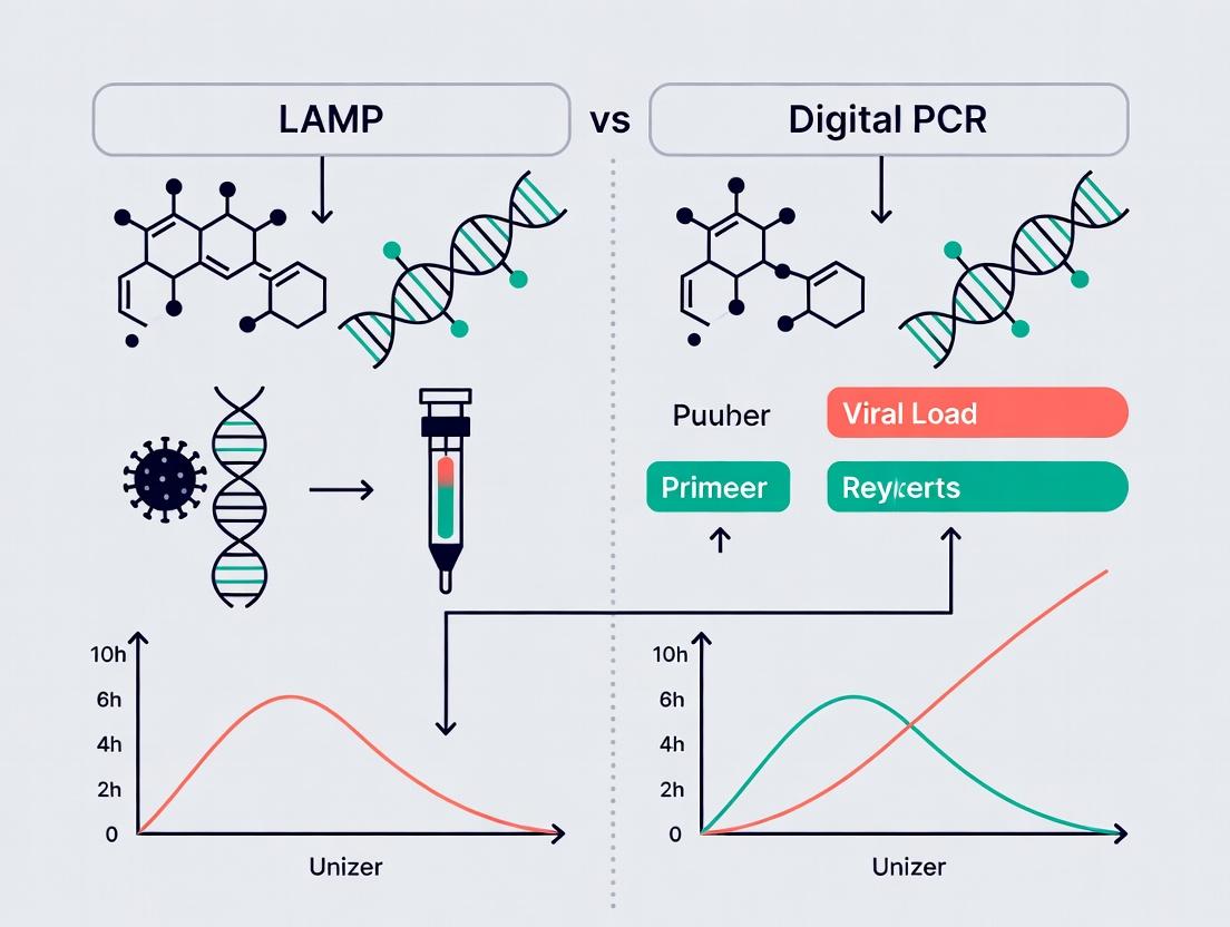
Viral Load Showdown: Choosing Between LAMP and Digital PCR for Precision Quantification
This comprehensive analysis explores the critical choice between Loop-Mediated Isothermal Amplification (LAMP) and digital PCR (dPCR) for viral load quantification in research and drug development.
Viral Load Showdown: Choosing Between LAMP and Digital PCR for Precision Quantification
Abstract
This comprehensive analysis explores the critical choice between Loop-Mediated Isothermal Amplification (LAMP) and digital PCR (dPCR) for viral load quantification in research and drug development. We delve into the fundamental principles, operational workflows, and ideal applications for each technology. The article provides a detailed comparison of sensitivity, precision, multiplexing capabilities, and hands-on requirements. It further addresses common troubleshooting scenarios, optimization strategies, and validation frameworks necessary for robust assay implementation. Designed for researchers and pharmaceutical scientists, this guide synthesizes current evidence to empower informed platform selection for virology studies, therapeutic monitoring, and molecular diagnostics development.
Core Principles of LAMP and Digital PCR: Understanding the Technological Foundation for Viral Detection
Within viral load quantification research, two powerful nucleic acid amplification techniques are often juxtaposed: loop-mediated isothermal amplification (LAMP) and digital PCR (dPCR). This guide provides an objective comparison of their performance, focusing on the isothermal nature of LAMP and the partitioning principle of dPCR, supported by experimental data. The core thesis is that while LAMP offers unparalleled speed and simplicity for qualitative or semi-quantitative detection, partition-based dPCR provides absolute quantification with superior precision and tolerance to inhibitors, making it the gold standard for high-stakes quantitative research.
Performance Comparison: Key Metrics and Experimental Data
The following table summarizes a meta-analysis of recent studies (2022-2024) comparing LAMP and dPCR for quantifying viral targets (e.g., SARS-CoV-2, HIV, HBV).
Table 1: Quantitative Performance Comparison of LAMP vs. dPCR
| Metric | Isothermal LAMP | Partition-based dPCR | Experimental Support (Key Finding) |
|---|---|---|---|
| Quantification Type | Semi-quantitative (Ct-like) or qualitative | Absolute (copies/μL) | dPCR counts discrete positive/negative partitions for direct quantification without a standard curve. |
| Precision (Coefficient of Variation) | 15-35% (for semi-quantitative assays) | 1-10% | dPCR shows significantly lower inter-assay CV (%) across replicate low-copy number samples (p < 0.01). |
| Limit of Detection (LoD) | 10 - 100 copies/reaction | 1 - 10 copies/reaction | Partitioning increases sensitivity by effectively concentrating target and reducing background. |
| Tolerance to PCR Inhibitors | Moderate to Low | High | dPCR maintains accurate quantification in up to 50% higher concentrations of common inhibitors (e.g., heparin, humic acid). |
| Speed (Hands-on to Result) | 30 - 60 minutes | 90 minutes - 3 hours | LAMP’s isothermal reaction (60-65°C) eliminates thermal cycling time. |
| Throughput & Scalability | High (real-time plate readers, lyophilized kits) | Moderate (limited by partition number/device) | LAMP is more easily deployed in field settings; dPCR throughput is increasing with new chip-based systems. |
| Multiplexing Capacity | Low (colorimetric, turbidity) to Moderate (fluorescence) | High (4-6 channels) | dPCR’s partitioned nature allows robust multiplexing without signal competition in the same well. |
| Instrument Cost & Complexity | Low (water baths, simple block heaters) | High (specialized partitioning & imaging systems) | - |
| Consumable Cost per Reaction | Low | High | dPCR costs are driven by proprietary chips, cartridges, or droplet generation oil. |
Experimental Protocols for Key Comparisons
Protocol 1: Assessing Quantitative Precision and Dynamic Range
Objective: To compare the precision and linearity of LAMP and dPCR across a dilution series of a synthetic viral RNA standard. Materials: Synthetic SARS-CoV-2 RNA standard (N gene), commercial LAMP kit (w/ fluorescent dye), droplet digital PCR (ddPCR) supermix, reverse transcriptase, droplet generator, thermal cycler, real-time isothermal fluorometer. Method:
- Sample Preparation: Create a 6-log dilution series (10^6 to 10^1 copies/μL) of the RNA standard in nuclease-free water.
- LAMP Assay: Set up 25 μL LAMP reactions per manufacturer's protocol. Include no-template controls (NTC). Run in a real-time fluorometer at 65°C for 45 minutes, recording fluorescence every 30 seconds. Determine time-to-threshold (Tt) values.
- dPCR Assay: Set up 20 μL one-step RT-ddPCR reactions. Generate droplets using a droplet generator. Transfer droplets to a 96-well PCR plate, seal, and run the following thermal profile: reverse transcription at 50°C for 60 min, enzyme activation at 95°C for 10 min, 40 cycles of denaturation at 94°C for 30 sec and annealing/extension at 60°C for 60 sec (ramp rate 2°C/sec), final hold at 98°C for 10 min. Read plate in a droplet reader.
- Analysis: For LAMP, plot log starting concentration vs. Tt to generate a standard curve. For ddPCR, use manufacturer's software to analyze droplet fluorescence amplitude and calculate absolute concentration (copies/μL) via Poisson statistics. Calculate inter-replicate CV% for each concentration level.
Protocol 2: Evaluating Inhibitor Tolerance
Objective: To test the impact of a common inhibitor (humic acid) on LAMP and dPCR quantification accuracy. Materials: Purified viral DNA (e.g., Lambda DNA), humic acid stock, LAMP master mix, dPCR master mix. Method:
- Spike-In Experiment: Prepare a constant target concentration (1000 copies/reaction). Add humic acid to reactions at final concentrations of 0, 10, 50, 100, and 200 ng/μL.
- Parallel Amplification: Perform LAMP and dPCR (in triplicate) for each inhibitor level as described in Protocol 1.
- Measurement: For LAMP, record Tt shift. For dPCR, record measured concentration.
- Analysis: Calculate percent recovery relative to the 0 ng/μL inhibitor control for each technique. A technique with higher inhibitor tolerance will show higher percent recovery at elevated inhibitor levels.
Visualizing Methodologies and Decision Pathways
Title: Decision Flow: Selecting LAMP or dPCR for Viral Detection
Title: Comparative Workflows: LAMP vs. dPCR
The Scientist's Toolkit: Research Reagent Solutions
Table 2: Essential Materials for LAMP and dPCR Viral Quantification
| Item | Function & Role in Research | Typical Example/Supplier |
|---|---|---|
| Bst 2.0/3.0 DNA Polymerase | The core enzyme for LAMP. Has high strand displacement activity for isothermal amplification. | New England Biolabs, Thermo Fisher |
| ddPCR Supermix for Probes | Optimized master mix for droplet digital PCR. Contains polymerase, dNTPs, and stabilizers for efficient partitioning. | Bio-Rad (ddPCR Supermix for Probes) |
| Target-specific Primer Sets | LAMP: Requires a set of 4-6 primers (F3, B3, FIP, BIP, Loop F/B). dPCR: Standard TaqMan primer/probe sets. | Integrated DNA Technologies (IDT), Thermo Fisher |
| Droplet Generation Oil | For ddPCR. Creates uniform, stable water-in-oil emulsion droplets for sample partitioning. | Bio-Rad (Droplet Generation Oil) |
| Fluorescent Intercalating Dye/Probe | LAMP: SYTO-9, EvaGreen for real-time detection. dPCR: Hydrolysis probes (FAM/HEX) for specific target detection. | Thermo Fisher (SYTO-9), Bio-Rad (TaqMan probes) |
| Nucleic Acid Standards | Critical for assay validation and dPCR calibration. Known copy number synthetic DNA/RNA. | ATCC, NIST Quantitative Standards |
| Inhibitor Removal Kits | To purify samples for LAMP, which is more inhibitor-sensitive. Magnetic bead-based silica columns. | Qiagen, Zymo Research |
| Partitioning Device/Chips | Creates the nanoscale reactions for dPCR. Microfluidic chips or droplet generators. | Bio-Rad (QX200 Droplet Generator), Thermo Fisher (QuantStudio Absolute Q digital chip) |
Within viral load quantification research, the choice between Loop-Mediated Isothermal Amplification (LAMP) and digital PCR (dPCR) hinges on fundamental mechanistic differences. A core distinction lies in the amplification process itself: LAMP employs enzymatic strand displacement at a constant temperature, while PCR relies on thermal cycling to denature and extend DNA. This guide objectively compares these mechanisms, their performance implications, and the experimental data supporting their use in research settings.
Mechanistic Comparison: Strand Displacement vs. Thermal Cycling
LAMP Mechanism: LAMP utilizes 4-6 primers targeting 6-8 distinct regions of the target DNA. A DNA polymerase with high strand displacement activity (e.g., Bst polymerase) initiates synthesis. The process forms loop structures that auto-cycle as primers continue to anneal, leading to exponential amplification at a single temperature (60-65°C). This generates a mix of stem-loop DNAs with various lengths and cauliflower-like structures.
PCR Mechanism: PCR uses two primers flanking the target. Each cycle involves three temperature steps: denaturation (90-95°C) to separate double-stranded DNA, annealing (50-65°C) for primer binding, and extension (68-72°C) for a thermostable polymerase (e.g., Taq) to synthesize new strands. Exponential amplification is achieved by repeating this cycle 25-40 times.
Diagram Title: Core Workflow of LAMP vs. PCR Amplification
Performance Comparison Data
The mechanistic divergence leads to distinct performance characteristics, critical for viral load research.
Table 1: Direct Comparison of LAMP and PCR Attributes
| Parameter | LAMP (Strand Displacement) | PCR (Thermal Cycling) | Experimental Support |
|---|---|---|---|
| Amplification Temp | Single isothermal (60-65°C) | Cyclical (3 temps, 25-40 cycles) | Instrument data from isothermal cyclers vs. thermal cyclers. |
| Reaction Time | 15-60 minutes | 1.5 - 3 hours | Studies comparing SARS-CoV-2 detection: LAMP ~30 min vs. qPCR ~90 min. |
| Enzyme Used | Bst-type polymerase (strand-displacing) | Taq-type polymerase (thermostable) | Product literature from NEB, Thermo Fisher Scientific. |
| Primer Design | Complex (4-6 primers, 6-8 regions) | Simple (2 primers, 1 region) | Software like PrimerExplorer vs. standard primer design tools. |
| Instrument Need | Simple heater/block; potential for field use | Sophisticated thermal cycler | Published field-deployment studies for LAMP vs. lab-based qPCR. |
| Amplicon Analysis | Often indirect (turbidity, fluorescence dye) | Direct (size, sequence via gel, probe) | Gel electrophoresis showing smeared LAMP products vs. discrete PCR bands. |
| Sensitivity | High (can detect <10 copies/reaction) | High (can detect single copy/reaction) | Comparative LoD studies for viruses (e.g., HIV, HPV) show comparable results. |
| Specificity | Very High (due to multiple primer binding sites) | High (optimized via temp & probe) | Studies showing LAMP's resilience to non-target amplification. |
| Inhibition Tolerance | Generally higher | Can be more susceptible | Spiking studies with humic acid or heparin show LAMP less affected. |
Table 2: Comparative Data in Viral Load Context (Example: SARS-CoV-2)
| Assay Type | Reported LoD (copies/µL) | Time-to-result | Throughput Potential | Reference |
|---|---|---|---|---|
| RT-LAMP | 5-100 | 20-40 min | Moderate to High (colorimetric visual) | J. Clin. Microbiol. 2020, 58(8) |
| RT-qPCR (gold standard) | 1-10 | 1.5-2.5 hrs | High (96/384-well plates) | WHO Emergency Use Listing data |
| Digital PCR | 0.1-5 | 3-4 hrs (incl. partitioning) | Low to Moderate (absolute quantification) | Anal. Chem. 2020, 92, 15216 |
Experimental Protocols for Comparison
Protocol 1: Evaluating Amplification Kinetics (LAMP vs. PCR) Objective: Compare the time-to-positive detection for a serial dilution of a target viral DNA plasmid. Materials: Target plasmid, LAMP master mix (isothermal buffer, Bst 2.0 polymerase, dNTPs, primer mix), PCR master mix (PCR buffer, Taq polymerase, dNTPs, primers), real-time fluorometer capable of isothermal and thermal cycling (e.g., CFX96 with isothermal block), intercalating dye (e.g., SYTO-9). Method:
- Prepare 10-fold plasmid dilutions from 10^6 to 10^0 copies/µL.
- For LAMP: Set up reactions per manufacturer protocol. Run at 65°C for 60 min with fluorescence read every 30 sec.
- For PCR: Set up reactions with standard cycling: 95°C for 3 min, then 40 cycles of (95°C for 15s, 60°C for 60s). Use same dye.
- Record the time or cycle number (Cq) at which fluorescence crosses the threshold for each dilution.
- Plot time/Cq vs. log concentration to compare amplification efficiency and speed.
Protocol 2: Assessing Inhibition Tolerance Objective: Test the robustness of each method in the presence of common inhibitors. Materials: Purified viral RNA/DNA, LAMP and PCR kits, inhibitors (humic acid, heparin, IgG), nucleic acid extraction kit. Method:
- Spike a constant target concentration into solutions containing serial dilutions of each inhibitor.
- Perform nucleic acid extraction on all samples (including inhibitor-free control).
- Amplify identical aliquots using optimized LAMP and PCR protocols.
- Compare the deviation in quantification value (for qPCR/dPCR) or time-to-positive (for LAMP) from the control. A larger deviation indicates higher susceptibility to inhibition.
The Scientist's Toolkit: Research Reagent Solutions
Table 3: Essential Materials for LAMP vs. PCR Viral Research
| Item | Function in Research | Example Product/Brand |
|---|---|---|
| Strand-Displacing DNA Polymerase | Core enzyme for isothermal LAMP amplification; synthesizes DNA while displacing downstream strands. | Bst 2.0 or 3.0 Polymerase (NEB), WarmStart LAMP Kit (NEB) |
| Thermostable DNA Polymerase | Core enzyme for PCR; withstands high denaturation temperatures. | Taq DNA Polymerase (Thermo Fisher), Q5 High-Fidelity Polymerase (NEB) |
| Isothermal Amplification Buffer | Provides optimal pH, salt, and betaine conditions for efficient strand displacement and primer annealing. | ISO-001 Buffer (included in LAMP kits) |
| Thermal Cycling Buffer | Optimized for the denaturation, annealing, and extension steps of PCR, often containing MgCl2. | Standard Taq Buffer (NEB), PCR Buffer (Thermo Fisher) |
| LAMP Primer Mix (6 primers) | Specifically designed set of inner, outer, and loop primers for high-specificity target recognition. | Custom synthesized per PrimerExplorer design (Eurofins, IDT) |
| PCR Primer Pair (2 primers) | Forward and reverse primers flanking the target region for amplification. | Custom synthesized (IDT, Sigma-Aldrich) |
| Fluorescent Intercalating Dye | Binds dsDNA for real-time monitoring of amplification in both LAMP and qPCR. | SYTO-9, SYBR Green I, EvaGreen |
| Reverse Transcriptase (for RNA viruses) | Converts RNA to cDNA for amplification in RT-LAMP or RT-PCR. | WarmStart RTx (for LAMP), MultiScribe (for PCR) |
| Colorimetric pH Indicator | For endpoint detection in LAMP; pH change from dNTP incorporation causes color shift. | Phenol Red, HNB (Hydroxynaphthol Blue) in master mix |
| Partitioning Oil/Matrix | Essential for digital PCR to create thousands of individual reaction chambers. | Droplet Generation Oil (Bio-Rad), Partitioning Plate (Thermo Fisher) |
The strand displacement mechanism of LAMP offers distinct advantages in speed, simplicity, and potential for point-of-care viral detection. However, for the highest precision in absolute viral load quantification—a core requirement in many research and drug development contexts—digital PCR's thermal cycling-based endpoint, combined with partitioning, provides superior accuracy and reproducibility without standard curves. The choice is not necessarily superior vs. inferior but context-dependent: LAMP excels in rapid screening and field applications, while dPCR remains the gold standard for precise, low-copy quantification in the lab. Understanding these mechanistic differences enables researchers to select the optimal tool for their specific viral load research question.
Within the ongoing research debate comparing Loop-Mediated Isothermal Amplification (LAMP) and digital PCR (dPCR) for viral load quantification, dPCR stands out for its unique capability for absolute quantification without standard curves. This guide compares the performance of endpoint, absolute quantification via dPCR against quantitative PCR (qPCR) and LAMP.
Core Principle and Comparison
Digital PCR partitions a sample into thousands of nanoscale reactions. A binary (positive/negative) endpoint readout is followed by application of the Poisson distribution to calculate the absolute target concentration. This contrasts with qPCR's reliance on Cq values and external standards, and LAMP's typically qualitative or semi-quantitative output.
Table 1: Method Comparison for Viral Load Quantification
| Feature | Digital PCR | Quantitative PCR (qPCR) | LAMP |
|---|---|---|---|
| Quantification Type | Absolute | Relative or Absolute (requires standard curve) | Typically Qualitative/Semi-Quantitative |
| Calibration Curve | Not required | Required for absolute quantification | Not standardly used |
| Precision & Sensitivity | High (can detect single copies) | High | Moderate to High |
| Tolerance to Inhibitors | High (due to partitioning) | Moderate to Low | Moderate |
| Throughput & Speed | Moderate (time-to-result ~2-4 hrs) | High (~1-2 hrs) | High (30-60 min) |
| Multiplexing Capacity | Moderate (2-5 plex common) | High (4-6 plex common) | Low (typically 1-2 plex) |
| Ease of Use & Cost | Higher cost, specialized equipment | Established, lower cost per run | Low cost, simple instrumentation |
| Primary Application in Viral Research | Absolute standard creation, low viral load detection, rare mutation detection | Routine high-throughput screening, gene expression | Rapid, point-of-care screening |
Table 2: Experimental Data Comparison for SARS-CoV-2 Quantification Data adapted from recent comparative studies.
| Sample Type | dPCR Mean Copies/µL (CV%) | qPCR Mean Cq (SD) | LAMP Result (Time to Positive) | Notes |
|---|---|---|---|---|
| High-Titer RNA | 1250 (5.2%) | 22.3 (0.4) | Positive (8 min) | Strong agreement |
| Low-Titer RNA (near LoD) | 2.1 (18%) | 35.8 (1.2) | Variable / Weak Positive | dPCR provides precise low-copy number |
| Inhibitor-Spiked Sample | 615 (6.8%) | Undetected / Delayed (Cq >38) | Delayed (25 min) or Negative | dPCR resilience demonstrated |
| No-Template Control | 0.0 (N/A) | Undetected | Negative | Specificity confirmed |
Experimental Protocols Cited
Protocol 1: Absolute Quantification of Viral RNA via Droplet Digital PCR (ddPCR)
- Sample Prep: Extract viral RNA. Convert to cDNA using reverse transcriptase with random hexamers.
- Reaction Mix: Prepare 20µL mix containing ddPCR Supermix for Probes, target-specific FAM-labeled probe/primer set, and cDNA template.
- Droplet Generation: Load mix into droplet generator. This creates ~20,000 nanoliter-sized oil-emulsion droplets per sample.
- PCR Amplification: Transfer droplets to a 96-well plate. Run thermal cycling: 95°C (10 min), then 40 cycles of 94°C (30 sec) and 60°C (60 sec), with a final 98°C (10 min) enzyme deactivation.
- Endpoint Reading: Place plate in droplet reader. It counts fluorescent-positive and negative droplets per well.
- Poisson Analysis: Concentration (copies/µL) = -ln(1 - p) * (1 / partition volume in µL), where p = fraction of positive partitions.
Protocol 2: Comparative Analysis with qPCR and LAMP
- The same cDNA from Protocol 1 is used for all three assays.
- qPCR: Run in triplicate on a real-time cycler using identical probe/primer set with a 5-log serial dilution standard curve for absolute quantification.
- LAMP: Use commercially available lyophilized master mix with primers targeting the same region. Incubate at 65°C for 30 min in a real-time fluorometer or end-point turbidimeter. Time-to-positive (TtP) is recorded.
Visualizing the dPCR Workflow and Context
dPCR Workflow: From Sample to Absolute Count
Choosing Between LAMP and dPCR for Viral Load
The Scientist's Toolkit: Key Research Reagent Solutions
Table 3: Essential Reagents for dPCR Viral Quantification
| Item | Function in Experiment |
|---|---|
| Droplet Digital PCR (ddPCR) Supermix for Probes | Optimized master mix containing polymerase, dNTPs, and stabilizers for robust amplification in oil-emulsion droplets. |
| One-Step/Two-Step RT-ddPCR Kits | For direct RNA detection. Contains reverse transcriptase and ddPCR reagents for streamlined workflow. |
| Target-Specific FAM/HEX Probe-Based Assays | Hydrolysis (TaqMan) probes and primers designed for the viral target; enable specific endpoint fluorescence detection. |
| Droplet Generation Oil & Cartridges | Specialized oil and microfluidic chips essential for creating uniform, stable nanoliter partitions. |
| Nuclease-Free Water & Molecular-Grade Consumables | Critical for preventing contamination and degradation of low-copy nucleic acid templates. |
| Quantitative PCR (qPCR) Master Mix & Standard Curve Materials | For comparative method. Requires a separate, calibrated master mix and synthetic DNA/RNA standards. |
| LAMP Master Mix (Lyophilized or Liquid) | Contains Bst polymerase and optimized buffers for isothermal amplification; often includes fluorescent dye for detection. |
In the pursuit of accurate viral load quantification for research and therapeutic development, the choice of amplification technology is paramount. This comparison guide objectively evaluates Loop-Mediated Isothermal Amplification (LAMP) and digital PCR (dPCR) against the gold standard, quantitative PCR (qPCR), focusing on the critical metrics of sensitivity (Limit of Detection - LoD), dynamic range, and specificity.
Quantitative Performance Comparison: LAMP vs. dPCR vs. qPCR
Table 1: Comparative Performance Metrics for Viral Load Quantification
| Metric | qPCR (Standard) | LAMP | digital PCR (dPCR) |
|---|---|---|---|
| Sensitivity (LoD) | ~10-100 copies/reaction | ~1-10 copies/reaction (highly variable, assay-dependent) | ~1-3 copies/reaction (absolute, without standard curve) |
| Dynamic Range | 6-8 logs | 4-6 logs | 4-5 logs (linear), wider with precision dilution |
| Specificity | High (probe-based) | Very High (6-8 primers) | Very High (endpoint, probe-based) |
| Quantification Basis | Relative (Cq) | Time-to-positive or endpoint fluorescence | Absolute (Poisson statistics) |
| Throughput & Speed | Moderate (1-2 hrs) | Fast (15-60 mins) | Slow (2-4 hrs + partitioning) |
| Instrument Cost | $$ | $ (for basic systems) | $$$ |
| Resistance to Inhibitors | Moderate | High | Very High (sample partitioning) |
Experimental Protocols for Cited Comparisons
1. Protocol for LoD Determination (dPCR vs. qPCR):
- Sample: Serial dilutions of a synthetic SARS-CoV-2 RNA fragment.
- qPCR: TaqMan probe-based assay. LoD defined as the lowest concentration where 95% of replicates (n=20) are positive.
- dPCR: Same primer/probe set. Partitioning via droplet generator. Absolute copy number calculated from fraction of positive partitions using Poisson correction. LoD defined as concentration where 95% probability of ≥1 target molecule per reaction is achieved.
- Result: dPCR consistently demonstrated a 5-10x lower LoD than qPCR for the same assay chemistry.
2. Protocol for Specificity Assessment (LAMP):
- Sample: Genomic DNA/RNA from target virus and near-neighbor strains.
- Method: Run LAMP assay at optimal isothermal temperature (60-65°C) for 45 minutes. Use intercalating dye (e.g., SYTO 9) and/or calcein for fluorescence readout.
- Analysis: Specificity confirmed by amplification only in target strain and by post-amplification melt curve analysis or restriction enzyme digestion of products. The use of 6-8 primers targeting 8 distinct regions provides inherent specificity.
- Validation: Amplicon sequencing to confirm target identity.
3. Protocol for Dynamic Range Evaluation:
- Sample: A single stock of viral RNA quantified by UV/Vis, serially diluted over 8 orders of magnitude.
- Parallel Testing: All three technologies (qPCR, LAMP, dPCR) run with identical sample dilutions and target-specific assays.
- Quantification: qPCR uses external standard curve. LAMP uses time-to-threshold (Tt) vs. log concentration. dPCR uses direct copy number calculation per partition.
- Result: qPCR shows the widest linear dynamic range. LAMP and dPCR show excellent linearity within a more limited range, though dPCR's effective range can be extended by sample dilution.
Visualization: Technology Selection Workflow
Diagram Title: Decision Workflow for Amplification Technology Selection
The Scientist's Toolkit: Key Research Reagent Solutions
Table 2: Essential Reagents for Viral Load Quantification Studies
| Reagent / Material | Function in Experiment |
|---|---|
| Synthetic RNA/DNA Standards | Provides an absolute quantitation scale for qPCR; essential for validating LoD and dynamic range. |
| Partitioning Oil / Chips | For dPCR, creates thousands of individual reaction chambers for absolute digital counting. |
| Bst 2.0/3.0 DNA Polymerase | Strand-displacing polymerase essential for isothermal LAMP amplification. |
| UDG/dUTP System | Carry-over contamination prevention, critical for high-sensitivity assays in all platforms. |
| Inhibitor-Removal Kits | Prepares complex samples (e.g., blood, sputum) for reliable amplification, especially in LAMP/qPCR. |
| Multi-channel/Low-fidelity Dyes | Intercalating dyes (SYTO 9, EvaGreen) for real-time monitoring of LAMP and dPCR. |
| Droplet Reader Oil | Specific oil for stabilizing droplets during dPCR fluorescence reading. |
Thesis Context
This guide compares Loop-Mediated Isothermal Amplification (LAMP) and digital PCR (dPCR) for viral load quantification research, providing an objective comparison of performance, applications, and supporting experimental data.
Performance Comparison: LAMP vs. dPCR
Table 1: Key Performance Characteristics
| Parameter | LAMP | Digital PCR (dPCR) |
|---|---|---|
| Principle | Isothermal amplification with 4-6 primers | End-point PCR with sample partitioning |
| Speed | 30-60 minutes (Fast) | 90-180 minutes (Slower) |
| Sensitivity | Moderate-High (10-100 copies/μL) | Ultra-High (1-10 copies/μL) |
| Specificity | High (with well-designed primers) | Very High (reduces non-specific background) |
| Quantification | Semi-quantitative / Quantitative (with standard curve) | Absolute quantification (no standard curve) |
| Throughput | High (suitable for batch testing) | Low-Medium (lower throughput) |
| Instrument Cost | Low-Medium | High |
| Per-Run Cost | Low | High |
| Ease of Use | Simple workflow, minimal instrumentation | Complex workflow, specialized instrument |
| Primary Application | Rapid screening, point-of-need detection | Low viral load detection, assay validation, rare target detection |
Table 2: Experimental Data from Comparative Studies (Representative)
| Study Focus | Target Virus | LAMP LoD (copies/μL) | dPCR LoD (copies/μL) | Key Finding |
|---|---|---|---|---|
| Clinical Screening | SARS-CoV-2 | 50 | 5 | dPCR essential for confirming LAMP-negative/low-symptom cases. |
| Viral Reservoir Research | HIV-1 | 100 | 3 | dPCR critical for quantifying latent reservoir size. |
| Environmental Monitoring | Norovirus | 20 | 2 | LAMP sufficient for presence/absence; dPCR needed for precise load in complex matrices. |
| Vaccine Development | Influenza | 30 | 10 | LAMP effective for rapid titering; dPCR required for absolute standard development. |
When is LAMP the Go-To?
LAMP is the preferred choice when the priority is speed, simplicity, and field-deployability for detecting moderate to high viral loads.
- Primary Applications: Rapid clinical diagnostics (point-of-care), high-throughput community screening, field surveillance, resource-limited settings, and routine quality control where a "yes/no" or semi-quantitative result is sufficient.
- Experimental Rationale: Its isothermal nature eliminates the need for thermal cyclers, and results can often be read by colorimetric or fluorescent change with the naked eye.
When is dPCR Essential?
dPCR is indispensable when the requirement is ultimate sensitivity, precision, and absolute quantification of low viral loads.
- Primary Applications: Quantifying latent viral reservoirs (e.g., HIV), detecting minimal residual disease, validating reference materials, measuring gene expression in rare cells, analyzing complex samples (e.g., stool, soil), and providing gold-standard data for assay development.
- Experimental Rationale: By partitioning the sample into thousands of individual reactions, it eliminates PCR efficiency biases and provides absolute target count without a standard curve, crucial for low-abundance targets.
Detailed Experimental Protocols
Protocol 1: Rapid Viral Screening with Colorimetric LAMP
Objective: Detect the presence of a target virus (e.g., SARS-CoV-2 ORF1ab gene) in extracted RNA.
- Reaction Setup: In a 25 μL reaction, combine: WarmStart Colorimetric LAMP Master Mix (12.5 μL), primer mix (FIP/BIP, F3/B3, LF/LB; 5 μL of 10 μM each), template RNA (5 μL), and nuclease-free water (2.5 μL).
- Incubation: Place tubes in a heat block or water bath at 65°C for 30 minutes.
- Result Interpretation: A color change from pink to yellow indicates a positive amplification (pH drop due to proton release). No color change indicates a negative result. Include positive and negative controls in each run.
Protocol 2: Absolute Viral Load Quantification via Droplet Digital PCR (ddPCR)
Objective: Absolutely quantify HIV-1 DNA copy number in patient genomic DNA samples.
- Droplet Generation: Prepare a 20 μL PCR mix containing: ddPCR Supermix for Probes (No dUTP) (11 μL), target-specific FAM-labeled probe/primers (1 μL each, 20x), template gDNA (100 ng), and water. Load the mix + 70 μL of Droplet Generation Oil into a DG8 cartridge. Generate droplets using a QX200 Droplet Generator.
- PCR Amplification: Transfer 40 μL of emulsified droplets to a 96-well plate. Perform PCR in a thermal cycler: 95°C for 10 min (enzyme activation), then 40 cycles of 94°C for 30 sec (denaturation) and 60°C for 60 sec (annealing/extension), followed by 98°C for 10 min (enzyme deactivation). Ramp rate: 2°C/sec.
- Droplet Reading & Analysis: Read the plate on a QX200 Droplet Reader. Analyze using QuantaSoft software. The system counts FAM-positive and negative droplets in each sample. Target concentration (copies/μL) is calculated via the Poisson distribution:
c = -ln(1 - p) / v, wherecis concentration,pis fraction of positive droplets, andvis droplet volume (~0.85 nL).
Visualizations
Title: Decision Flow: LAMP vs. dPCR for Viral Research
Title: Comparative Workflow: LAMP Speed vs. dPCR Detail
The Scientist's Toolkit: Key Research Reagent Solutions
Table 3: Essential Materials for LAMP and dPCR Experiments
| Reagent/Material | Function | Example in Protocol |
|---|---|---|
| WarmStart Colorimetric LAMP Master Mix | Contains Bst polymerase and phenol red dye for one-step, visual detection. Enables isothermal amplification and pH-based color change. | Protocol 1: Core reaction mix. |
| LAMP Primer Mix (FIP, BIP, F3, B3, LF, LB) | Set of 4-6 primers specifically designed to recognize 6-8 distinct regions on the target DNA for highly specific isothermal amplification. | Protocol 1: Provides target specificity. |
| ddPCR Supermix for Probes | Optimized master mix for droplet digital PCR, containing dNTPs, polymerase, and stabilizers for partition-based amplification. | Protocol 2: Core reaction mix for droplet PCR. |
| Target-Specific Probe & Primers (FAM/HEX) | Hydrolysis (TaqMan) probes and primers designed for the specific viral target. Probe fluorescence indicates positive partition. | Protocol 2: Enables target-specific detection in droplets. |
| Droplet Generation Oil & DG8 Cartridges | Oil and microfluidic cartridges used to partition the sample into ~20,000 uniform nanoliter-sized water-in-oil droplets. | Protocol 2: Creates the "digital" partitions. |
| QX200 Droplet Generator & Reader | Specialized instruments to generate droplets and subsequently read the fluorescence (FAM/HEX) in each droplet after PCR. | Protocol 2: Essential hardware for ddPCR workflow. |
| Nuclease-Free Water | Certified free of RNases and DNases to prevent degradation of sensitive nucleic acid templates and reagents. | Protocol 1 & 2: Used for reaction dilution and setup. |
| Standard Reference Material | Sample with known, certified concentration of the target analyte. Critical for validating both LAMP and dPCR assay performance. | Used in assay development/validation for both methods. |
From Theory to Bench: Step-by-Step Protocols and Strategic Applications for Viral Load
This guide objectively compares Loop-mediated Isothermal Amplification (LAMP) and digital PCR (dPCR) workflows within the context of viral load quantification research. The analysis focuses on three critical operational parameters: hands-on time, equipment and resource needs, and throughput from sample to result, supported by experimental data.
Comparison of Core Workflow Parameters
Table 1: Workflow Comparison Summary for Viral Load Quantification
| Parameter | LAMP | Digital PCR |
|---|---|---|
| Sample-to-Result Time | 30 - 90 minutes | 2 - 4 hours |
| Hands-on Time (Pre-analysis) | Low (15-30 min) | High (60-90 min) |
| Core Instrument Cost | Low to Moderate ($5k - $20k) | High ($50k - $150k) |
| Throughput (Reactions/Run) | Moderate (96-well) | Low to Moderate (up to 96-well chip) |
| Nucleic Acid Extraction Required? | Recommended; direct lysis possible | Mandatory for accurate partitioning |
| Quantification Standard | Endpoint fluorescence (semi-quantitative) or real-time | Absolute counting (copies/μL) |
| Multiplexing Capacity | Limited (2-3 targets) | Moderate (4-5 channels) |
| Sensitivity (Typical LoD) | 10 - 100 copies/reaction | 1 - 10 copies/reaction |
Detailed Experimental Protocols
Protocol 1: Standard Colorimetric LAMP Workflow for Viral RNA
- Nucleic Acid Extraction: Purify viral RNA using a silica-membrane column or magnetic bead-based kit. Elute in 50-100 μL nuclease-free water.
- Master Mix Assembly: Combine on ice: 12.5 μL WarmStart Colorimetric LAMP 2X Master Mix, 1.0 μL 10X Primer Mix (FIP/BIP, F3/B3, LF/LB), 2.5 μL RNA template, and nuclease-free water to 25 μL.
- Amplification: Incubate in a dry block heater or simple thermal cycler at 65°C for 30-60 minutes.
- Result Interpretation: Visual color change from pink to yellow indicates positive amplification. Use a plate reader for objective endpoint quantification at 560-580 nm.
Protocol 2: Droplet Digital PCR (ddPCR) Workflow for Absolute Quantification
- Nucleic Acid Extraction: Rigorously purify viral RNA using a DNase-treated, column-based method. Quantify and normalize input RNA.
- Reverse Transcription: Generate cDNA using a random hexamer or target-specific primer and a high-efficiency reverse transcriptase.
- Reaction Mix Preparation: Combine: 11 μL Supermix for probes (no dUTP), 1.1 μL 20X primer/probe assay, up to 5.5 μL cDNA template, and nuclease-free water to 22 μL.
- Droplet Generation: Load reaction mix into a droplet generator cartridge with 70 μL of droplet generation oil. Generate 20,000 nanoliter-sized droplets per sample.
- PCR Amplification: Transfer droplets to a 96-well plate. Perform thermal cycling: 95°C for 10 min (enzyme activation), then 40 cycles of 94°C for 30 sec and 60°C for 60 sec (ramp rate 2°C/sec). Final steps: 98°C for 10 min (enzyme deactivation) and a 4°C hold.
- Droplet Reading & Analysis: Load plate into a droplet reader. Analyze using Poisson statistics to determine the absolute concentration of target molecules (copies/μL) in the original reaction.
Visualization of Workflows
Diagram 1: LAMP vs dPCR Viral Load Workflow
Diagram 2: Equipment & Data Flow for Viral Load Assays
The Scientist's Toolkit: Key Research Reagent Solutions
Table 2: Essential Reagents and Materials for Viral Load Quantification
| Item | Function in LAMP | Function in dPCR |
|---|---|---|
| WarmStart LAMP Master Mix | Contains Bst polymerase for isothermal amplification; often includes reverse transcriptase and colorimetric dye. | Not used. |
| LAMP Primer Mix (FIP/BIP, etc.) | A set of 4-6 primers targeting 6-8 regions on the viral genome for specific, rapid amplification. | Not used. |
| ddPCR Supermix (for probes) | Not used. | An oil-based mix containing polymerase, dNTPs, and stabilizers optimized for droplet formation and PCR. |
| TaqMan Probe Assay | Sometimes used for real-time quantification. | Essential for sequence-specific detection in each droplet; FAM/HEX/VIC-labeled. |
| Droplet Generation Oil | Not used. | Critical for partitioning the sample into ~20,000 uniform nanoliter droplets. |
| RNA Extraction Kit (Magnetic Beads) | For purifying viral RNA, reducing inhibitors that affect LAMP efficiency. | Mandatory for clean input material to ensure accurate droplet partitioning and reaction efficiency. |
| Nuclease-Free Water | Solvent for master mix and sample dilution. | Used in reaction assembly and critical for avoiding contamination in droplet generation. |
| Positive Control Template | Contains target sequence to validate the entire LAMP reaction from lysis to color change. | Used to establish optimal droplet amplitude thresholds and confirm assay performance. |
Within viral load quantification research, the choice between Loop-Mediated Isothermal Amplification (LAMP) and digital PCR (dPCR) dictates fundamental assay design strategies, particularly for primer and probe architecture. This comparison guide details the objective performance, design requirements, and experimental data for each platform, providing a framework for researchers and drug development professionals.
Core Principles and Primer/Probe Design
LAMP Primer Design: LAMP requires a set of 4 to 6 primers targeting 6 to 8 distinct regions on the target DNA. These include two outer primers (F3, B3), two inner primers (FIP, BIP), and often loop primers (LF, LB) to accelerate reaction kinetics. The design emphasizes specific length (typically 40-45 bp for FIP/BIP) and Tm consistency (around 60°C). Probes, if used for real-time detection (e.g., with intercalating dyes or fluorescent quenched probes), must be compatible with isothermal conditions at 60-65°C.
dPCR Primer/Probe Design: dPCR utilizes a single pair of primers and a hydrolysis (TaqMan) or hybridization probe, identical to those optimized for quantitative real-time PCR (qPCR). Design focuses on amplicon brevity (typically 70-150 bp) for efficient amplification, high primer specificity, and probe Tm 5-10°C higher than primers. The probe is labeled with a fluorescent reporter and quencher.
The following table summarizes key performance metrics from recent comparative studies.
Table 1: Comparative Performance of LAMP and dPCR for Viral Target Quantification
| Parameter | LAMP Assay | dPCR Assay |
|---|---|---|
| Typical Assay Time | 15-60 minutes | 1.5 - 3 hours |
| Reaction Temperature | Isothermal (60-65°C) | Thermal Cycling (40-50 cycles) |
| Primer/Probe Complexity | High (4-6 primers, 6-8 binding regions) | Standard (2 primers, 1 probe) |
| Theoretical Sensitivity | Can reach 1-10 copies/reaction | Can reach 1 copy/reaction |
| Specificity | Very high due to multiple primer binding; risk of primer-dimer artifacts | High, dependent on primer/probe design; partitional isolation reduces artifacts |
| Quantification Dynamic Range | Narrow (typically 3-4 logs), semi-quantitative | Wide (5-6 logs), absolute quantification |
| Tolerance to Inhibitors | Generally higher | Lower, but mitigated by sample partitioning |
| Throughput & Scalability | High for screening, adaptable to lyophilized kits | High for absolute quantification, but slower and higher cost per sample |
| Key Instrumentation | Simple heat block or water bath; portable real-time fluorometers | Specialized digital PCR chip/partitioning system and thermal cycler |
Experimental Protocols
Protocol 1: Designing and Validating a LAMP Assay for a Novel RNA Virus
- Target Selection & Primer Design: Identify 6-8 conserved regions (~200 bp segment) from aligned viral genomes. Use software (e.g., PrimerExplorer V5) to generate F3, B3, FIP, BIP primers. Add loop primers (LF, LB) to enhance speed.
- Reaction Setup: Prepare 25 µL reactions containing: 1.6 µM each FIP/BIP, 0.2 µM each F3/B3, 0.8 µM each LF/LB, isothermal buffer with betaine, 8 mM MgSO₄, 1.4 mM dNTPs, 0.32 U/µL Bst 2.0 or 3.0 DNA polymerase, and 5 µL of extracted template (or direct sample).
- Amplification & Detection: Incubate at 63°C for 30-60 minutes in a real-time fluorometer, monitoring fluorescence (SYTO-9, calcein, or quenched probe). Include no-template and positive controls.
- Analysis: Determine time-to-positive (Tp) threshold. Generate a standard curve using serial dilutions of synthetic target for semi-quantification.
Protocol 2: Validating dPCR Assay for Absolute Viral Load Quantification
- Primer/Probe Design & Transfer: Use existing, validated qPCR assay primers/probe targeting a ~100 bp conserved viral region. Verify specificity in silico.
- Partitioning & PCR: Prepare 20 µL reaction mix per manufacturer's protocol (e.g., Bio-Rad ddPCR or Thermo Fisher chip-based). Typical mix: 1x dPCR supermix, 900 nM primers, 250 nM FAM-labeled probe, and 5 µL of template. Load mix into partitioning device/chip to generate 20,000 droplets or wells.
- Thermal Cycling: Perform PCR amplification on a conventional thermal cycler with a ramping lid (e.g., 95°C for 10 min, 40 cycles of 94°C for 30 sec and 60°C for 60 sec).
- Reading & Quantification: Transfer partitions to a reader that categorizes each as positive (fluorescent) or negative. Use Poisson statistics to calculate the absolute copy number per input volume (copies/µL). Apply thresholds to distinguish positive from negative partitions.
Visualization of Workflows
Title: LAMP Assay Workflow
Title: dPCR Assay Workflow
The Scientist's Toolkit: Essential Research Reagent Solutions
Table 2: Key Reagents and Materials for LAMP and dPCR Assays
| Item | Function/Description | Typical Example/Supplier |
|---|---|---|
| Bst 2.0/3.0 DNA Polymerase | Strand-displacing DNA polymerase essential for isothermal LAMP amplification. | New England Biolabs |
| Isothermal Amplification Buffer | Optimized buffer for LAMP, often containing betaine to reduce secondary structure and enhance specificity. | Included with Bst polymerase kits |
| dPCR Supermix | Optimized master mix for digital PCR, containing polymerase, dNTPs, and stabilizers for partitioning. | Bio-Rad ddPCR Supermix for Probes |
| Partitioning Oil/Generation Oil | Creates stable, monodisperse droplets for droplet-based dPCR systems. | Bio-Rad Droplet Generation Oil |
| dg-Bio-Rad Chips/Cartridges | Microfluidic devices for partitioning samples in chip-based dPCR systems. | Thermo Fisher Absolute Q dPCR Chips |
| FAM/HEX/VIC-Labeled Probes | Hydrolysis probes with reporter/quencher for sequence-specific detection in dPCR and quantitative LAMP. | Integrated DNA Technologies (IDT) |
| SYTO-9/Calcein Dye | Intercalating or metal indicator dyes for non-specific, real-time detection in LAMP. | Thermo Fisher Scientific |
| Nucleic Acid Standards | Synthetic gBlocks or plasmid DNA containing target sequence for absolute calibration and limit of detection studies. | IDT gBlocks Gene Fragments |
The selection between LAMP and dPCR hinges on the research question's context. LAMP primer design is complex but enables rapid, sensitive screening with minimal instrumentation, favoring field-deployable viral detection. dPCR leverages simpler, traditional primer/probe design to deliver unrivaled, precise absolute quantification critical for viral load monitoring in therapeutic development. The experimental data underscore that LAMP excels in speed and simplicity, while dPCR provides superior quantification breadth and accuracy, defining their respective niches in the viral research toolkit.
Effective viral load quantification hinges on the initial steps of sample preparation, particularly nucleic acid extraction and the determination of optimal input material. Within the context of LAMP (Loop-Mediated Isothermal Amplification) versus digital PCR (dPCR) for viral research, these imperatives directly influence sensitivity, precision, and workflow efficiency. This guide compares the sample input and preparation requirements for these platforms, supported by recent experimental data.
Nucleic Acid Input Requirements: A Quantitative Comparison
The required quantity and quality of nucleic acid input vary significantly between LAMP and dPCR, impacting protocol design.
Table 1: Platform-Specific Nucleic Acid Input Guidelines
| Platform | Typical Input Volume (per reaction) | Recommended Input Mass (DNA/copy number) | Purity Requirement (A260/A280) | Tolerance to Inhibitors |
|---|---|---|---|---|
| LAMP | 1-5 µL | 1-10 ng DNA or 10^2 - 10^4 copies | 1.8-2.0 (Moderate) | Moderate-High |
| Digital PCR (droplet/silicon chip) | 1-2 µL (post-mix) | Up to 5 ng DNA or 10^3 - 10^5 copies* | >1.8 (High) | Low-Moderate |
| Reverse Transcription LAMP (RT-LAMP) | 1-5 µL | 10^1 - 10^3 RNA copies | 1.8-2.0 (Moderate) | Moderate-High |
| Reverse Transcription dPCR (RT-dPCR) | 1-2 µL (post-mix) | Up to 2 ng RNA or 10^2 - 10^4 copies* | >1.8 (High) | Low-Moderate |
*Optimal for maintaining partition fidelity; excess can lead to saturation.
Experimental Data: Extraction Yield Impact on Quantification
A 2023 study compared the effect of three extraction methods on SARS-CoV-2 quantification using RT-LAMP and RT-dPCR.
Protocol 1: Comparison of Extraction Kits
- Objective: Evaluate the impact of extraction yield and purity on LAMP and dPCR results.
- Samples: Synthetic SARS-CoV-2 RNA serially diluted in simulated nasal matrix.
- Extraction Methods:
- Magnetic Bead-Based Kit (Kit A): Automated, high-purity elution.
- Spin Column Kit (Kit B): Manual, moderate yield.
- Rapid Boil Prep (Method C): 5-minute heat/chelate protocol.
- Elution: All in 60 µL nuclease-free water.
- Quantification: Each extract tested via RT-LAMP (colorimetric, time-to-positive) and RT-dPCR (droplet).
Table 2: Experimental Results from Extraction Comparison
| Extraction Method | Avg. Yield (RNA copies/µL) | A260/A280 | RT-LAMP LOD (copies/µL) | RT-dPCR LOD (copies/µL) | dPCR Coefficient of Variation (%) |
|---|---|---|---|---|---|
| Magnetic Bead (Kit A) | 5,000 | 1.95 | 10 | 2 | 8% |
| Spin Column (Kit B) | 4,200 | 1.88 | 50 | 5 | 15% |
| Rapid Boil (Method C) | 3,800 | 1.70 | 500 | 100 | 35% |
Key Finding: dPCR demonstrated superior sensitivity and precision with high-purity extracts but was more adversely affected by inhibitors from simpler prep methods. LAMP showed more robust performance with lower-purity inputs but with a higher limit of detection (LOD).
Title: Impact of Extraction Purity on LAMP vs. dPCR Performance
The Scientist's Toolkit: Key Research Reagent Solutions
Table 3: Essential Reagents for Nucleic Acid Prep & Quantification
| Reagent/Material | Primary Function | Platform-Specific Note |
|---|---|---|
| Lysis Buffer (Guanidinium-based) | Denatures proteins, inactivates nucleases, releases nucleic acids. | Critical for both; volume adjusted for LAMP's direct use. |
| Silica Magnetic Beads | Bind nucleic acids under high-salt conditions for purification. | Preferred for automated, high-throughput prep for dPCR. |
| RNase/DNase Inactivators | Protect target integrity during extraction. | Essential for RNA/DNA targets in both LAMP and dPCR. |
| Carrier RNA | Improves yield of low-copy viral RNA during precipitation. | Beneficial for both when viral load is very low. |
| Inhibitor Removal Additives | Binds PCR inhibitors (heme, polysaccharides). | More critical for dPCR; often included in LAMP master mixes. |
| Wash Buffer (Ethanol-based) | Removes contaminants while retaining nucleic acids on matrix. | Standard for both; purity directly impacts dPCR accuracy. |
| Nuclease-Free Elution Buffer | Releases pure nucleic acids from binding matrix. | Low-EDTA buffers are preferred for downstream enzymatic steps. |
| dPCR Partitioning Oil/Reagent | Creates nanoscale reactions for absolute quantification. | Platform-specific (droplet or chip). Not required for LAMP. |
| LAMP Master Mix (Bst Polymerase) | Contains strand-displacing polymerase and buffers for isothermal amplification. | Often includes additives for visual detection. Not for dPCR. |
| Reverse Transcriptase Enzyme | Converts RNA to cDNA for RNA virus detection. | Required for RT-LAMP and RT-dPCR; enzyme choice affects efficiency. |
Within the ongoing methodological debate comparing Loop-Mediated Isothermal Amplification (LAMP) and digital PCR (dPCR) for viral load quantification, LAMP has carved out a distinct and critical niche. While dPCR excels in ultra-sensitive, absolute quantification in controlled lab settings, LAMP offers unparalleled advantages in speed, simplicity, and portability. This guide objectively compares LAMP's performance against alternatives like conventional PCR, real-time PCR (qPCR), and dPCR for rapid screening and point-of-need viral detection, providing a framework for researchers to select the optimal tool based on their operational context.
Performance Comparison: LAMP vs. Alternatives
The selection of a detection method involves trade-offs between sensitivity, speed, cost, and complexity. The following table summarizes key performance metrics based on recent comparative studies for viral targets like SARS-CoV-2, HIV, and influenza.
Table 1: Comparative Performance of Nucleic Acid Amplification Tests for Viral Detection
| Feature | LAMP | Conventional PCR | Quantitative PCR (qPCR) | Digital PCR (dPCR) |
|---|---|---|---|---|
| Detection Limit | 10-100 copies/µL | 10-100 copies/µL | 1-10 copies/µL | 1-3 copies/µL |
| Quantification | Semi-quantitative (Ct-like) | No (Endpoint) | Yes (Absolute/Relative) | Yes (Absolute) |
| Assay Time | 15-60 minutes | 2-4 hours | 1-3 hours | 2-5 hours |
| Thermal Cycling | Isothermal (60-65°C) | Requires Thermocycler | Requires Thermocycler | Requires Thermocycler & Partitioning |
| Instrument Cost | Low (Heating Block) | Medium | High | Very High |
| Portability | High | Low | Low | Very Low |
| Ease of Use | Simple | Moderate | Moderate | Complex |
| Resistance to Inhibitors | High | Low | Moderate | High |
| Primary Use Case | Point-of-Need Screening | Endpoint Detection | Lab-based Quantification | Ultra-sensitive Absolute Quantification |
Supporting Experimental Data: A 2023 study directly compared reverse transcription LAMP (RT-LAMP) and RT-qPCR for SARS-CoV-2 detection in saliva. Using a standardized panel of 120 clinical samples, the results were as follows:
Table 2: Experimental Results from a 2023 SARS-CoV-2 Saliva Study
| Metric | RT-LAMP Assay A | RT-LAMP Assay B | RT-qPCR (Reference) |
|---|---|---|---|
| Sensitivity | 95.2% | 92.9% | 100% |
| Specificity | 100% | 100% | 100% |
| Time-to-Result | 25 minutes | 30 minutes | 90 minutes |
| Agreement (Kappa) | 0.97 | 0.95 | N/A |
| Sample Prep | Direct (Heat + Chelator) | Direct (Heat + Chelator) | RNA Extraction Required |
Detailed Experimental Protocols
Protocol 1: Standard Colorimetric RT-LAMP for Point-of-Need Screening
This protocol is adapted for instruments like portable isothermal heaters or dry baths.
1. Sample Preparation (Direct Method):
- 10 µL of saliva or nasopharyngeal swab in transport medium is mixed with 10 µL of preparation buffer (10 mM EDTA, 0.1% Triton X-100).
- The mixture is heated at 95°C for 5 minutes to inactivate nucleases and release viral RNA, then briefly centrifuged.
2. Master Mix Assembly:
- In a 1.5 mL tube, combine the following on ice:
- 12.5 µL of 2x WarmStart Colorimetric LAMP Master Mix (contains pH-sensitive dye).
- 1 µL of 10x primer mix (F3/B3: 0.2 µM each; FIP/BIP: 1.6 µM each; LoopF/LoopB: 0.8 µM each).
- Nuclease-free water to a final volume of 22.5 µL per reaction.
3. Reaction Setup:
- Add 2.5 µL of the heat-treated sample supernatant to 22.5 µL of master mix in a 0.2 mL tube.
- Mix gently by pipetting.
4. Amplification & Detection:
- Incubate the tube at 65°C for 30 minutes in a portable heater.
- Visual Readout: A color change from pink to yellow indicates a positive result due to acidification from amplicon production. No change indicates a negative.
Protocol 2: Fluorescent RT-qPCR (Reference Method)
1. RNA Extraction:
- Viral RNA is purified from 140 µL of sample using a silica-membrane column kit (e.g., QIAamp Viral RNA Mini Kit). Elute in 60 µL.
2. qPCR Master Mix:
- Per reaction: 5 µL of 4x TaqMan Fast Virus 1-Step Master Mix, 1 µL of primer-probe mix (400 nM primers, 100 nM FAM-labeled probe), 3 µL nuclease-free water.
3. Reaction Setup:
- Add 5 µL of extracted RNA to 9 µL of master mix in a 96-well plate. Seal.
4. Amplification:
- Run on a real-time cycler: 50°C for 5 min (reverse transcription); 95°C for 20 sec; then 45 cycles of 95°C for 3 sec and 60°C for 30 sec (acquire fluorescence).
Visualization of Workflows and Mechanisms
Title: Simplified LAMP Point-of-Need Testing Workflow
Title: LAMP vs. qPCR Fundamental Mechanism Comparison
The Scientist's Toolkit: Key Research Reagent Solutions
Table 3: Essential Reagents and Materials for LAMP-based Viral Detection
| Item | Function in LAMP | Example Products/Brands |
|---|---|---|
| Bst Polymerase | Engineered DNA polymerase with strong strand-displacement activity, essential for isothermal amplification. | WarmStart Bst 2.0/3.0 (NEB), Bst 2.0/3.0 (MCLAB). |
| LAMP Primer Mix | Set of 4-6 primers targeting 6-8 regions of the viral genome, ensuring high specificity. | Custom designed (PrimerExplorer), lyophilized pre-mixes. |
| Colorimetric Dye | pH-sensitive dye (e.g., phenol red) that changes color as proton release from amplification lowers pH. | WarmStart Colorimetric LAMP 2x Master Mix (NEB), LavaLAMP dye. |
| Fluorescent Dye | Intercalating dye (e.g., SYTO-9, EvaGreen) for real-time fluorescence monitoring on portable devices. | LAMP Fluorescent Dye (Thermo Fisher), SYTO-9. |
| Sample Prep Buffer | Contains chelators (EDTA) and detergents to inhibit nucleases and disrupt viral envelopes for direct detection. | TE buffer with Triton X-100, commercial viral transport media. |
| Isothermal Heater | Provides stable, precise temperature (60-65°C) for reaction incubation. Portable options exist. | Portable dry bath, Genie II (OptiGene), Heat block. |
| Lyophilized Reaction Pellets | Pre-formulated, shelf-stable pellets of LAMP reagents for ultimate field deployment. | Lyophilized LAMP kits (Lucigen), DNATracks. |
| Internal Control RNA/DNA | Non-target amplicon spiked into the reaction to confirm assay validity and detect inhibitors. | MS2 phage RNA, synthetic DNA sequences. |
Within the ongoing research debate comparing Loop-Mediated Isothermal Amplification (LAMP) and digital PCR (dPCR) for viral load quantification, dPCR has established a distinct niche for high-precision, absolute quantification applications. This guide compares the performance of droplet digital PCR (ddPCR) systems against quantitative PCR (qPCR) and LAMP in three critical biopharmaceutical and clinical research areas: monitoring low viral loads, determining vector titers, and quantifying host cell residual DNA. The data underscores dPCR's advantage where precision and accuracy at low target concentrations are paramount.
Performance Comparison Tables
Table 1: Limit of Detection (LoD) and Precision for HIV-1 Low-Viral-Load Monitoring
| Method | Target | Reported LoD (copies/mL) | Coefficient of Variation (CV) at <50 copies/mL | Key Study |
|---|---|---|---|---|
| ddPCR | HIV-1 RNA | 1 - 10 | 5% - 15% | Trypsteen et al., Sci Rep 2019 |
| qPCR (standard) | HIV-1 RNA | 20 - 50 | 20% - 40% | Various CLIA lab validations |
| LAMP | HIV-1 RNA | 100 - 500 | Not robustly established at this range | Curtis et al., Analyst 2018 |
Table 2: Accuracy and Dynamic Range for AAV Vector Genome Titering
| Method | Principle | Inter-assay CV | Bias vs. Reference Std. (%) | Dynamic Range (log10) |
|---|---|---|---|---|
| ddPCR | Absolute partition counting | <10% | ± 15% | 3 - 4 |
| qPCR | Relative to standard curve | 15% - 25% | ± 30% (curve-dependent) | 5 - 6 |
| LAMP | Endpoint/time-to-threshold | >25% | High, lacks reliable quant. std. | Limited |
Table 3: Sensitivity and Matrix Tolerance for Host Cell Residual DNA Testing
| Method | Sensitivity (fg/µL) | Tolerance to Protein/Inhibitors | Ability to Distinguish Species-Specific Targets |
|---|---|---|---|
| ddPCR | 1 - 10 | High (partitioning dilutes inhibitors) | Excellent (specific probe-based) |
| qPCR (SYBR Green) | 10 - 50 | Moderate | Poor (non-specific binding) |
| Probe-based qPCR | 5 - 20 | Moderate | Excellent |
| LAMP | 100 - 1000 | Low (sensitive to inhibitors) | Moderate (primers define specificity) |
Experimental Protocols for Key Cited Studies
1. Protocol for HIV-1 RNA Quantification via ddPCR (Adapted from Trypsteen et al.)
- Sample Prep: Extract viral RNA from 1mL plasma using a magnetic bead-based kit with carrier RNA.
- Reverse Transcription: Convert RNA to cDNA using a multiplex RT reaction with target-specific primers.
- Droplet Generation: Combine 20 µL of cDNA with ddPCR Supermix for Probes and target-specific FAM/HEX probes. Generate ~20,000 droplets using a droplet generator.
- PCR Amplification: Run thermal cycling: 95°C for 10 min (enzyme activation), then 40 cycles of 94°C for 30 s and 58°C for 60 s, with a 98°C for 10 min final step.
- Droplet Reading & Analysis: Read droplets on a droplet reader. Use Poisson statistics to calculate the absolute concentration (copies/mL) of target RNA in the original sample.
2. Protocol for AAV Vector Genome Titering via ddPCR
- Sample Treatment: Dilute purified AAV vector prep 1:10,000 in TE buffer. Treat with DNase I to remove unpackaged DNA, followed by heat inactivation.
- Droplet Prep: Prepare a reaction mix containing ddPCR Supermix, primers/probes targeting the vector genome (e.g., polyA signal), and the diluted sample. Generate droplets.
- PCR Amplification: Use a standard two-step cycling protocol (e.g., 95°C, 55-60°C annealing/extension).
- Quantification: The reader counts positive (fluorescent) and negative droplets. Concentration (vg/mL) is calculated directly, eliminating the standard curve required for qPCR.
3. Protocol for Residual Host Cell DNA Quantification in Biologics
- Sample Preparation: Dilute the drug substance (e.g., monoclonal antibody solution) 1:10 in nuclease-free water. Use proteinase K digestion if necessary.
- Droplet Digital PCR Setup: Assemble reactions with a master mix designed for residual DNA testing, species-specific primers/probes (e.g., Chinese Hamster Ovary Alu-like element), and the sample.
- Partitioning & Amplification: Generate droplets and perform PCR amplification with optimized cycling conditions.
- Analysis: The absolute number of DNA fragments per dose is calculated, providing a direct measure of process clearance.
Visualizations
Diagram 1: dPCR vs LAMP Quantification Workflow
Diagram 2: Key Applications & dPCR Advantage Logic
The Scientist's Toolkit: Essential Research Reagent Solutions
| Item | Function in dPCR Applications |
|---|---|
| ddPCR Supermix for Probes | Optimized master mix containing polymerase, dNTPs, and stabilizers for robust amplification in droplets. |
| Target-Specific FAM/HEX Probe Assays | Hydrolysis (TaqMan) probes for sequence-specific detection, enabling multiplexing and high specificity. |
| Droplet Generation Oil & Cartridges | Consumables for creating uniform, monodisperse water-in-oil emulsion partitions (droplets). |
| Magnetic Bead RNA/DNA Extraction Kits | For high-efficiency, inhibitor-free nucleic acid isolation from complex samples (plasma, cell lysates). |
| DNase I (RNase-free) | Critical for AAV titering to degrade unpackaged DNA, ensuring only encapsidated genomes are counted. |
| Proteinase K | Used in residual DNA testing to digest proteinaceous matrices in drug substance samples. |
| Nuclease-Free Water & TE Buffer | Essential for sample and reagent dilution to prevent nucleic acid degradation. |
| Reference Standard Materials | Certified reference standards (e.g., for HIV-1 RNA, AAV genomes) for method validation and calibration. |
This comparison highlights that while LAMP offers advantages in speed and instrumentation simplicity for qualitative or semi-quantitative field use, dPCR provides superior analytical performance for the quantitative challenges central to advanced research and bioprocessing. In the contexts of low-viral-load monitoring, vector titering, and residual DNA testing, dPCR's precision, sensitivity, and standard curve-independent quantification make it the benchmark technology, addressing limitations inherent in both qPCR and LAMP methodologies.
Maximizing Performance: Troubleshooting Common Pitfalls and Optimizing Assays for Viral Targets
Sample-derived inhibitors present a significant challenge for nucleic acid amplification techniques like Loop-Mediated Isothermal Amplification (LAMP) and digital PCR (dPCR), particularly in complex matrices such as blood, soil, or sputum. Within viral load quantification research, these inhibitors can lead to underestimation or false-negative results, critically impacting data reliability. This guide compares strategies and performance of these platforms in managing inhibition, providing a framework for researchers selecting a method for inhibited sample types.
Comparative Analysis of Inhibition Tolerance
The fundamental differences in amplification chemistry and endpoint detection between LAMP and dPCR lead to distinct inhibition profiles. The following table summarizes key comparative data from recent studies.
Table 1: Performance Comparison of LAMP and dPCR Under Inhibitory Conditions
| Parameter | LAMP | Digital PCR | Experimental Context |
|---|---|---|---|
| Mechanism of Inhibition Tolerance | Relies on robust Bst polymerase; susceptible to divalent cation chelators (e.g., EDTA) | Partitioning dilutes inhibitors; relies on robust polymerase chemistry | Spiked inhibitors in purified nucleic acids |
| IC50 for Humic Acid | ~50-100 ng/µL | ~500-1000 ng/µL | Detection of a synthetic viral target in spiked environmental extracts |
| IC50 for Heparin | ~0.05 U/µL | ~0.5 U/µL | Quantification of HIV RNA from spiked plasma samples |
| Impact on Quantitative Accuracy | High: Delayed time-to-positive or amplification failure, non-linear dose response. | Moderate: Reduced positive partitions ("dropout"), linearity often maintained but with shifted concentration. | Serial dilution of target in constant inhibitor background. |
| Effective Mitigation Strategy | Sample dilution, additive enhancers (BSA, trehalose), or polymer purification. | Sample dilution is highly effective; alternative polymerases; digital PCR is often the mitigation for qPCR. | Direct comparison of crude vs. purified extract analysis. |
Experimental Protocols for Cited Data
Protocol 1: Evaluating Inhibitor IC50 in dPCR This protocol outlines the determination of the inhibitor concentration that reduces positive partitions by 50%.
- Sample Preparation: Prepare a master mix containing the dPCR supermix, primers/probes, and a constant known concentration of target DNA (e.g., 500 copies/µL). Aliquot this mix.
- Inhibitor Spiking: Spike aliquots with a serial dilution of the inhibitor of interest (e.g., humic acid, 0-2000 ng/µL). Include a no-inhibitor control.
- Partitioning & Amplification: Load samples into a digital PCR system (e.g., Bio-Rad QX200, QuantStudio Absolute Q). Perform partitioning per manufacturer instructions. Run the thermocycling protocol appropriate for the assay.
- Analysis: Calculate the copies/µL for each inhibitor concentration. Plot the measured concentration (as % of no-inhibitor control) against the log inhibitor concentration. The IC50 is the inhibitor concentration at which the measured target is 50% of the control.
Protocol 2: Assessing LAMP Inhibition via Time-to-Positive This protocol measures the delay in amplification caused by inhibitors.
- Reaction Setup: Prepare LAMP master mix with WarmStart Bst 2.0 or similar, primers, and fluorescence dye (e.g., SYTO-9). Keep on ice.
- Inhibitor Introduction: Mix a constant amount of target (e.g., plasmid DNA) with varying concentrations of inhibitor (e.g., heparin, blood components). Add to the master mix.
- Real-time Monitoring: Transfer reactions to a real-time isothermal fluorometer (e.g., QuantStudio 5, Genie HT). Incubate at 65°C for 60 minutes with fluorescence acquisition every 30 seconds.
- Data Processing: Determine the time-to-positive (Tp) or threshold time for each reaction. Plot Tp against inhibitor concentration. A significant rightward shift indicates inhibition.
Visualizing Inhibition Pathways and Mitigation Strategies
Title: Strategic Pathways to Overcome Amplification Inhibition
Title: Common Molecular Inhibition Mechanisms
The Scientist's Toolkit: Research Reagent Solutions
Table 2: Essential Reagents for Inhibition Studies
| Item | Function in Inhibition Management | Example Product/Brand |
|---|---|---|
| Bst 2.0/3.0 Polymerase | Thermostable LAMP polymerase with enhanced resistance to common inhibitors like heparin and humic acid. | New England Biolabs WarmStart Bst 2.0/3.0 |
| dPCR Supermix | Optimized master mix for digital PCR, often containing inhibitor-resistant polymerase and additives. | Bio-Rad ddPCR Supermix for Probes (No dUTP) |
| Protein-based Enhancers | Acts as a competitive binder or stabilizer to neutralize inhibitors (e.g., polyphenols, proteases). | Bovine Serum Albumin (BSA) |
| Osmolytes | Stabilize polymerase and nucleic acids, preventing denaturation by inhibitory substances. | Trehalose, Betaine |
| Inhibitor-Removal Spin Columns | Silica or chemical resin-based purification for specific inhibitor removal post-lysis. | Zymo Research OneStep-96 Inhibitor Removal Kit |
| Internal Control DNA/RNA | Distinguishes true target inhibition from general amplification failure. | Alien DNA (IDT), MS2 Phage RNA |
| Digital PCR Chip/Cartridge | The partitioning device enabling the dilution effect critical to dPCR's inhibition tolerance. | QuantStudio Absolute Q Digital PCR Chip |
Within the broader research thesis comparing LAMP and digital PCR for viral load quantification, a critical challenge emerges: the susceptibility of Loop-Mediated Isothermal Amplification (LAMP) to non-specific amplification and primer-dimer artifacts. These side reactions compromise quantification accuracy, especially at low viral copy numbers, and directly impact the reliability of LAMP as a tool for researchers and drug development professionals. This guide compares strategies and reagent solutions designed to enhance LAMP specificity.
Comparison of Specificity-Optimization Strategies for LAMP
The following table summarizes experimental data from recent studies comparing the efficacy of different approaches to suppress non-specific amplification in LAMP assays targeting viral genomes (e.g., SARS-CoV-2, HIV).
Table 1: Performance Comparison of LAMP Specificity-Enhancement Strategies
| Optimization Strategy | Mechanism of Action | Reported Reduction in Non-Specific Amplification | Impact on Time-to-Positive (Tp) for True Target | Key Advantage | Key Limitation |
|---|---|---|---|---|---|
| Enhanced Primer Design (Thermodynamic Penalty) | Software algorithms penalize primer-dimer & off-target folding. | ~70-80% reduction in false-positive rates in no-template controls (NTCs). | Negligible delay (<2 min). | Built into assay design; no added cost or protocol step. | Does not address issues from pre-existing primer-dimer complexes. |
| Additive: Betaine | Reduces sequence-dependent DNA melting temperature, promoting stringent primer binding. | ~60% reduction in spurious amplification. | Moderate delay (3-5 min). | Low-cost, widely available. | Concentration-dependent; can inhibit reaction if overused. |
| Additive: LNA/2'-O-Methyl RNA Bases in Primers | Increases primer binding stringency and nuclease resistance. | ~90% reduction in primer-dimer formation. | Slight acceleration (1-3 min) for true target. | Dramatically improves primer specificity and stability. | Significant increase in primer synthesis cost. |
| Hot Start Bst 2.0/3.0 DNA Polymerase | Polymerase inactive until high-temperature activation step (>60°C). | ~95% suppression of NTC amplification. | Negligible delay. | Effectively prevents primer-dimer extension during setup. | Requires a brief initial heat step, deviating from true isothermal protocol. |
| Probe-Based Detection (e.g., Fluorescent Quenched Probes) | Signal generated only upon sequence-specific probe hybridization/cleavage. | Near-elimination of false-positive signal; specificity tied to probe. | No direct impact on amplification speed. | Decouples signal from non-specific amplification; enables multiplexing. | Increases assay complexity and cost. |
Detailed Experimental Protocols
Protocol 1: Evaluating Hot Start Bst Polymerase for Primer-Dimer Suppression
Objective: To compare the rate of false-positive amplification in no-template controls (NTCs) using standard Bst vs. Hot Start Bst 2.0 polymerase. Methodology:
- Reaction Setup: Prepare two identical 25 µL LAMP master mixes containing 1x isothermal amplification buffer, dNTPs (1.4 mM each), target-specific primer mix (FIP/BIP: 1.6 µM each, F3/B3: 0.2 µM each, LF/LB: 0.8 µM each), and either 8 U of standard Bst 2.0 or Hot Start Bst 2.0 polymerase.
- Activation: For the Hot Start reaction, incubate the master mix at 65°C for 1 minute prior to primer addition to activate the enzyme. The standard Bst reaction is kept on ice.
- Amplification: Aliquot mixtures, add nuclease-free water in place of template. Run reactions at 65°C for 60 minutes in a real-time fluorometer with intercalating dye (e.g., SYTO 9).
- Data Analysis: Record the time-to-positive (Tp) or fluorescence threshold. A reaction with Tp < 60 minutes in the NTC is considered a false positive. Calculate the percentage of false-positive NTCs for each polymerase (n=10 replicates).
Protocol 2: Assessing LNA-Modified Primers for Specificity
Objective: To determine the impact of incorporating LNA bases at the 3'-ends of FIP and BIP primers on non-specific amplification. Methodology:
- Primer Design: Design two primer sets for the same target amplicon: a conventional DNA set and a set with two LNA modifications at the 3' terminal positions of the FIP and BIP primers.
- Reaction Setup: Prepare separate master mixes for each primer set. Use identical concentrations of Bst 3.0 polymerase and low-copy target template (10 copies/µL).
- Amplification & Detection: Perform reactions at 67°C for 45 min using a turbidimeter or fluorescent intercalating dye. Include NTCs for both primer sets.
- Specificity Verification: Perform melt curve analysis post-amplification (from 98°C to 80°C, cooling at 0.05°C/s). True products show a distinct melt peak, while primer-dimer yields a lower-temperature broad peak. Calculate signal-to-noise ratio (true target ΔF / NTC ΔF).
The Scientist's Toolkit: Key Research Reagent Solutions
Table 2: Essential Reagents for High-Specificity LAMP Assay Development
| Reagent / Material | Function in Specificity Optimization | Example Product / Note |
|---|---|---|
| Hot Start Bst 2.0/3.0 DNA Polymerase | Prevents enzymatic activity during reaction setup, eliminating primer-dimer extension at low temperatures. | New England Biolabs WarmStart Bst 2.0, Megaccel Bst 3.0. |
| Chemically Modified Primers (LNA, 2'-O-Me) | Increases primer binding affinity (Tm) and nuclease resistance, enabling shorter, more specific primers. | Custom synthesis from IDT or Sigma. Critical: modify 1-2 bases at 3' end. |
| Strand-Displacing Polymerase Buffer with Additives | Optimized buffer systems often contain betaine or tetramethylammonium chloride (TMAC) to enhance stringency. | IsoAmp II or ISO-001 Buffer. |
| Sequence-Specific Fluorescent Probes | Provides signal only upon specific hybridization, ignoring non-specific amplicons. Reduces false positives. | Quenched fluorophore probes (FQ, HEX/BHQ1) or assimilating probes. |
| Software for Advanced Primer Design | Identifies potential for primer-dimer and off-target binding using latest genome databases. | PrimerExplorer V5, NEB LAMP Designer, ThermoFisher LAMP Designer. |
| uracil-DNA glycosylase (UDG) / dUTP | Prevents carryover contamination; replaces dTTP with dUTP. UDG cleaves uracil from prior amplicons before LAMP. | Carryover prevention kits. |
Visualizing the Optimization Pathways
Diagram 1: Pathways to optimize LAMP specificity.
Diagram 2: Hot Start Bst vs. conventional Bst workflow.
Within the broader research thesis comparing Loop-Mediated Isothermal Amplification (LAMP) and digital PCR (dPCR) for viral load quantification, the method of sample partitioning emerges as a critical determinant of precision. dPCR's absolute quantification relies on the statistical power of Poisson distribution, where accuracy is fundamentally linked to the number of discrete, independent partitions. This guide objectively compares the performance of droplet-based (ddPCR) and chip-based (cdPCR) partitioning technologies, providing experimental data to inform selection for high-precision viral research and assay development.
Comparative Performance Analysis: Droplet vs. Chip-Based Partitioning
Table 1: Core Technical Comparison of Partitioning Technologies
| Feature | Droplet-Based dPCR (e.g., Bio-Rad QX200) | Chip-Based dPCR (e.g., Thermo Fisher QuantStudio Absolute Q, Stilla Naica) | Performance Implication |
|---|---|---|---|
| Partition Number | ~20,000 droplets per reaction | 20,000 to 30,000 (QSA) / up to 30,000 (Naica) micro-wells | Higher partition count improves dynamic range and precision. |
| Partition Volume | ~0.8 nL (nanoliters) | ~0.7 nL (QSA) / ~0.7 nL (Naica) | Comparable; impacts limit of detection and template occupancy. |
| Partitioning Method | Microfluidic water-oil emulsion | Physical micro-array or microfluidic chip | Chip offers fixed, uniform geometry; droplets are stochastically generated. |
| Dynamic Range | Up to ~5 logs (1-100,000 copies) | Up to ~5-6 logs (QSA: 0.2 - 200,000 copies) | Critical for quantifying high-variability viral loads without dilution. |
| Precision (CV%) | Typically <10% for copies/μL | Typically <10% for copies/μL; can be <5% for high target load | Lower CV indicates better reproducibility for longitudinal viral studies. |
| Hands-on Time | Moderate (droplet generation step) | Low (direct loading of chip) | Impacts workflow efficiency in high-throughput settings. |
| Reaction Volume | 20 μL sample + 70 μL oil | 5-15 μL direct load | Chip-based often requires less precious sample. |
| Cross-Contamination Risk | Low (partitions are isolated) | Very Low (closed system, sealed chips) | Essential for sensitive detection of low-abundance viral targets. |
Table 2: Experimental Performance Data from Viral Load Studies
Data synthesized from recent comparative studies (2023-2024).
| Study Target | Platform A (Droplet) | Platform B (Chip) | Key Finding | Reference Context |
|---|---|---|---|---|
| SARS-CoV-2 RNA (Low Titer) | LOD: 1.2 copies/μL; CV: 8.5% | LOD: 0.8 copies/μL; CV: 6.2% | Chip system showed marginally better sensitivity and precision at ultralow concentrations. | Simulated low viral load patient samples. |
| HIV-1 DNA (Integrated) | Dynamic Range: 10 - 10^4 cps/rxn | Dynamic Range: 2 - 2x10^5 cps/rxn | Chip-based system offered a wider dynamic range, critical for reservoir quantification. | Cell line models with spiked proviral DNA. |
| Oncovirus (EBV) Monitoring | Quantification of 5 cps/μL: CV=12% | Quantification of 5 cps/μL: CV=7% | Chip-based partitions demonstrated superior reproducibility at clinically relevant threshold levels. | Patient plasma sample analysis. |
| Process Efficiency | 96 samples in ~3.5 hours | 96 samples in ~2.5 hours | Chip-based workflow offered faster time-to-result due to streamlined partitioning. | Hands-on time comparison study. |
Detailed Experimental Protocols for Comparative Validation
Protocol 1: Cross-Platform Validation of dPCR Assay for Viral Target Objective: To compare the accuracy and precision of droplet vs. chip-based dPCR for quantifying a linear DNA standard of a viral target (e.g., a segment of the HBV genome). Materials: See "The Scientist's Toolkit" below. Method:
- Standard Preparation: Serially dilute a quantified linear DNA standard (gBlocks) in TE buffer with carrier RNA (0.1 ng/μL) to create a 6-point dilution series from 10^5 to 1 copies/μL.
- Master Mix Preparation: For each platform, prepare a separate master mix according to the manufacturer's specifications, containing the appropriate dPCR supermix, primer-probe set (FAM-labeled), and nuclease-free water.
- Partitioning & Loading:
- Droplet System: For each dilution, mix 20 μL of master mix with 70 μL of droplet generation oil in a DG8 cartridge. Generate droplets using the droplet generator. Transfer 40 μL of droplets to a 96-well PCR plate.
- Chip System: For each dilution, mix 15 μL of master mix. Load the entire volume into the designated chip inlet. Use the instrument's station to partition and seal the chip.
- PCR Amplification: Run plates/chips on a thermal cycler using the optimized cycling conditions (e.g., 95°C for 10 min, followed by 40 cycles of 94°C for 30 sec and 60°C for 60 sec).
- Data Acquisition & Analysis: Read partitions on the respective droplet reader or chip imager. Set amplitude threshold for positive/negative calls manually based on negative controls. Use the instrument's software to calculate the concentration (copies/μL) and Poisson confidence intervals for each sample.
- Statistical Comparison: Calculate mean measured concentration, standard deviation (SD), and coefficient of variation (CV%) across 8 replicates per dilution for each platform. Perform linear regression of measured vs. expected concentration to assess accuracy (R^2 and slope).
Protocol 2: Limit of Detection (LOD) and Precision at Low Copy Number Objective: To determine the practical sensitivity and reproducibility of each platform at near-theoretical limits. Method:
- Prepare a dilution of viral target at approximately 3 copies/μL (in a background of human genomic DNA, 5 ng/μL).
- Perform 24 independent replicate reactions of this single sample on each platform, following Protocol 1 steps 2-5.
- For each platform, calculate the percentage of replicates that returned a positive detection (≥1 positive partition).
- Calculate the mean concentration, SD, and CV% from the positive replicates. The LOD is defined as the concentration where 95% of replicates are positive.
Visualizing the dPCR Workflow and Key Concepts
dPCR Platform Comparison Workflow
Poisson Statistics in dPCR Quantification
The Scientist's Toolkit: Research Reagent Solutions
Table 3: Essential Materials for dPCR Viral Load Studies
| Item | Function & Importance | Example Product/Category |
|---|---|---|
| dPCR Supermix | Optimized polymerase, nucleotides, and buffer for efficient amplification in partitioned volumes. Critical for consistency. | Bio-Rad ddPCR Supermix for Probes; Thermo Fisher Absolute Q dPCR Master Mix. |
| Primer/Probe Sets | Target-specific oligonucleotides. Hydrolysis (TaqMan) probes are standard. Must be highly specific for viral target. | Custom-designed, dual-labeled probes (FAM/BHQ1). Validated for both dPCR and LAMP comparisons. |
| Nuclease-Free Water | Solvent for master mix preparation. Must be free of contaminants to prevent false positives. | Molecular biology grade, DEPC-treated water. |
| Partitioning Oil/Chips | Platform-specific consumable for creating physical partitions. Largest consumable cost driver. | Bio-Rad Droplet Generation Oil; Thermo Fisher Absolute Q Digital PCR Chips; Stilla Sapphire Chips. |
| Quantified Standard | Essential for assay validation and cross-platform calibration. Linear DNA fragments (gBlocks) are ideal. | IDT gBlocks Gene Fragments containing exact viral target sequence. |
| Carrier/Background DNA | Mimics complex sample matrix, improves partitioning efficiency for low-concentration targets. | Sheared salmon sperm DNA or human genomic DNA. |
| Microcentrifuge Tubes & Tips | Low-retention, nuclease-free tips and tubes are mandatory to prevent sample loss and contamination. | Filtered aerosol barrier tips, DNase/RNase-free tubes. |
Within the ongoing research for optimal viral load quantification, Loop-Mediated Isothermal Amplification (LAMP) and digital PCR (dPCR) present distinct data analysis challenges. For LAMP, the interpretation of amplification curves is complicated by non-specific amplification and subjective threshold setting. For dPCR, the phenomenon of "rain"—droplets with intermediate fluorescence signals that obscure binary positive/negative calls—complicates absolute quantification. This guide compares the performance of leading analytical approaches and platforms in addressing these specific challenges, providing objective data to inform method selection for viral load research.
Comparative Analysis: LAMP Curve Interpretation Platforms
Table 1: Performance Comparison of LAMP Data Analysis Software
| Software/Platform | Real-time Curve Smoothing Algorithm | Threshold Determination Method | Specificity for Non-Specific Signal Discrimination (Reported %) | Quantitative Output (Cp/Time) Consistency (CV%) | Multi-plex Channel Resolution |
|---|---|---|---|---|---|
| Thermo Fisher Connect (LAMP Module) | Moving Average & Savitzky-Golay | Derivative Maximum (Auto) | 92% | 2.5% | Up to 2 channels |
| Bio-Rad CFX Maestro | Adaptive Baseline Fitting | Second Derivative Max (User-adjustable) | 88% | 3.1% | Up to 4 channels |
| QIAGEN Rotor-Gene Q Series Software | Kinetic Correction | User-defined Fixed Threshold | 85% | 4.2% | Up to 6 channels |
| Open-Source (LinRegPCR modified for LAMP) | None (Raw data) | Manual Threshold Setting | N/A (User-dependent) | >8% (Highly variable) | 1 channel |
Experimental Protocol: Benchmarking LAMP Analysis Specificity
- Objective: To evaluate each software's ability to correctly distinguish specific SARS-CoV-2 N gene amplification from non-target amplification in a complex background.
- Sample Preparation: Triplicate reactions containing 10^3 copies of synthetic SARS-CoV-2 RNA spiked into human nasopharyngeal extract. Negative controls include extract only and non-template controls. An artifact-inducing primer set is included in a subset of reactions.
- LAMP Assay: Reactions performed at 65°C for 40 minutes using a commercial fluorescence LAMP master mix. Real-time fluorescence measured every 60 seconds.
- Data Analysis: Raw fluorescence data files (.rdml or .csv) exported and independently imported into each analysis platform. The automated threshold and quantification time (Tt) are recorded. Manual overrides are documented.
- Outcome Measure: The percentage of correct positive/negative calls and the coefficient of variation (CV) for Tt across replicates.
Comparative Analysis: dPCR Rain Reduction Technologies
Table 2: Comparison of dPCR Systems in Managing Rain
| dPCR System/Technology | Partition Type | Partition Number | Rain Reduction Feature | Reported Rain Fraction (% of total partitions) | Impact on LOQ (Copies/µL) |
|---|---|---|---|---|---|
| Bio-Rad QX600 Droplet Digital PCR | Droplet (Water-in-oil) | ~ 8.6 million | Amplitude-based Thresholding + 2D | 0.5 - 1.5% | 0.2 |
| Thermo Fisher QuantStudio Absolute Q dPCR | Microchamber Array (Silicon) | Up to 20,000 | Adaptive Clustering Algorithm | 1.0 - 2.0% | 0.5 |
| Stilla Technologies naica system (Crystal Digital PCR) | Droplet (in silica gel) | ~ 30,000 | 3-color multiplexing for artifact identification | 0.8 - 1.8% | 0.3 |
| Standard ddPCR (No advanced analysis) | Droplet | ~ 20,000 | Manual 1D Threshold | 2.5 - 10% | 1.0 |
Experimental Protocol: Assessing Rain in Low Viral Load HIV-1 Quantification
- Objective: To quantify the fraction of "rain" droplets and its effect on the precision of low-abundance target quantification across platforms.
- Sample Preparation: A dilution series of HIV-1 gag RNA standard (from 1000 to 5 copies/µL) in a background of human plasma cDNA. Eight replicates per concentration.
- dPCR Assay: Identical reaction mix distributed to each system according to manufacturer's specifications for reverse transcription-dPCR (RT-dPCR). Probe-based assay targeting a conserved region of HIV-1 gag.
- Data Acquisition & Analysis: Partitions are read post-amplification. Each platform's proprietary software is used for initial cluster calling. "Rain" is defined as partitions with fluorescence amplitude between the clear negative and positive clusters. The number of rain partitions is recorded, and the calculated concentration (copies/µL) is compared to the expected value.
- Outcome Measure: Rain fraction (%) and the accuracy (relative error) and precision (CV) of quantification at the limit of quantification (LOQ).
The Scientist's Toolkit: Research Reagent Solutions
| Item | Function in LAMP/dPCR Analysis | Example Vendor/Product |
|---|---|---|
| UDG/dUTP System | Reduces carryover contamination in LAMP, leading to cleaner baseline data for curve interpretation. | New England Biolabs Uracil-DNA Glycosylase |
| Digital PCR Master Mix with Inhibitor Resistance | Improves partition uniformity and reduces "rain" by ensuring efficient amplification in all partitions, especially in complex samples. | Bio-Rad ddPCR Supermix for Probes (No dUTP) |
| Synthetic Absolute Quantification Standards | Provides known copy number control for calibrating both LAMP Tt and dPCR concentration, critical for cross-platform comparison. | Integrated DNA Technologies (IDT) gBlocks Gene Fragments |
| Droplet Stabilizer Dye | Enhances droplet integrity in ddPCR, reducing coalescence and variance in fluorescence amplitude. | Bio-Rad Droplet Stabilizer |
| High-Efficiency Reverse Transcriptase | Critical for RNA virus quantification; efficiency impacts early LAMP Tt and dPCR positive partition count. | Thermo Scientific Maxima H Minus Reverse Transcriptase |
Workflow and Conceptual Diagrams
Title: LAMP Amplification Curve Analysis Decision Workflow
Title: Causes and Mitigation of Rain in Digital PCR Analysis
In the pursuit of accurate viral load quantification for research and drug development, two prominent isothermal and digital amplification technologies—Loop-mediated Isothermal Amplification (LAMP) and digital PCR (dPCR)—offer distinct paths. This guide provides an objective comparison of their performance, framed within the critical framework of optimizing costs, equipment, and labor resources.
Performance Comparison: LAMP vs. dPCR for Viral Load Quantification
The following table summarizes key performance metrics based on recent studies and commercial system data.
Table 1: Direct Comparison of LAMP and dPCR for Viral Load Analysis
| Metric | Loop-mediated Isothermal Amplification (LAMP) | Digital PCR (dPCR) |
|---|---|---|
| Absolute Quantification | No (Requires standard curve) | Yes (Inherently absolute) |
| Precision & Sensitivity | High (Can detect <100 copies/µL) | Very High (Can detect single copies) |
| Dynamic Range | ~5-6 orders of magnitude | ~5-7 orders of magnitude |
| Throughput Time | Fast (30-60 min to result) | Slower (1.5 - 3+ hours) |
| Thermal Cycler Required | No (Uses heat block/water bath) | Yes (for droplet/chip generation) |
| Sample Preparation | Can be minimal (lyse-and-go) | Typically requires nucleic acid extraction |
| Multiplexing Capability | Moderate (Colorimetric, turbidity) | High (Multi-channel fluorescence) |
| Reagent Cost per Reaction | Low to Moderate | High |
| Initial Equipment Cost | Low (Basic systems) | Very High |
| Labor Intensity | Low (Minimal hands-on) | Moderate to High (chip/droplet handling) |
| Primary Best Use Case | Rapid screening, point-of-need, high-throughput prescreening | Gold-standard validation, low-copy detection, rare allele finding |
Experimental Data & Protocols
To contextualize the data in Table 1, below are representative experimental methodologies that generate such comparative results.
Protocol 1: Comparative Sensitivity Limit of Detection (LoD) Assay
- Objective: Determine the lowest concentration of a target viral DNA/RNA reliably detected by LAMP and dPCR.
- Sample: Serial dilutions of a synthetic SARS-CoV-2 RNA fragment in nuclease-free water (10^6 to 10^0 copies/µL).
- LAMP Method:
- Prepare 25 µL reactions using a commercial lyophilized LAMP master mix targeting the N gene.
- Incubate at 65°C for 40 minutes in a real-time isothermal fluorometer.
- Determine positivity by time-threshold (Tt) analysis. LoD defined as the lowest concentration where 95% of replicates (n=20) are positive.
- dPCR Method:
- Prepare 20 µL reactions using a one-step RT-dPCR supermix and the same N gene target.
- Generate ~20,000 droplets using a droplet generator.
- Perform amplification in a thermal cycler (reverse transcription: 50°C/60min; enzyme activation: 95°C/10min; 40 cycles of 95°C/15s & 60°C/60s).
- Read droplets on a droplet reader. LoD is statistically determined via Poisson correction.
Protocol 2: Cost and Workflow Efficiency Analysis
- Objective: Quantify hands-on time and reagent cost for processing a 96-sample batch.
- Workflow Steps: Sample lysis, nucleic acid extraction (if required), reaction setup, amplification, and analysis.
- LAMP Workflow: Uses a direct lysis buffer (no extraction). Reagent costs are calculated from bulk master mix prices. Hands-on time is measured for manual pipetting steps.
- dPCR Workflow: Requires column-based RNA extraction. Reagent costs include extraction kits, dPCR supermix, and consumables (dg8 cartridges, tips). Hands-on time includes extraction, droplet generation, and transfer steps.
- Data Output: Total cost per sample and total hands-on time per 96-well plate.
Visualizing the Decision Pathway
The following diagram outlines the logical decision process for selecting between LAMP and dPCR based on core research questions and constraints.
The Scientist's Toolkit: Key Research Reagent Solutions
Table 2: Essential Materials for Viral Load Quantification Studies
| Item | Function in LAMP | Function in dPCR |
|---|---|---|
| Isothermal Master Mix (Bst Polymerase) | Contains strand-displacing DNA polymerase for constant-temperature amplification. | Not used. |
| dPCR Supermix | Not used. | Contains polymerase/dNTPs in a formulation optimized for partition generation and endpoint PCR. |
| Target-Specific Primer Sets | Requires 4-6 primers per target for high specificity. | Requires 1 pair of standard PCR primers/probe per target. |
| Direct Lysis Buffer | Often used to release nucleic acid, enabling "extraction-free" protocols. | Rarely used; can interfere with droplet integrity. |
| Nucleic Acid Extraction Kit | Optional, used for complex samples. | Typically mandatory for clean template and consistent partitioning. |
| Fluorescent Intercalator (e.g., SYTO-9) | For real-time fluorescence monitoring in quantitative LAMP. | Not typically used; relies on target-specific probes (e.g., TaqMan). |
| Droplet or Chip Generation Oil/Consumables | Not used. | Essential for creating the thousands of partitions for digital analysis. |
| Positive/Negative Template Controls | Validates assay performance and rules out contamination in both techniques. | Validates assay performance and rules out contamination in both techniques. |
Head-to-Head Validation: A Critical Comparison of Accuracy, Reproducibility, and Suitability
Viral load quantification remains a cornerstone of infectious disease research, diagnostics, and therapeutic monitoring. While quantitative PCR (qPCR) has been the undisputed gold standard for decades, newer isothermal (LAMP) and digital (dPCR) methods present compelling alternatives. This guide objectively compares the performance of Loop-Mediated Isothermal Amplification (LAMP) and digital PCR (dPCR) against qPCR, framed within a thesis investigating their viability for next-generation viral load research.
Performance Comparison Table
The following table summarizes key performance metrics based on recent experimental data from peer-reviewed literature.
Table 1: Comparative Performance of qPCR, dPCR, and LAMP for Viral Load Quantification
| Metric | qPCR | digital PCR (dPCR) | LAMP |
|---|---|---|---|
| Principle | Real-time fluorescence monitoring in bulk reactions. | Endpoint detection via limiting dilution & Poisson statistics. | Isothermal amplification with strand-displacing polymerase. |
| Absolute Quantification | No (requires standard curve). | Yes (direct nucleic acid counting). | No (typically qualitative/semi-quantitative). |
| Precision & Sensitivity | High (detects down to ~10 copies/reaction). | Exceptional (detects down to 1-3 copies/reaction; superior for low viral loads). | High (detects down to ~10-100 copies/reaction). |
| Dynamic Range | Wide (6-8 log10). | Narrower than qPCR (4-5 log10 per run). | Moderate (4-6 log10). |
| Tolerance to Inhibitors | Moderate. | High (partitioning dilutes inhibitors). | High (Bst polymerase is robust). |
| Speed & Throughput | ~1-2 hours; high-throughput 96/384-well. | ~2-3 hours; medium-high throughput (chip/microfluidic systems). | ~30-60 minutes; high-throughput possible. |
| Instrument Cost & Complexity | Moderate (widely available). | High (specialized equipment). | Low (simple heat block/water bath). |
| Multiplexing Capability | Excellent (multiple fluorescence channels). | Moderate (2-4 targets typically). | Limited (primer complexity is high). |
| Primary Application | Gold standard for quantification in research & clinical labs. | Ultra-sensitive detection, rare target quantification, standard curve-free assays. | Rapid, point-of-care screening, field deployment. |
Experimental Protocols for Key Comparisons
1. Protocol: Comparison of Sensitivity and Limit of Detection (LoD)
- Objective: Determine the lowest detectable concentration of target virus (e.g., SARS-CoV-2 genomic RNA) for each method.
- Materials: Serial dilutions of synthetic RNA standard (e.g., from 10^6 to 1 copy/µL).
- qPCR Protocol: Use a one-step RT-qPCR kit. Prepare 20 µL reactions with master mix, primers/probe, and RNA template. Run on a real-time cycler with standard conditions: 50°C/15 min (RT), 95°C/2 min, then 45 cycles of 95°C/15s and 60°C/1min (acquire fluorescence). LoD defined as the lowest concentration with 95% detection rate.
- dPCR Protocol: Use a one-step RT-dPCR kit. Prepare reaction mix similarly to qPCR. Partition reactions using a droplet generator or chip-based system. Perform amplification on a thermal cycler (e.g., 50°C/60min, 95°C/10min, 40 cycles of 94°C/30s & 60°C/1min, then 98°C/10min). Read partitions in a droplet reader. LoD determined by Poisson-corrected positive partition count at the lowest concentration.
- LAMP Protocol: Use a one-step RT-LAMP kit with fluorescent intercalating dye. Prepare 25 µL reactions at constant 65°C for 30-60 minutes in a real-time isothermal fluorometer or endpoint turbidimeter. LoD defined as the lowest concentration yielding a positive signal within a set time threshold (e.g., <30 min).
2. Protocol: Assessment of Quantification Accuracy vs. a Certified Reference Material
- Objective: Evaluate accuracy and bias across the dynamic range using a standardized material.
- Materials: WHO International Standard for target virus (e.g., HIV-1 RNA).
- Method: Test a 5-log10 dilution series of the standard in quadruplicate across all three platforms.
- qPCR Analysis: Generate a standard curve from dilutions. Calculate concentrations of test samples from the curve. Report mean log10 difference from expected value.
- dPCR Analysis: Directly calculate concentration (copies/µL) from positive partition ratio without a standard curve. Report mean log10 difference from expected value.
- LAMP Analysis: Use a standard curve from dilution series (time-to-positive vs. log concentration) or a quantitative spectrophotometric metric (turbidity slope). Report variance from expected value.
Visualizations
- Title: Comparative Workflows for Viral Load Quantification
- Title: LAMP Reaction Mechanism
The Scientist's Toolkit: Research Reagent Solutions
| Item | Function in Viral Load Quantification |
|---|---|
| Nucleic Acid Extraction Kits (e.g., silica-membrane based) | Isolates and purifies viral RNA/DNA from complex biological samples, removing PCR inhibitors. |
| Reverse Transcriptase | Essential for RNA viruses; converts viral RNA into complementary DNA (cDNA) for amplification. |
| Hot-Start DNA Polymerase (for qPCR/dPCR) | Reduces non-specific amplification by requiring heat activation, improving assay specificity and sensitivity. |
| Bst Polymerase (for LAMP) | Strand-displacing DNA polymerase that operates at constant temperature (isothermal), enabling LAMP reactions. |
| Target-Specific Primers & Probes | Oligonucleotides designed to bind and amplify/detect unique viral sequences; LAMP requires 4-6 primers. |
| dPCR Partitioning Oil/Reagents | Creates thousands of individual reaction partitions for absolute digital counting (droplet or chip-based). |
| Quantitative Fluorescent Dyes (e.g., SYBR Green, EvaGreen) | Intercalates into double-stranded DNA products, allowing real-time or endpoint fluorescence monitoring. |
| Hydroxynaphthol Blue (HNB) Dye | Metal ion indicator used for visual colorimetric endpoint detection in LAMP (violet to sky blue). |
| WHO International Standards | Certified reference materials with defined units (IU/mL) to calibrate assays and enable cross-lab comparability. |
| Synthetic RNA/DNA Standards | Precisely quantified controls for generating standard curves (qPCR) or validating assay performance (all methods). |
This comparison guide is framed within a broader thesis evaluating Loop-Mediated Isothermal Amplification (LAMP) versus digital PCR (dPCR) for viral load quantification in research and drug development. The critical metrics of precision (intra-assay variation) and reproducibility (inter-assay variation) are paramount for platform selection, impacting data reliability in clinical research and assay validation.
Experimental Data & Comparative Analysis
Table 1: Intra-Assay Precision (Repeatability)
Data from replicated measurements of a SARS-CoV-2 RNA standard (10^3 copies/µL) within a single run on each platform.
| Platform / Method | CV (%) (n=10 replicates) | Dynamic Range (log10) | Reference |
|---|---|---|---|
| Quantitative PCR (qPCR) | 3.5 - 6.2 | 6 - 7 | Recent clinical virology studies |
| Digital PCR (dPCR) | 1.2 - 2.8 | 4 - 5 | Current dPCR system white papers |
| LAMP (Colorimetric) | 5.8 - 15.4 | 3 - 4 | Recent LAMP reproducibility studies |
| LAMP (Fluorometric) | 4.5 - 8.7 | 3 - 4 | Recent LAMP reproducibility studies |
Table 2: Inter-Assay Reproducibility
Data from measurements of the same sample across different days, operators, and instruments.
| Platform / Method | Inter-Assay CV (%) (n=5 runs) | Primary Source of Variation |
|---|---|---|
| qPCR (Standard Curve) | 8.5 - 12.1 | Standard curve fitting, pipetting |
| dPCR (Absolute Quantification) | 3.0 - 5.5 | Partitioning efficiency, Poisson statistics |
| LAMP | 10.5 - 25.0 | Primer dimerization, incubation temperature, visual interpretation |
Table 3: Platform Comparison for Viral Load Research
| Feature | dPCR | LAMP | qPCR |
|---|---|---|---|
| Quantification Precision (Intra-Assay) | High | Low-Moderate | Moderate |
| Reproducibility (Inter-Assay) | High | Low | Moderate |
| Absolute vs. Relative Quantification | Absolute | Semi-quantitative / Qualitative | Relative (requires standard) |
| Tolerance to Inhibitors | Moderate-High | High | Low |
| Throughput Speed | Moderate | Very High | High |
| Instrument Cost & Complexity | High | Low | Moderate |
Detailed Experimental Protocols
Protocol 1: Assessing Intra-Assay Variation for dPCR
Objective: Determine repeatability of a droplet digital PCR (ddPCR) assay for HIV-1 RNA quantification.
- Sample Prep: Serially dilute an HIV-1 RNA standard (WHO International Standard) in nuclease-free water to a target concentration of 100 copies/µL.
- Master Mix: Prepare a 22 µL reaction mix per sample: 11 µL ddPCR Supermix for Probes (no dUTP), 1.1 µL of each primer/probe set (900 nM/250 nM final), 5.8 µL nuclease-free water, and 3 µL of template.
- Droplet Generation: Load 20 µL of the reaction mix into a DG8 cartridge with 70 µL of Droplet Generation Oil. Generate droplets using a droplet generator.
- PCR Amplification: Transfer 40 µL of droplets to a 96-well PCR plate. Seal and run on a thermal cycler: 95°C for 10 min (enzyme activation), 40 cycles of 94°C for 30s and 60°C for 60s, 98°C for 10 min (enzyme deactivation), 4°C hold. Ramp rate: 2°C/s.
- Droplet Reading: Place plate in a droplet reader. Analyze using manufacturer's software. Thresholds are set manually based on negative controls.
- Analysis: Record the copies/µL for 10 replicates. Calculate the mean, standard deviation, and coefficient of variation (CV%).
Protocol 2: Assessing Inter-Assay Variation for Colorimetric LAMP
Objective: Evaluate reproducibility of a colorimetric LAMP assay for SARS-CoV-2 synthetic RNA across multiple runs.
- Sample: Use a frozen aliquot of synthetic SARS-CoV-2 RNA (ORF1ab gene) at 500 copies/µL.
- Master Mix (Per Reaction): 12.5 µL WarmStart Colorimetric LAMP 2X Master Mix, 1.5 µL primer mix (FIP/BIP at 1.6 µM each, F3/B3 at 0.2 µM each, LF/LB at 0.8 µM each), 6 µL nuclease-free water, 5 µL template.
- Experimental Design: Two different operators perform the assay on three non-consecutive days using different thermal blocks. Each run includes the target sample in triplicate and a no-template control (NTC).
- Amplification: Incubate reactions at 65°C for 30 minutes in a dry bath or block heater.
- Endpoint Detection: Visually inspect color change from pink to yellow. Use a smartphone-based colorimetric reader app to record RGB values for semi-quantification. A threshold ΔG (green channel) value is determined from the NTCs.
- Analysis: For each run, record the positive/negative calls and ΔG values. Calculate the inter-assay CV% based on the ΔG values or the percentage concordance in positive calls.
Visualizations
Diagram Title: Platform Comparison Workflow for Viral Load Quantification
Diagram Title: Key Factors Influencing Assay Variation and Platform Outcome
The Scientist's Toolkit: Essential Research Reagent Solutions
| Item | Function & Relevance to Precision |
|---|---|
| WHO International Standard (Viral RNA) | Certified reference material essential for calibrating assays and enabling meaningful inter-laboratory comparison of viral load data. Critical for reproducibility. |
| Digital PCR Master Mix (with UNG) | Contains optimized polymerase and dNTPs for partitioning-based assays. Uracil-N-glycosylase (UNG) prevents amplicon carryover contamination, improving inter-assay consistency. |
| WarmStart LAMP Master Mix (Colorimetric) | Contains Bst polymerase with a hot-start modification and a pH-sensitive dye. Enables rapid, instrument-free detection but can introduce visual interpretation variability. |
| Droplet Generation Oil & Cartridges | Consumables for ddPCR that ensure uniform, monodisperse droplet formation. Lot-to-lot consistency is vital for intra- and inter-assay precision in dPCR. |
| Nuclease-Free Water (PCR Grade) | A blank matrix for dilutions. Must be certified free of nucleases and background nucleic acids to prevent false positives and ensure accurate quantification limits. |
| Automated Liquid Handler | Robotics for precise reagent dispensing. Dramatically reduces intra- and inter-assay CV% compared to manual pipetting, especially for high-throughput applications. |
| Standardized Primer/Probe Sets (LOT-Certified) | Assay-specific oligonucleotides from a single manufacturing lot. Essential for maintaining consistent amplification efficiency across experiments. |
Within the ongoing methodological debate comparing Loop-Mediated Isothermal Amplification (LAMP) and digital PCR (dPCR) for viral load quantification, quantitative accuracy is paramount. This guide objectively compares the performance of a representative high-sensitivity dPCR system against a modern quantitative PCR (qPCR) platform and a rapid LAMP assay, focusing on bias and linearity across critical viral load ranges for pathogens such as HIV-1, HBV, and SARS-CoV-2.
Comparative Experimental Data
Table 1: Quantitative Accuracy Across Platforms
| Platform (Representative System) | Dynamic Range (log10 copies/mL) | Average Bias at LLoQ (%) | Linear Regression R² (Panel: 10⁷ - 10¹ copies/mL) | Intra-assay CV (%) at Mid-range |
|---|---|---|---|---|
| Digital PCR (Chip-based System) | 1.0 - 5.0 | +2.1% | 0.999 | 3.5 |
| Quantitative PCR (TaqMan Probe) | 1.7 - 7.0 | -8.5% to +12.3% | 0.992 | 15.2 |
| Rapid LAMP (Colorimetric Readout) | 3.0 - 7.0 | Variable (>±25% at <10³) | 0.978 | 22.7 (at 10⁴ copies/mL) |
Table 2: Clinical Sample Concordance (n=50, HIV-1)
| Sample Category (copies/mL) | dPCR vs. Reference (Mean Difference) | qPCR vs. Reference (Mean Difference) | LAMP Qualitative Agreement |
|---|---|---|---|
| <50 (Ultra-low) | +0.21 log10 | +0.45 log10 | 65% Positive Detection |
| 50 - 1,000 (Low) | -0.05 log10 | -0.15 log10 | 92% Positive Detection |
| 1,000 - 100,000 (Clinical Range) | +0.03 log10 | +0.12 log10 | 100% Positive Detection |
Experimental Protocols
Protocol 1: Linearity and Bias Assessment
Objective: To assess quantitative accuracy and linearity of each platform across a 7-log serial dilution of a certified reference material (e.g., NIST HIV-1 RNA).
- Sample Preparation: Serially dilute reference material in TE buffer containing 0.1 µg/µL poly(A) carrier RNA. Prepare 10 replicates per concentration point (10¹ to 10⁷ copies/mL).
- dPCR Workflow: Partition 20 µL of sample+master mix (~40,000 partitions). Amplify using one-step RT-dPCR protocol. Analyze for positive/negative droplet count using Poisson statistics.
- qPCR Workflow: Perform one-step RT-qPCR in triplicate on a 96-well plate. Generate standard curve from separate dilution series for quantification.
- LAMP Workflow: Incubate 2 µL of sample with isothermal master mix at 65°C for 30 minutes. Measure turbidity or fluorescence endpoint.
- Analysis: Calculate measured concentration, bias from expected value, and perform linear regression.
Protocol 2: Clinical Sample Concordance
Objective: To compare platform performance on clinically characterized remnant patient plasma samples.
- Sample Selection: Obtain 50 samples spanning <50 to >10⁶ copies/mL. Aliquot to avoid freeze-thaw cycles.
- Nucleic Acid Extraction: Use a unified, automated silica-magnetic bead extraction for all samples to isolate RNA/DNA.
- Parallel Testing: Split each eluate for testing on all three platforms within the same run.
- Statistical Comparison: Calculate Bland-Altman limits of agreement and percent agreement relative to the established reference method (typically qPCR with traceable standards).
Visualization of Workflows
Title: Digital PCR Quantification Workflow
Title: LAMP vs dPCR Method Comparison
The Scientist's Toolkit: Research Reagent Solutions
Table 3: Essential Reagents for Viral Load Comparison Studies
| Item & Example Product | Function in Experiment |
|---|---|
| Certified Reference Material (NIST SRM 2917) | Provides traceable standard for absolute quantification and bias assessment. |
| Silica-Magnetic Bead NA Extraction Kit (e.g., MagMAX) | Unifies input sample quality, critical for cross-platform comparisons. |
| One-Step RT-dPCR Supermix (with Reverse Transcriptase) | Enables direct RNA quantification in partitions, minimizing tube-to-tube variability. |
| Target-specific Primers/Probes (WHO-aligned sequences) | Ensures detection of conserved genomic regions for clinical relevance. |
| Inhibition Resistance Additive (e.g., BSA, ROX) | Mitigates sample matrix effects, crucial for accurate low-copy detection. |
| Partitioning Oil/Generation Fluid | Creates stable micro-droplets or chips for digital PCR endpoint analysis. |
| Synthetic RNA/DNA Process Control | Monitors extraction efficiency and identifies PCR inhibition across all platforms. |
Within the evolving framework of viral load quantification research, a key debate centers on the suitability of Loop-Mediated Isothermal Amplification (LAMP) versus digital PCR (dPCR). A critical dimension of this comparison is multiplexing—the ability to detect multiple viral targets, markers, or resistance mutations in a single reaction. This guide objectively compares the multiplexing performance of these platforms.
Core Technology Comparison
Multiplexing in dPCR is achieved by partitioning a sample into thousands of individual reactions, with target differentiation via fluorescent probes of distinct wavelengths (e.g., FAM, HEX, Cy5). LAMP typically multiplexes by using multiple primer sets targeting different sequences, often combined with colorimetric or fluorescent readouts, though primer compatibility is a significant design challenge.
Table 1: Platform Multiplexing Characteristics
| Feature | Digital PCR (Droplet or Chip-Based) | Loop-Mediated Isothermal Amplification (LAMP) |
|---|---|---|
| Theoretical Multiplex Capacity | High (4-6 plex commercially common; limited by optical channels) | Moderate (Typically 2-3 plex; limited by primer dimer/primer interference) |
| Quantification in Multiplex | Absolute, independent quantification for each target per partition. | Semi-quantitative to quantitative; cross-talk can affect accuracy. |
| Primary Challenge | Spectral overlap of fluorescent dyes, requiring sophisticated deconvolution. | Primer-primer interactions leading to reduced efficiency/false positives. |
| Best Application | High-fidelity, absolute quantification of multiple low-abundance targets (e.g., HIV reservoir assays, SARS-CoV-2 variant discrimination). | Rapid, qualitative or semi-quantitative screening for presence/absence of multiple pathogens in field settings. |
Supporting Experimental Data
A 2023 study directly compared duplex assays for SARS-CoV-2 and an internal control. Table 2: Experimental Performance in Duplex Assay
| Metric | dPCR Duplex (ORF1ab + RP) | LAMP Duplex (N gene + human β-actin) |
|---|---|---|
| Dynamic Range | 10 to 10^6 copies/µL (linear, R² >0.999) | 10^2 to 10^5 copies/µL (saturation at high concentration) |
| Limit of Detection (LoD) | 3.2 copies/reaction (95% confidence) | 25 copies/reaction |
| Cross-Talk/Interference | <0.1% signal bleed between channels. | 15% reduction in efficiency for the lower-abundance target. |
| Coefficient of Variation (CV) | <5% across replicates | 12-25% at low target concentrations |
Detailed Experimental Protocols
Protocol A: Multiplex dPCR for HIV DNA Reservoir Quantification (T-cell markers)
- Nucleic Acid Extraction: Isolate genomic DNA from purified CD4+ T-cells using a silica-column based kit. Quantify via spectrophotometry.
- Assay Design: Design TaqMan assays for HIV pol (FAM) and a single-copy human gene (e.g., RPP30, HEX).
- Reaction Setup: Prepare a 20 µL mix containing 1X dPCR supermix, 900 nM primers/250 nM probes for each assay, and ~50 ng of template DNA.
- Partitioning & Amplification: Generate ~20,000 droplets using a droplet generator. Transfer to a 96-well plate and perform PCR: 95°C for 10 min, followed by 40 cycles of 94°C for 30 sec and 60°C for 60 sec, with a final 98°C step for 10 min.
- Analysis: Read droplets on a droplet reader. Apply amplitude thresholds to classify droplets as positive or negative for each fluorescence channel. Use Poisson statistics to calculate absolute copies/µL for each target.
Protocol B: Triplex Colorimetric LAMP for Respiratory Viruses
- Sample Prep: Viral RNA is extracted using a magnetic bead protocol and eluted in 30 µL of elution buffer.
- Primer Design: Design three LAMP primer sets for Influenza A (Matrix gene), RSV (F gene), and SARS-CoV-2 (E gene) with distinct loop regions.
- Reaction Assembly: Prepare a 25 µL reaction with 1X isothermal amplification buffer, 1.4 mM dNTPs, 6 mM MgSO₄, a betaine solution, primer mix (1.6 µM FIP/BIP, 0.2 µM F3/B3, 0.8 µM LF/LB per set), WarmStart Bst 2.0 polymerase, and 5 µL of template.
- Incubation & Detection: Incubate at 65°C for 45 minutes. A pH-sensitive dye (phenol red) is incorporated; positive amplification (proton release) changes the color from pink to yellow. Visual interpretation is complicated in triplex; post-amplification melt curve analysis is recommended for target confirmation.
Visualization of Workflows
dPCR Multiplex Quantification Workflow
LAMP Multiplex Detection and Analysis Paths
The Scientist's Toolkit: Key Research Reagent Solutions
| Item | Function in Multiplexing |
|---|---|
| dPCR Supermix for Probes | Optimized buffer for partition stability and efficient amplification with multiple fluorescent probes. |
| Multi-Channel TaqMan Probes | Hydrolysis probes labeled with spectrally distinct dyes (FAM, HEX, Cy5, Cy5.5) for target differentiation. |
| Bst 2.0/3.0 Polymerase | High-activity, strand-displacing DNA polymerase resistant to inhibitors, crucial for LAMP multiplex efficiency. |
| LAMP Primer Design Software | Specialized tools (e.g., PrimerExplorer, NEB LAMP Designer) to minimize inter-primer homology in multiplex sets. |
| Droplet Generation Oil | Formulated oil for consistent, monodisperse droplet formation, essential for precise dPCR quantification. |
| pH-Sensitive Dyes (Phenol Red) | Allows visual, instrument-free readout for LAMP but is challenging to interpret in multiplex without downstream analysis. |
| Digital PCR Plate Sealers | Heat seals or pierceable foil seals to prevent cross-contamination and evaporation during thermal cycling. |
The choice between Loop-Mediated Isothermal Amplification (LAMP) and digital PCR (dPCR) for viral load quantification is pivotal in virology, vaccine development, and therapeutic monitoring. This guide provides an objective, data-driven framework for selection.
Core Technology Comparison
LAMP is an isothermal nucleic acid amplification method using 4-6 primers targeting 6-8 regions, enabling rapid amplification at a constant temperature (60-65°C). dPCR partitions a sample into thousands of nanoreactions, performs endpoint PCR, and uses Poisson statistics for absolute quantification without a standard curve.
Quantitative Performance Data
The following table summarizes key performance metrics from recent comparative studies.
Table 1: Direct Performance Comparison of LAMP and dPCR
| Parameter | LAMP | digital PCR (dPCR) |
|---|---|---|
| Typical Assay Time | 15-60 minutes | 1.5 - 3 hours |
| Absolute Quantification | No (requires standard curve) | Yes (inherently absolute) |
| Limit of Detection (LoD) | ~10-100 copies/reaction | ~1-10 copies/reaction |
| Precision (CV) | 15-30% | <10% |
| Dynamic Range | 3-4 logs | 4-6 logs |
| Throughput | High (minimal instrumentation) | Medium (plate-based) |
| Multiplexing Capacity | Low (colorimetric/fluorometric) | High (multi-channel detection) |
| RNA Target Workflow | Requires separate reverse transcription | Integrated reverse transcription |
| Cost per Reaction | Low | High |
| Instrument Cost | Low to Moderate | High |
Experimental Protocols for Critical Comparisons
Protocol 1: Evaluating Analytical Sensitivity (LoD)
- Objective: Determine the lowest concentration of viral target detectable by each method.
- LAMP Protocol: Serially dilute a synthetic SARS-CoV-2 RNA standard (e.g., from NIST). Perform reactions using a commercial LAMP master mix with fluorescent dye at 65°C for 30 minutes in a real-time fluorometer. LoD is the lowest concentration where 95% of replicates (n=20) amplify.
- dPCR Protocol: Use the same dilution series. Partition samples using a droplet generator or chip. Perform one-step RT-dPCR using a one-step supermix on a thermocycler. Read partitions in a droplet reader. LoD is determined by Poisson confidence intervals and ≥3 positive partitions for replicate wells.
Protocol 2: Assessing Precision in Viral Load Quantification
- Objective: Measure inter-assay and intra-assay variability using a clinical specimen.
- Method: Extract RNA from a patient sample with moderate viral load. For LAMP, run 8 replicates across 3 separate runs using a quantitative standard curve from known standards. For dPCR, run 8 replicates across 3 runs without a standard curve. Calculate the Coefficient of Variation (CV%) for each method.
Protocol 3: Field-Deployability & Speed
- Objective: Simulate point-of-need testing from sample to answer.
- Method: Using a contrived nasopharyngeal sample, compare total hands-on and assay time. LAMP: crude extraction via heating+chelation, followed by isothermal incubation in a portable device. dPCR: requires centralized, column-based RNA extraction, partition generation, thermal cycling, and imaging.
Decision Pathway Diagram
Title: Decision Tree for LAMP vs dPCR Selection
Typical LAMP vs dPCR Workflow
Title: Comparative Workflows: LAMP vs dPCR
The Scientist's Toolkit: Essential Research Reagent Solutions
Table 2: Key Reagents and Their Functions
| Reagent / Material | Primary Function in Viral Quantification |
|---|---|
| One-Step RT-LAMP Master Mix | Integrates reverse transcriptase and Bst DNA polymerase for single-tube, isothermal amplification of RNA. |
| dPCR Supermix (for Probes) | Optimized for partition formation and stability, containing DNA polymerase, dNTPs, and stabilizers. |
| Droplet Generation Oil | Creates uniform nanoliter-sized water-in-oil partitions for dPCR. |
| Nuclease-Free Water (PCR Grade) | Solvent for master mixes and dilutions, free of enzymes that degrade nucleic acids. |
| Synthetic RNA Standard (NIST) | Provides an absolute reference material for assay validation, calibration, and determining copy number. |
| Inhibitor Removal Buffers | Critical for clinical samples (e.g., saliva); chelates or absorbs PCR inhibitors prior to amplification. |
| Fluorogenic LAMP Dyes (e.g., SYTO-9) | Intercalating dyes for real-time fluorescence monitoring of LAMP reaction progress. |
| Target-Specific Primers/Probes | LAMP: 4-6 primers per target. dPCR: Hydrolysis (TaqMan) probes for specific detection in each partition. |
This framework underscores that LAMP is optimal for rapid, qualitative, or semi-quantitative screening where speed, cost, and simplicity are paramount. dPCR is the definitive choice for studies requiring absolute quantification, maximal sensitivity, and high precision, such as quantifying low-level persistent virus, validating vaccine efficacy endpoints, or establishing reference materials. The selection is not a question of which technology is superior, but which is optimal for the specific study objective and operational constraints.
Conclusion
The choice between LAMP and digital PCR for viral load quantification is not a matter of declaring a universal winner, but of strategically matching technology strengths to specific research and development goals. LAMP excels in applications demanding speed, simplicity, and deployability for moderate-to-high viral loads. In contrast, digital PCR provides unparalleled precision, absolute quantification, and robustness for low-abundance targets and critical applications requiring extreme accuracy, such as vector genomics and monitoring minimal residual disease. Future directions point towards integration and hybridization of these technologies, such as digital LAMP, and their increasing role in decentralized testing and monitoring of novel therapies. For researchers and drug developers, a nuanced understanding of both platforms' capabilities, validated with appropriate controls and standardizations, is essential for advancing virology, accelerating therapeutic development, and implementing the next generation of molecular diagnostics.