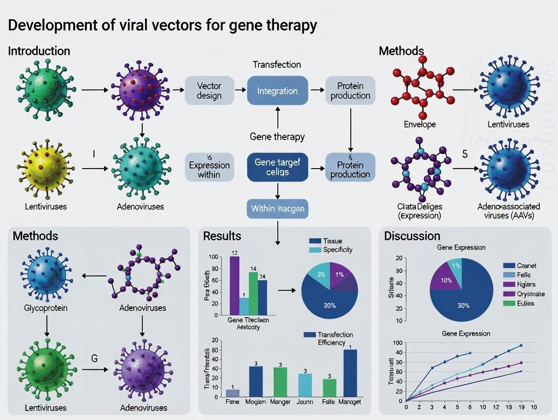
Viral Vectors for Gene Therapy: A Comprehensive Guide to Engineering, Application, and Safety in Modern Medicine
This article provides a detailed technical overview of viral vector development for gene therapy, targeting researchers and drug development professionals.
Viral Vectors for Gene Therapy: A Comprehensive Guide to Engineering, Application, and Safety in Modern Medicine
Abstract
This article provides a detailed technical overview of viral vector development for gene therapy, targeting researchers and drug development professionals. It explores the fundamental biology of major vector systems (AAV, Lentivirus, Adenovirus), their engineering methodologies, and key therapeutic applications. The content further addresses critical challenges in vector optimization, manufacturing, and immunogenicity, while presenting established and emerging strategies for validation, safety assessment, and comparative analysis. The goal is to serve as a current, actionable resource for navigating the complex landscape of translating viral vector technology into safe and effective clinical treatments.
Viral Vector Fundamentals: From Natural Pathogens to Engineered Gene Delivery Vehicles
The development of effective viral vectors is predicated on repurposing the innate efficiency of viral infection to deliver therapeutic genetic cargo. This principle leverages millions of years of viral evolution, optimizing for cell entry, genome delivery, and, in some cases, genomic integration. Within the broader thesis on viral vector development, this document details practical applications and protocols for utilizing leading viral vector systems, focusing on Adeno-Associated Virus (AAV) and Lentivirus (LV) as primary models for in vivo and ex vivo gene therapy, respectively.
Recent advancements highlight critical trends: the engineering of novel AAV capsids with enhanced tissue tropism and reduced immunogenicity, and the development of self-inactivating (SIN) lentiviral vectors with improved safety profiles. Quantitative comparisons of key vector parameters are essential for experimental design.
Table 1: Quantitative Comparison of Primary Viral Vector Systems
| Parameter | Adeno-Associated Virus (AAV) | Lentivirus (LV) | Adenovirus (AdV) |
|---|---|---|---|
| Packaging Capacity | ~4.7 kb | ~8 kb | ~8-36 kb |
| Integration Profile | Predominantly episomal; rare targeted integration | Stable integration (into active genes) | Non-integrating |
| Transduction Efficiency | High in permissive tissues | High in dividing & non-dividing cells | Very high in vitro & in vivo |
| In Vivo Immune Response | Moderate (capsid/transgene-specific) | Low for SIN vectors | Very high (highly immunogenic) |
| Peak Expression Onset | 1-4 weeks | 2-7 days (post-integration) | 1-3 days |
| Expression Durability | Months to years (in non-dividing cells) | Long-term (stable integration) | Transient (weeks) |
Experimental Protocols
Protocol: Production and Purification of Recombinant AAV Vectors (Serotype 9) via PEI Transfection
Objective: To generate high-titer, research-grade recombinant AAV9 vectors using a triple-plasmid transfection method in HEK293T cells.
Materials (Research Reagent Solutions):
- Cell Line: HEK293T cells (ATCC CRL-3216). Function: Provides adenoviral helper functions (E1, E2a, E4, VA RNA) and a permissive platform for AAV replication.
- Plasmids:
- pAAV-Transgene: ITR-flanked vector plasmid containing your gene of interest.
- pAAV-RC9: AAV Rep/Cap plasmid providing serotype 9 capsid proteins and replication enzymes.
- pAdDeltaF6: Adenoviral helper plasmid supplying essential helper functions without wild-type adenovirus contamination.
- Transfection Reagent: Polyethylenimine (PEI) Max, linear, 40 kDa (Polysciences). Function: Forms cationic complexes with plasmid DNA for efficient cell entry.
- Lysis Buffer: 150 mM NaCl, 50 mM Tris-HCl, pH 8.5. Function: Releases viral particles from harvested cells.
- Purification Reagent: Iodixanol (OptiPrep Density Gradient Medium). Function: Forms density gradients for ultracentrifugation-based separation of full AAV capsids from empty capsids and cellular debris.
- Concentration Device: 100 kDa Molecular Weight Cut-off (MWCO) Amicon Ultra centrifugal filter. Function: Concentrates and buffer-exchanges purified viral stock.
- Quantification Kit: AAVpro Titration Kit (Takara Bio) or equivalent qPCR-based kit. Function: Accurately determines viral genome titer (vg/mL).
Methodology:
- Day 0: Cell Seeding: Seed fifteen 15-cm plates with 6x10^6 HEK293T cells/dish in DMEM + 10% FBS. Incubate at 37°C, 5% CO2 overnight (~70% confluency target).
- Day 1: PEI Transfection: For each plate, prepare DNA/PEI complexes in 2 mL serum-free DMEM:
- Plasmid Mix: 7.5 µg pAAV-Transgene, 5.5 µg pAAV-RC9, 10 µg pAdDeltaF6.
- Dilute PEI Max (1 mg/mL stock) to 75 µL in serum-free DMEM.
- Combine diluted PEI with plasmid mix, vortex, incubate 15 min at RT.
- Add complex dropwise to cells. Refresh media after 6-8 hours.
- Day 3-5: Harvest: At 72 hours post-transfection, detach cells using scrapers. Pellet cells and media separately by centrifugation (500 x g, 10 min). Retain both pellets and media supernatant.
- Cell Lysis & Nuclease Treatment: Resuspend cell pellets in lysis buffer, freeze-thaw 3x (dry ice/37°C water bath). Pool with supernatant, treat with Benzonase (50 U/mL, 37°C, 1 hr) to digest unpackaged nucleic acids. Clarify by centrifugation (4,000 x g, 30 min).
- Iodixanol Gradient Ultracentrifugation: Prepare a discontinuous gradient (15%, 25%, 40%, 60% iodixanol) in ultracentrifuge tubes. Layer clarified lysate on top. Centrifuge at 350,000 x g (avg), 18°C, 2 hours (Beckman Coulter Type 70 Ti rotor).
- Collection & Concentration: Collect the opaque 40-60% interface containing purified AAV. Concentrate and exchange into PBS-MK (PBS with 1 mM MgCl2, 2.5 mM KCl) using a 100 kDa MWCO centrifugal filter. Aliquot and store at -80°C.
- Titer Determination: Perform qPCR using the AAVpro Titration Kit following manufacturer's instructions. Use primers/probe specific to your transgene or a universal ITR sequence.
Protocol: Generation of VSV-G Pseudotyped Third-Generation Lentiviral Vectors
Objective: To produce high-titer, replication-incompetent lentiviral vectors using a four-plasmid, third-generation system for transduction of dividing and non-dividing cells.
Materials (Research Reagent Solutions):
- Plasmids (Third-Generation System):
- Transfer Plasmid (pRRL-SIN-Transgene): Contains LTRs, Ψ packaging signal, WPRE, and your transgene in a self-inactivating (SIN) configuration.
- Packaging Plasmid (pMDLg/pRRE): Provides Gag and Pol polyproteins.
- Rev-Encoding Plasmid (pRSV-Rev): Supplies Rev protein for nuclear export of unspliced viral RNA.
- Envelope Plasmid (pMD2.G): Expresses VSV-G glycoprotein for broad tropism and particle stability.
- Transfection Reagent: PEI Max, as above.
- Concentration Reagent: Lenti-X Concentrator (Takara Bio). Function: A polymer solution that precipitates viral particles for easy low-speed pelleting.
- Quantification Kit: Lenti-X qRT-PCR Titration Kit (Takara Bio). Function: Quantifies viral RNA genomes to determine functional titer (TU/mL).
Methodology:
- Day 0-1: Seed HEK293T cells as in Protocol 2.1.
- Day 1: Transfection: For each 15-cm plate, prepare:
- Plasmid Mix: 10 µg Transfer Plasmid, 7.5 µg pMDLg/pRRE, 3 µg pRSV-Rev, 5 µg pMD2.G.
- PEI Mix: 75 µL PEI Max (1 mg/mL).
- Form complexes in serum-free DMEM, incubate 15 min, add to cells. Refresh media after 6-8 hours.
- Day 2 & 3: Media Exchange & Collection: At 24h post-transfection, replace media with fresh, pre-warmed media. At 48h and 72h post-transfection, collect supernatant containing viral particles. Filter through a 0.45 µm PES filter.
- Concentration: Pool filtered supernatants. Add 1 volume of Lenti-X Concentrator to 3 volumes of supernatant, mix, incubate at 4°C overnight. Centrifuge at 1500 x g for 45 min at 4°C. Resuspend pellet in 1/100th original volume in sterile PBS or desired buffer. Aliquot and store at -80°C.
- Titer Determination: Transduce HEK293T cells with serial dilutions of vector. After 72 hours, extract genomic DNA and perform qPCR (using the Lenti-X kit) for the integrated proviral sequence (e.g., WPRE). Compare to a standard curve to calculate transducing units per mL (TU/mL).
Visualization
Title: Viral Vector Production Workflow
Title: AAV Cellular Entry & Trafficking Pathway
The Scientist's Toolkit: Essential Research Reagent Solutions
Table 2: Key Reagents for Viral Vector Development & Titration
| Reagent/Solution | Function & Application | Key Consideration |
|---|---|---|
| HEK293T Cell Line | Robust producer cell line providing essential adenoviral (E1) helper functions for AAV and LV production. | Maintain low passage number; ensure high viability (>95%) for transfection. |
| Polyethylenimine (PEI) Max | Cationic polymer for transient, high-efficiency plasmid transfection in suspension or adherent cultures. | pH and molecular weight (40kDa) are critical for efficiency and low cytotoxicity. |
| Iodixanol (OptiPrep) | Density gradient medium for ultracentrifugation-based purification of AAV. Separates full vs. empty capsids. | High purity grade is essential; gradients must be prepared with precision. |
| Lenti-X Concentrator | Chemical precipitant for gentle, low-speed centrifugal concentration of lentiviral particles from supernatant. | Faster than ultracentrifugation; may reduce recovery for some pseudotypes. |
| Benzonase Nuclease | Degrades unpackaged nucleic acids (host & plasmid) during vector purification, reducing viscosity and improving purity. | Critical for reducing immune triggers and meeting regulatory guidelines. |
| AAVpro Titration Kit (qPCR) | Quantifies DNase-resistant viral genome titer (vg/mL) using standardized primers/probes (e.g., for ITR). | Includes essential DNase step to remove unencapsidated DNA. |
| Lenti-X qRT-PCR Titration Kit | Quantifies functional vector titer (TU/mL) by measuring integrated proviral DNA in transduced cells. | Provides a standardized, rapid method vs. traditional colony counting. |
| Anti-AAV Capsid Antibodies | Used in ELISA to determine physical particle titer and assess capsid integrity/immunoreactivity. | Distinguishes between full and empty capsids when paired with genome titer. |
Within the broader thesis on the Development of viral vectors for gene therapy research, selecting the appropriate vector system is a foundational decision. Each vector offers distinct capabilities and limitations in transduction efficiency, cargo capacity, immunogenicity, and persistence. These application notes provide a comparative overview and practical protocols for the major viral vector platforms.
Table 1: Key Characteristics of Major Viral Vectors
| Vector | Genome | Cargo Capacity | Tropism | Integration | Expression Duration | Immunogenicity | Primary Applications |
|---|---|---|---|---|---|---|---|
| Adeno-Associated Virus (AAV) | ssDNA | ~4.7 kb | Broad (serotype-dependent) | Predominantly episomal | Long-term in post-mitotic cells | Low (capsid-specific) | In vivo gene therapy, clinical therapies |
| Lentivirus (LV) | RNA (retrovirus) | ~8 kb | Broad (pseudotyping) | Integration into host genome | Stable, long-term | Moderate (pre-existing immunity rare) | Ex vivo cell engineering, basic research |
| Adenovirus (AdV) | dsDNA | ~8-36 kb (gutless) | Broad (CAR-dependent) | Episomal | Transient (weeks) | High (innate & adaptive) | Vaccines, oncolytic therapy, transient high-level expression |
| Gamma-retrovirus (RV) | RNA (retrovirus) | ~8 kb | Dividing cells only | Integration | Stable, long-term | Moderate | Historical ex vivo use (e.g., CAR-T) |
| Herpes Simplex Virus (HSV) | dsDNA | >30 kb | Neurons, broad | Episomal (latent) | Long-term in neurons | High | Neurological gene delivery, large cargo delivery |
Application Notes & Protocols
Protocol: Production and Purification of Recombinant AAV Serotype 9
Application: Generating high-titer, research-grade AAV9 for in vivo delivery to central nervous system and muscle tissues.
Materials (Research Reagent Solutions):
- Plasmids: pAAV-CAG-GFP (transgene), pAAV9 (rep/cap), pAdDeltaF6 (helper).
- Cells: HEK293T cells (ATCC CRL-3216).
- Transfection Reagent: Polyethylenimine (PEI Max, linear, MW 40,000).
- Lysis Buffer: 150 mM NaCl, 50 mM Tris-HCl, pH 8.5.
- Purification Reagents: Iodixanol density gradient medium, Benzonase nuclease.
- Buffers: PBS-MK (PBS with 1 mM MgCl2 and 2.5 mM KCl).
- Concentration Device: 100 kDa molecular weight cut-off centrifugal filter.
Methodology:
- Cell Culture: Seed fifteen 15-cm dishes with HEK293T cells to reach 70-80% confluency at transfection.
- Transfection: For each dish, prepare a DNA-PEI complex mix in serum-free DMEM: 10 µg pAAV-CAG-GFP, 15 µg pAAV9, and 20 µg pAdDeltaF6. Add PEI at a 3:1 ratio (PEI:total DNA). Incubate 15 min, add dropwise to cells.
- Harvest: 72 hours post-transfection, collect cells and media. Pellet cells via centrifugation (500 x g, 10 min).
- Lysis & Nuclease Treatment: Resuspend cell pellet in lysis buffer. Freeze-thaw cycled (3x). Add Benzonase (50 U/mL), incubate at 37°C for 30 min.
- Iodixanol Gradient Centrifugation: Layer clarified lysate atop a discontinuous iodixanol gradient (15%, 25%, 40%, 60%) in quick-seal tubes. Ultracentrifuge at 350,000 x g, 2.5 h, 18°C.
- Collection & Dialysis: Extract the opaque 40% fraction. Concentrate and buffer exchange into PBS-MK using a centrifugal filter. Aliquot and store at -80°C.
- Titration: Quantify vector genome titer (vg/mL) via qPCR against a standard curve.
Protocol: Generation of VSV-G Pseudotyped Lentivirus forEx VivoTransduction
Application: Stable gene delivery and integration into dividing and non-dividing cells, such as primary T cells or stem cells.
Materials (Research Reagent Solutions):
- Plasmids: pLVX-EF1α-mCherry-Puro (transfer), psPAX2 (packaging), pMD2.G (VSV-G envelope).
- Cells: HEK293T/17 cells (high transfectability).
- Transfection Reagent: Calcium Phosphate Transfection Kit or PEI.
- Concentration Reagent: Lenti-X Concentrator (Takara Bio).
- Culture Media: High-glucose DMEM with 10% FBS; transduction medium may require polybrene (4-8 µg/mL).
Methodology:
- Cell Seeding: Seed HEK293T cells in 10-cm dishes 24h prior to reach ~60% confluency.
- Calcium Phosphate Transfection: Per dish, combine 10 µg pLVX, 7.5 µg psPAX2, and 2.5 µg pMD2.G in 0.1X TE. Add 2M CaCl2, mix, then add dropwise to 2X HBS with vortexing. Incubate mixture 20 min, then add to cells.
- Media Change: Replace media 8-16 hours post-transfection.
- Virus Harvest: Collect supernatant at 48 and 72 hours post-transfection. Filter through a 0.45 µm PES filter.
- Concentration: Mix filtered supernatant with Lenti-X Concentrator (1:3 ratio). Incubate >30 min at 4°C. Centrifuge at 1500 x g for 45 min. Resuspend pellet in 1/100th volume of PBS or media.
- Transduction: Incubate target cells with lentiviral supernatant in the presence of polybrene. Centrifuge at 600 x g for 60-90 min (spinoculation) to enhance efficiency. Assay expression after 48-72 hours.
Visualization of Key Concepts
Decision Flowchart for Viral Vector Selection
AAV Vector Genome Packaging in Producer Cell
The Scientist's Toolkit: Essential Research Reagents
Table 2: Key Reagents for Viral Vector Research & Development
| Reagent / Material | Function / Purpose | Example/Note |
|---|---|---|
| HEK293T/HEK293 Cells | Standard production cell line; expresses SV40 T-antigen (293T) for plasmid amplification. | ATCC CRL-3216. Provides adenoviral E1 functions. |
| Polyethylenimine (PEI) | Cationic polymer for transient transfection of packaging plasmids. | Cost-effective, high efficiency for HEK293 cells. |
| Calcium Phosphate | Chemical transfection method; alternative to PEI for lentivirus production. | Requires precise pH control of HBS buffer. |
| Iodixanol | Density gradient medium for ultracentrifugation-based purification of AAV and LV. | Preferred over CsCl for better particle recovery and bioactivity. |
| Lenti-X Concentrator | Solution for precipitating lentivirus from culture supernatant. | PEG-based, simplifies concentration without ultracentrifugation. |
| Polybrene | Cationic polymer that reduces charge repulsion between virions and cell membrane. | Enhances transduction efficiency; can be cytotoxic. |
| Benzonase Nuclease | Degrades unpackaged nucleic acids and host cell DNA/RNA during purification. | Reduces viscosity and improves purity. |
| qPCR Standards | Plasmid or linear DNA fragment containing the target sequence for vector genome titration. | Essential for accurate and reproducible titer determination. |
| VSV-G Envelope Plasmid | Provides pantropic viral envelope for pseudotyping LV and broadening tropism. | pMD2.G is a common plasmid. |
| Transduction Enhancers | Small molecules or agents that improve vector uptake (e.g., for AAV). | Includes HDAC inhibitors (Valproic Acid) for some cell types. |
Application Notes
Viral vectors are engineered tools designed to deliver genetic material into cells for gene therapy research. Their development is central to the thesis that optimizing each component enhances transduction efficiency, specificity, and safety. The core components are:
- Capsid: The protein shell that determines tropism, immunogenicity, and physical stability. Engineering capsids (e.g., AAV serotype swapping, peptide insertions) is a primary strategy to evade pre-existing immunity and target specific tissues.
- Genome: The viral genetic backbone. In recombinant vectors, essential replication genes are removed to create space for the transgene and regulatory elements, rendering the vector replication-incompetent.
- Promoter: Drives expression of the transgene. Choice is critical for defining the level, specificity, and duration of expression (e.g., constitutive CMV vs. tissue-specific synapsin promoters).
- Transgene: The therapeutic or reporter gene of interest. Its design includes the coding sequence and often incorporates regulatory elements like polyadenylation signals and WPRE for enhanced expression.
Recent data (2023-2024) highlights trends in AAV and lentiviral vectors, which dominate clinical applications.
Table 1: Quantitative Comparison of Common Viral Vector Components
| Vector Type | Typical Capsid (Serotype/Viral Envelope) | Genome Capacity | Promoter Examples (Size) | Primary Transgene Applications (2023-24 Clinical Trials) |
|---|---|---|---|---|
| Adeno-Associated Virus (AAV) | AAV1, AAV2, AAV5, AAV8, AAV9, AAVrh74, engineered variants (e.g., AAV-LK03) | ~4.7 kb | CAG (~1.8 kb), mini-CMV (~0.6 kb), SYN1 (~0.5 kb), liver-specific TBG (~0.2 kb) | Hemophilia B (FIX), Spinal Muscular Atrophy (SMN1), Leber's Congenital Amaurosis (RPE65) |
| Lentivirus (LV) | VSV-G (pseudotyped), other glycoproteins (e.g., Rabies-G) | ~8-10 kb | EF1α (~1.2 kb), PGK (~0.5 kb), inducible Tet-On systems | CAR-T Cell Engineering, β-thalassemia (β-globin), X-linked Adrenoleukodystrophy (ABCD1) |
| Adenovirus (AdV) | Hexon, fiber, penton proteins (many serotypes) | ~8-36 kb (gutted) | CMV (~0.6 kb), tumor-specific promoters | Oncolytic virotherapy, Vaccine platforms (e.g., COVID-19) |
Experimental Protocols
Protocol 1: Evaluating Capsid Tropism viaIn VivoBiodistribution
Objective: Quantify viral vector genome copies in target and off-target tissues following systemic administration to assess capsid targeting efficiency.
Materials:
- Purified viral vector (e.g., AAV9 vs. AAV-PHP.eB)
- Experimental animal model (e.g., C57BL/6 mouse)
- qPCR reagents (SYBR Green master mix, primers for vector genome)
- Tissue homogenizer
- DNA extraction kit
Procedure:
- Administration: Systemically administer 1x10^11 vector genomes (vg) per animal via tail vein injection (n=5 per capsid group).
- Tissue Collection: At 14 days post-injection, euthanize animals and collect target tissues (e.g., brain, liver, heart, skeletal muscle) and control tissues (e.g., spleen, kidney).
- DNA Extraction: Homogenize 20-50 mg of each tissue. Extract total genomic DNA using a commercial kit. Elute in 50 µL nuclease-free water.
- qPCR Setup: Design primers targeting a conserved region of the vector genome (e.g., polyA signal). Prepare a standard curve using a plasmid containing the vector genome (serial dilutions from 10^7 to 10^1 copies/µL).
- Quantification: Perform qPCR in triplicate for each tissue sample. Calculate vg per diploid genome using the formula: (vg/µL from qPCR) / (Genomic DNA (ng/µL) / (6.6 pg/diploid genome)).
Protocol 2: Assessing Promoter Activity via Luciferase Reporter Assay
Objective: Compare the strength and specificity of different promoters in vitro.
Materials:
- HEK293 cells (constitutive) and differentiated SH-SY5Y cells (neuronal model)
- Viral vectors encoding firefly luciferase under test promoters (e.g., CMV, CAG, SYN1)
- Dual-Luciferase Reporter Assay System
- Microplate luminometer
Procedure:
- Cell Seeding: Seed cells in a 24-well plate at 70% confluency.
- Transduction: Transduce cells at an MOI of 10^4 vg/cell in triplicate. Include a non-transduced control.
- Incubation: Incubate for 48-72 hours.
- Lysis & Assay: Lyse cells per manufacturer's instructions. Transfer 20 µL of lysate to a white-walled plate. Inject 50 µL of Luciferase Assay Reagent II, measure firefly luminescence immediately.
- Normalization (Optional): For co-transduction with a Renilla luciferase control vector, inject Stop & Glo reagent and measure Renilla luminescence.
- Analysis: Compare relative light units (RLUs) across promoter groups in each cell type to determine absolute strength and cell-type specificity.
Visualizations
Diagram 1: In Vivo Capsid Tropism Analysis Workflow
Diagram 2: Functional Relationships of Vector Components
The Scientist's Toolkit: Research Reagent Solutions
Table 2: Essential Materials for Viral Vector Research
| Item | Function in Research |
|---|---|
| Plasmid DNA Systems (pAAV, pLV Helper, Transgene Constructs) | Backbone for vector genome production. Contains ITRs (AAV) or LTRs (LV), promoter, transgene, and selection markers. |
| Packaging Cell Lines (HEK293T/293AAV, Sf9 insect cells) | Provide essential viral genes (rep/cap for AAV, gag/pol/rev for LV) in trans for recombinant vector production. |
| Purification Kits/Resins (Iodixanol gradients, AVB Sepharose, Ion Exchange) | Isolation of high-purity, high-titer viral vectors from crude lysate or supernatant. Critical for in vivo studies. |
| Titer Quantification Kits (qPCR-based, ELISA for capsid) | Accurate measurement of physical (vg/mL) and functional (IU/mL) vector titers for dose standardization. |
| Cell Type-Specific Media & Supplements (Primary neuron media, cytokine mixes) | Maintenance of target cells for in vitro transduction efficiency and specificity assays. |
| In Vivo Delivery Equipment (Stereotaxic frames, Tail vein infusion pumps) | Precise administration of vector to target tissues (brain, systemic circulation) in animal models. |
Historical Milestones and Breakthroughs in Viral Vector Gene Therapy
Application Notes and Protocols
A. Introduction and Historical Context The development of viral vectors is the cornerstone of modern gene therapy, enabling the stable introduction, silencing, or editing of genetic material in target cells. Framed within the thesis on the Development of viral vectors for gene therapy research, this document outlines critical historical milestones and provides detailed protocols that have emerged from these breakthroughs. The evolution from first-generation adenoviral vectors to sophisticated, engineered AAV capsids and lentiviral platforms reflects a trajectory of increasing safety, specificity, and efficacy.
B. Key Historical Milestones and Quantitative Data Summary Table 1: Historical Milestones in Viral Vector Gene Therapy
| Year | Milestone Event | Vector Type | Key Outcome/Therapeutic Area | Significance |
|---|---|---|---|---|
| 1990 | First approved human gene therapy trial (ADA-SCID) | Retrovirus (MoMLV) | Treatment of severe combined immunodeficiency. | Proof-of-concept for ex vivo gene correction. |
| 1999 | Death of Jesse Gelsinger in an OTCD trial | Adenovirus (first-gen) | Ornithine transcarbamylase deficiency. | Highlighted acute immune toxicity, pivoting field to safety. |
| 2003-09 | Success in X-linked SCID, ADA-SCID trials | Gamma-retrovirus | Restoration of immune function. | Demonstrated curative potential, but revealed insertional oncogenesis risk. |
| 2012 | Approval of Glybera (alipogene tiparvovec) in EU | AAV (serotype 1) | Lipoprotein lipase deficiency. | First approved gene therapy in Western world. |
| 2016 | Marketing authorization for Strimvelis (ADA-SCID) | Gamma-retrovirus | ADA-SCID. | First ex vivo stem cell gene therapy approved in EU. |
| 2017 | FDA approval of Luxturna (voretigene neparvovec) | AAV (serotype 2) | RPE65 mutation-associated retinal dystrophy. | First in vivo gene therapy approved in US. |
| 2019-22 | FDA approvals for Zolgensma (onasemnogene abeparvovec) & Hemgenix (etranacogene dezaparvovec) | AAV (serotype 9 & AAVhu37) | Spinal muscular atrophy & Hemophilia B. | Validated systemic AAV delivery for CNS/liver diseases. |
| 2023-24 | FDA approvals for Lyfgenia & Casgevy (ex vivo) | Lentivirus | Sickle cell disease / β-thalassemia. | First approved therapies using CRISPR/Cas9-modified lentiviral vectors. |
Table 2: Quantitative Comparison of Major Viral Vector Classes
| Parameter | Adenovirus (Ad5) | Adeno-Associated Virus (AAV2) | Lentivirus (HIV-1 based) | Gamma-retrovirus (MoMLV) |
|---|---|---|---|---|
| Max. Capacity | ~8-36 kb | ~4.7 kb | ~8-10 kb | ~8 kb |
| Integration | Episomal | Predominantly episomal | Integrating | Integrating |
| Tropism | Broad (CAR receptor) | Broad (requires specific serotype) | Broad (pseudotyping possible) | Broad (infects dividing cells) |
| Titer (vg/mL) | 10^10 - 10^12 | 10^12 - 10^14 | 10^8 - 10^9 (transducing units) | 10^6 - 10^7 (transducing units) |
| Immune Response | High (innate & adaptive) | Moderate (capsid/transgene-specific) | Moderate | Low (ex vivo) |
| Oncogenic Risk | Low | Very Low | Moderate (integration profile) | High (preferential integration near promoters) |
C. Detailed Experimental Protocols
Protocol 1: Production and Purification of Recombinant AAV Vectors via PEI-Mediated Triple Transfection (HEK293T Cells) This protocol is derived from modern scalable methods enabling clinical-grade vector production.
1. Materials:
- HEK293T cells
- Polyethylenimine (PEI), linear, 25 kDa
- Plasmid triad: (1) AAV rep/cap plasmid, (2) AAV ITR-flanked transgene plasmid, (3) pAdDeltaF6 helper plasmid
- DMEM, high glucose, serum-free or with 5% FBS
- 150 mM NaCl (sterile)
- Benzonase nuclease
- MgCl₂ (1 M stock)
- OptiPrep density gradient medium
- PBS-MK buffer (PBS with 1 mM MgCl₂ and 2.5 mM KCl)
- Amicon Ultra-15 centrifugal filters (100K MWCO)
2. Procedure: Day 1: Seed HEK293T cells at 70-80% confluency in cell factory stacks or multi-layer flasks. Day 2: Prepare transfection mix. For 1 cell factory, combine the three plasmids at a 1:1:1 molar ratio in 150 mM NaCl. Add PEI at a 3:1 PEI:total DNA ratio (w/w). Vortex and incubate 15 min at RT. Add mixture dropwise to cells. Day 3: Replace medium with serum-free DMEM. Day 5-6: Harvest cells and media. Pellet cells via centrifugation (2,000 x g, 10 min). Resuspend cell pellet in PBS-MK. Perform 3-5 freeze-thaw cycles (liquid N₂/37°C water bath). Add Benzonase (50 U/mL) and MgCl₂ (final 1 mM). Incubate 1 hr at 37°C. Clarify lysate by centrifugation (4,000 x g, 30 min). Purification: Load clarified supernatant onto an iodixanol step gradient (15%, 25%, 40%, 60% in PBS-MK). Centrifuge in a fixed-angle rotor at 350,000 x g for 1 hr. Collect the 40% fraction containing purified AAV. Concentrate and buffer exchange into final formulation buffer using Amicon centrifugal filters. Titrate via qPCR (ITR-specific primers).
Protocol 2: Ex Vivo T-Cell Transduction for CAR-T Therapy Using Lentiviral Vectors This protocol outlines a key application stemming from lentiviral vector development.
1. Materials:
- Patient-derived or donor T-cells
- RetroNectin-coated plates
- Lentiviral vector supernatant (VSV-G pseudotyped, ~1x10^8 TU/mL)
- IL-2 and IL-7 cytokines
- Anti-CD3/CD28 activation beads
- X-VIVO 15 serum-free medium
2. Procedure: Day 1: T-Cell Activation. Isolate PBMCs via Ficoll density centrifugation. Isolate T-cells using a negative selection kit. Activate T-cells with anti-CD3/CD28 beads (bead:cell ratio 3:1) in X-VIVO 15 + 5% human AB serum + IL-2 (100 IU/mL) + IL-7 (10 ng/mL). Day 2: Transduction. Coat non-tissue culture plate with RetroNectin (10 µg/mL). Block with 2% BSA. Add lentiviral supernatant, spin at 2,000 x g for 2 hr (spinoculation). Remove viral supernatant. Plate activated T-cells on coated plate at 1x10^6 cells/mL in fresh medium with cytokines. Day 3-4: Replace medium with fresh cytokine-containing medium. Day 5-14: Expansion. Maintain cells at 0.5-1x10^6 cells/mL, feeding every 2-3 days. Remove activation beads on Day 7. Monitor CAR expression by flow cytometry using a target antigen-Fc fusion protein. Harvest cells when sufficient expansion and transduction efficiency (>30%) are achieved for infusion or cryopreservation.
D. Visualizations
AAV Production via Triple Transfection
Ex Vivo CAR-T Cell Generation Workflow
E. The Scientist's Toolkit: Research Reagent Solutions
Table 3: Essential Research Reagents for Viral Vector R&D
| Reagent/Material | Primary Function & Application | Key Considerations |
|---|---|---|
| Polyethylenimine (PEI), 25kDa | Cationic polymer for transient transfection of plasmid DNA into packaging cells (e.g., HEK293T). | Cost-effective, scalable. Ratio optimization (DNA:PEI) critical for yield/viability. |
| pAdDeltaF6 Helper Plasmid | Provides essential adenoviral helper functions (E2A, E4, VA RNA) for AAV replication, without wild-type AAV generation. | Standard for triple transfection; reduces RCA risk. |
| RetroNectin (Recombinant Fibronectin Fragment) | Enhances retroviral/lentiviral transduction efficiency by co-localizing viral particles and target cells (e.g., T-cells). | Critical for clinical-grade ex vivo transduction protocols. |
| Iodixanol (OptiPrep) | Density gradient medium for ultracentrifugation-based purification of AAV and other viral vectors. | Inert, iso-osmotic, allows high recovery of bioactive vectors. |
| Benzonase Nuclease | Digests unpackaged nucleic acids (plasmid DNA, cellular RNA/DNA) in crude lysates, reducing viscosity and improving purity. | Essential for downstream processing and column-based purification. |
| Anti-CD3/CD28 Activation Beads | Mimic antigen presentation, providing Signal 1 & 2 for robust primary T-cell activation prior to transduction. | Magnetically removable; defined, serum-free alternative to OKT3/feeder cells. |
| AAV rep/cap Plasmid Library | Engineered capsid variant libraries for directed evolution or screening of novel tissue tropisms. | Enables development of next-generation vectors with enhanced targeting. |
| VSV-G Expression Plasmid | Pseudotyping envelope for lentiviral vectors to confer broad tropism and enhance particle stability. | Enables high-titer production via ultracentrifugation concentration. |
Key Advantages and Inherent Limitations of Viral vs. Non-Viral Delivery Methods
Within the broader thesis on the Development of Viral Vectors for Gene Therapy Research, selecting the appropriate gene delivery vehicle is a foundational decision. The choice between viral and non-viral methods dictates the efficiency, durability, safety, and applicability of a therapeutic intervention. This application note provides a comparative analysis, protocols for key evaluation experiments, and a toolkit for researchers to guide method selection and optimization.
Comparative Analysis: Viral vs. Non-Viral Vectors
Table 1: Key Advantages and Inherent Limitations of Major Delivery Systems
| Feature | Viral Vectors (e.g., AAV, Lentivirus, Adenovirus) | Non-Viral Vectors (e.g., LNPs, Polymers, Electroporation) |
|---|---|---|
| Transduction Efficiency | Very High (often >70% in permissive cells) | Low to Moderate (typically 1-50%, highly cell-dependent) |
| Transgene Capacity | Small to Moderate (AAV: ~4.7 kb; Lentivirus: ~8 kb; Adenovirus: ~8-36 kb) | Large (>10 kb, limited mainly by packaging) |
| Immunogenicity | High Inherent Risk. Pre-existing & de novo immune responses can limit re-administration & cause toxicity. | Generally Low. Cationic lipids/polymers can cause dose-related inflammation. |
| Integration Profile | Variable. Lentivirus: semi-random integration. AAV: predominantly episomal (risks exist). | Typically Non-integrating (episomal), reducing insertional mutagenesis risk. |
| Manufacturing & Cost | Complex, time-intensive, high cost-of-goods ($100k-$1M per GMP batch). | Relatively simple, scalable, lower cost (potentially <$100k per batch). |
| Delivery Route Versatility | Established for in vivo direct administration (e.g., IV, intracranial). | Rapidly advancing for systemic in vivo delivery (e.g., mRNA-LNPs). |
| Repeat Dosing Feasibility | Low (neutralizing antibodies often prevent re-administration). | High (less immunogenic, allowing repeated treatments). |
| Speed to Clinic | Long development & safety assessment timelines (vectorology, producer lines). | Faster from design to GMP production (modular platforms). |
Data synthesized from current industry reports (2023-2024) and peer-reviewed literature.
Experimental Protocols for Vector Evaluation
Protocol 3.1: Parallel In Vitro Transduction Efficiency & Cytotoxicity Assay
Purpose: To directly compare the gene delivery performance and cellular toxicity of a candidate viral and non-viral vector.
Materials: See "Scientist's Toolkit" (Section 5).
Procedure:
- Cell Seeding: Seed HEK293T or target primary cells in a 96-well plate at 70% confluence. Incubate overnight.
- Vector Preparation:
- Viral (e.g., AAV): Prepare serial dilutions in complete medium (e.g., 1e3 to 1e5 vg/cell).
- Non-Viral (e.g., LNP): Complex plasmid DNA (pDNA) or mRNA encoding reporter (e.g., eGFP, Luciferase) at optimal N/P ratio. Dilute in serum-free medium.
- Transduction/Transfection: Aspirate medium from cells. Apply 100 µL of each vector dilution per well (n=6). Include untreated and mock-treated controls.
- Incubation: Incubate for 48-72 hours at 37°C, 5% CO₂.
- Analysis:
- Efficiency: Harvest cells. Analyze % of eGFP+ cells and mean fluorescence intensity (MFI) via flow cytometry. For luciferase, perform lysate assay.
- Cytotoxicity: Perform an MTT or CellTiter-Glo assay on the same wells according to manufacturer instructions.
- Calculation: Normalize reporter signal to cell viability for each dose. Plot dose-response curves.
Protocol 3.2: In Vivo Biodistribution and Persistence Study
Purpose: To assess tissue tropism, transgene expression kinetics, and durability of viral vs. non-viral vectors in a murine model.
Procedure:
- Vector Administration: Divide mice (n=5-8/group) into three cohorts: 1) AAV (e.g., AAV9, 1e11 vg/mouse, IV), 2) LNP-mRNA (5 µg mRNA, IV), 3) Saline control.
- Longitudinal Imaging: For luciferase reporters, administer D-luciferin (150 mg/kg, IP) and image using an IVIS system at days 1, 3, 7, 14, 28, and 56 post-injection.
- Terminal Biodistribution: At selected endpoints (e.g., day 7 for LNP, day 56 for AAV), euthanize animals. Harvest organs (liver, spleen, heart, lung, kidney, brain).
- Tissue Analysis:
- qPCR for Vector Genomes: Extract total DNA. Perform qPCR with vector-specific primers to quantify vector genome copies per µg genomic DNA.
- mRNA/Protein Analysis: Extract RNA for RT-qPCR of transgene mRNA. Homogenize tissue for Western blot or ELISA to quantify protein expression.
- Immunogenicity Assessment: Collect serum at endpoints. Perform ELISA to measure anti-capsid (AAV) or anti-protein antibodies.
Visualizations
Title: Vector Evaluation Workflow
Title: Immune Pathways in Viral Vector Response
The Scientist's Toolkit
Table 2: Essential Research Reagents & Materials
| Item | Function & Application | Example Vendor(s) |
|---|---|---|
| High-Titer Viral Vector Stocks | In vitro and in vivo functional studies; require precise titer (vg/mL). | Vigene Biosciences, Vector Biolabs, academic core facilities. |
| Cationic Lipid/Nanoparticle Kits | For formulating pDNA or mRNA; enables rapid non-viral screening. | Thermo Fisher (Lipofectamine), Precision NanoSystems (NanoAssemblr). |
| Reporter Plasmid/mRNA | Encodes measurable protein (e.g., eGFP, Luciferase) to quantify delivery efficiency. | Addgene (plasmids), Trilink BioTechnologies (cleanCap mRNA). |
| Cell Viability Assay Kit | Quantifies cytotoxicity (e.g., MTT, ATP-based) post-transduction/transfection. | Promega (CellTiter-Glo), Abcam (MTT). |
| In Vivo Imaging System (IVIS) | Enables longitudinal, non-invasive tracking of bioluminescent reporter expression. | PerkinElmer, Spectral Instruments Imaging. |
| qPCR Reagents & Probes | For quantifying vector genome biodistribution and transgene mRNA levels. | Bio-Rad, Thermo Fisher (TaqMan). |
| ELISA Kits | Quantifies transgene protein expression and anti-vector antibody responses. | R&D Systems, Abcam. |
| Next-Generation Sequencing (NGS) Service | Assesses vector integration sites (lentivirus) or off-target effects (CRISPR). | Illumina, PacBio. |
Engineering and Deploying Viral Vectors: From Bench-Side Design to Clinical Application
Within the broader thesis on the Development of Viral Vectors for Gene Therapy Research, the design and construction of plasmid DNA is the foundational molecular step. These plasmids serve as the genetic blueprints for viral vector production, encoding the therapeutic transgene, regulatory elements, and, in split-packaging systems, the essential viral genes required for particle assembly. This Application Note details current strategies, protocols, and considerations for constructing plasmids for Adeno-Associated Virus (AAV) and Lentivirus (LV) production, the two most prominent vector systems in clinical gene therapy.
Core Plasmid Components for Viral Vector Systems
Effective plasmid design hinges on the precise assembly of functional genetic cassettes. The required components differ between AAV and Lentiviral systems, primarily due to their distinct replication and packaging mechanisms.
Table 1: Essential Components of AAV and Lentiviral Packaging Plasmids
| Component | Function in AAV System | Function in Lentiviral System | Common Design Features |
|---|---|---|---|
| Therapeutic Transgene Cassette | Flanked by AAV Inverted Terminal Repeats (ITRs). Contains promoter, gene of interest, poly-A signal. | Incorporated into the transfer plasmid. Contains promoter, gene of interest, WPRE, poly-A signal. | Strong, cell-type-specific promoters (e.g., CAG, SYN1); optimized codon usage; inclusion of introns for enhanced expression. |
| Replication (Rep) & Capsid (Cap) Genes | Provided in trans on a helper plasmid. Rep78/68, Rep52/40, and VP1/2/3 proteins. | Not applicable. | Serotype-specific cap gene (e.g., AAV2, AAV5, AAV9) determines tropism. Can be mutated for enhanced targeting (e.g., tyrosine). |
| Adenoviral Helper Functions | Provided in trans on a helper plasmid (E4, E2a, VA RNA). | Not applicable. | Essential for AAV replication in production cells (e.g., HEK293). |
| Viral Structural & Enzymatic Proteins | Not applicable. | Provided in trans on 2nd/3rd generation packaging plasmids: Gag, Pol, Rev. | 3rd generation splits gag/pol and rev onto separate plasmids for enhanced safety. |
| Envelope Glycoprotein | Not applicable. | Provided in trans on a separate plasmid (e.g., VSV-G). | Pseudotyping determinant (e.g., VSV-G for broad tropism, Rabies-G for neuronal targeting). |
| Packaging Signal (Ψ) | Contained within the ITR sequences. | Located in the 5' UTR of the transfer plasmid. Essential for genomic RNA incorporation into virions. | Must be present in cis on the transfer plasmid. |
Diagram 1: AAV vs. Lentiviral Vector Plasmid Maps
Key Cloning Strategies & Methodologies
Protocol 2.1: Golden Gate Assembly for Modular Plasmid Construction
Objective: Assemble multiple DNA fragments (e.g., promoter, transgene, poly-A) into a recipient viral backbone in a single, seamless reaction. Rationale: Golden Gate uses Type IIS restriction enzymes (e.g., BsaI, BsmBI) which cut outside their recognition sequence, generating unique, user-defined 4bp overhangs. This allows for scarless, directional, and one-pot assembly of multiple fragments.
Materials:
- DNA fragments with appropriate overhangs (PCR-amplified or synthesized).
- Recipient viral backbone (e.g., plasmid containing AAV ITRs or LV LTRs).
- Type IIS Restriction Enzyme (e.g., BsaI-HFv2, NEB).
- T4 DNA Ligase (high concentration).
- T4 DNA Ligase Buffer.
- Nuclease-free water.
- Thermocycler.
Procedure:
- Design: Design all fragments so their terminal overhang sequences are complementary and unique within the assembly. The recipient backbone must have compatible terminal overhangs.
- Setup Reaction:
- In a 0.2 mL tube, mix on ice:
- 50 ng recipient backbone.
- 10-20 ng of each insert fragment (equimolar ratio recommended).
- 1.5 µL 10x T4 DNA Ligase Buffer.
- 0.5 µL BsaI-HFv2 (10 U/µL).
- 0.5 µL T4 DNA Ligase (400 U/µL).
- Nuclease-free water to 15 µL.
- In a 0.2 mL tube, mix on ice:
- Incubation: Place tube in a thermocycler. Run the following program:
- 37°C for 5 minutes (digestion).
- 16°C for 5 minutes (ligation).
- Repeat cycles 1-2, 25-50 times.
- Final digestion: 37°C for 5-15 minutes.
- Heat inactivation: 80°C for 5 minutes.
- Hold at 4°C.
- Transformation: Transform 2-5 µL of the reaction into competent E. coli (e.g., NEB Stable or DH5α). Plate on selective media.
- Validation: Screen colonies by colony PCR and/or diagnostic restriction digest. Confirm final plasmid by Sanger sequencing across all junctions.
Diagram 2: Golden Gate Assembly Workflow
Protocol 2.2: Bacterial Artificial Chromosome (BAC) Recombineering for Large Plasmid Modification
Objective: Seamlessly modify large (>10 kb) viral packaging plasmids (e.g., AAV Rep/Cap or LV Gag/Pol plasmids) without using restriction enzymes, which are often limited in large constructs. Rationale: Recombineering utilizes bacteriophage homologous recombination proteins (e.g., RecE/RecT or Redα/Redβ) expressed in E. coli to integrate linear dsDNA or ssDNA oligonucleotides with short (50bp) homologies.
Materials:
- E. coli strain harboring the large target plasmid and a recombineering system (e.g., SW105, EL250).
- Electrocompetent cells prepared from the above strain.
- Linear dsDNA cassette (PCR product) or ssDNA oligo containing the desired modification flanked by 50bp homology arms.
- Electroporator and 1mm cuvettes.
- Luria-Bertani (LB) broth and agar plates with appropriate antibiotics.
- Arabinose (for inducing recombinase expression in some systems).
Procedure:
- Induction: Grow a culture of the BAC-containing recombineering strain to mid-log phase (OD600 ~0.4-0.6). Induce recombinase expression as required (e.g., add 10 mM L-arabinose, heat shock at 42°C for Red system).
- Electrocompetent Cell Preparation: Chill culture on ice, wash cells 2-3 times with ice-cold 10% glycerol. Concentrate to a final volume of ~50-100 µL.
- Electroporation: Mix 50-100 ng of linear dsDNA cassette or 100 pmol of ssDNA oligo with 50 µL of competent cells. Electroporate at 1.8 kV, 200Ω, 25µF. Immediately add 1 mL of pre-warmed SOC broth.
- Recovery & Selection: Recover cells at 32-34°C (permissive temperature for some systems) for 1-2 hours. Plate on selective agar. For cassette integration, use antibiotic selection. For point mutations (ssDNA oligo), screen colonies by PCR/RFLP.
- Verification: Isolate plasmid DNA from candidate clones. Verify modifications by analytical restriction digest, PCR, and Sanger sequencing.
The Scientist's Toolkit: Research Reagent Solutions
Table 2: Essential Reagents for Viral Vector Plasmid Construction
| Reagent / Kit | Supplier Examples | Primary Function in Context |
|---|---|---|
| Type IIS Restriction Enzymes (BsaI, BsmBI) | New England Biolabs (NEB), Thermo Fisher | Core enzyme for Golden Gate assembly; enables scarless, multi-fragment cloning. |
| High-Efficiency Cloning Competent E. coli (e.g., NEB Stable, Stbl3) | NEB, Invitrogen | Essential for stable propagation of large plasmids and repeats (e.g., AAV ITRs, Lentiviral LTRs) which are prone to recombination in standard strains. |
| Gibson Assembly Master Mix | NEB | Enables isothermal, single-reaction assembly of multiple overlapping DNA fragments; useful for building cassettes prior to Golden Gate. |
| Site-Directed Mutagenesis Kit (Q5) | NEB | Introduction of point mutations (e.g., tyrosine mutations in AAV cap, promoter tweaks) into plasmid backgrounds. |
| BAC Recombinering Kit (e.g., Counter-Selection BAC Modification Kit) | GeneBridges | Streamlined system for modifying large viral genomic or packaging plasmids via homologous recombination. |
| Plasmid Purification Kits (Mini, Midi, Maxi) | Qiagen, Macherey-Nagel | High-purity, endotoxin-free plasmid preparation is critical for high-efficiency transfection in viral vector production. |
| Sanger Sequencing Service (with custom primers) | Genewiz, Eurofins | Mandatory validation of all cloned junctions, homology arms, and open reading frames post-construction. |
Packaging System Workflow & Considerations
Table 3: Transfection-Based Production System Plasmid Ratios
| Vector System | Standard Plasmid Tri-Transfection (HEK293 cells) | Plasmid Ratio (Mass-based) | Rationale for Ratio |
|---|---|---|---|
| AAV (Serotype 2/9) | 1. Vector Plasmid (ITR-Transgene)2. Helper Plasmid (Ad helper functions)3. Packaging Plasmid (Rep2/CapX) | 1:1:1 (equal mass) | Optimizes co-transfection efficiency and stoichiometric expression of all components for efficient AAV replication/packaging. |
| Lentivirus (3rd Gen, VSV-G) | 1. Transfer Plasmid (Ψ-Transgene)2. Packaging Plasmid (Gag/Pol)3. Envelope Plasmid (VSV-G)4. Rev Plasmid (if separate) | 3:2:1 (or 4:2:1:1 if Rev separate) | Favors incorporation of transfer plasmid genomic RNA; excess envelope plasmid ensures efficient pseudotyping. |
Diagram 3: Plasmid to Viral Vector Production Pipeline
Robust plasmid design and reliable construction protocols are non-negotiable prerequisites for the generation of high-titer, clinically relevant viral vectors. The adoption of modern, seamless cloning techniques like Golden Gate assembly, coupled with robust systems for large plasmid modification, directly enhances the speed and fidelity of the gene therapy development pipeline. These plasmids, when co-transfected in optimized ratios, form the core of transient production systems that translate molecular design into functional therapeutic vectors, enabling subsequent in vitro and in vivo research within this thesis.
This application note details the critical upstream production workflow for viral vector development, framed within the broader thesis on the Development of viral vectors for gene therapy research. Efficient and scalable upstream processes—encompassing host cell line selection, nucleic acid delivery, and viral propagation—are fundamental to producing high-titer, high-quality vectors for pre-clinical and clinical research. The protocols herein are designed for researchers, scientists, and drug development professionals aiming to establish robust, reproducible production platforms.
Application Note: Host Cell Line Selection for Viral Vector Production
The choice of host cell line is the foundational decision in upstream process development. Key quantitative parameters for comparison include doubling time, maximum cell density, transfection efficiency, suspension adaptation, and permissiveness for viral replication.
Table 1: Quantitative Comparison of Common Production Cell Lines
| Cell Line | Typical Doubling Time (hrs) | Max Viable Density (cells/mL) | Transfection Efficiency (%) | Common Viral Vector | Key Advantage |
|---|---|---|---|---|---|
| HEK 293T | 20-24 | 4-6 x 10^6 | >90% (Transient) | Lentivirus, AAV, Adenovirus | High transfectability, robust AAV production |
| HEK 293F | 22-26 | 5-7 x 10^6 | 70-85% (Transient) | AAV, Lentivirus | Serum-free suspension growth, scalable |
| Sf9 | 18-24 | 8-10 x 10^6 | N/A (Baculovirus) | AAV (Baculovirus system) | High yield, defined insect cell system |
| CAP-T | 30-36 | 1-2 x 10^7 | N/A (Stable) | Retrovirus, Lentivirus | Inducible, stable packaging cell line |
Experimental Protocol 1.1: Cell Line Screening for Productivity
- Objective: To evaluate multiple candidate cell lines for specific viral vector (e.g., AAV8) productivity.
- Materials: Seed cultures of HEK293T, HEK293F, and Sf9 cells; respective growth media; transfection reagents (for HEK lines); baculovirus stock (for Sf9); plasmid DNA (Rep/Cap, ITR-flanked transgene, helper).
- Method:
- Expand all cell lines to log phase in appropriate conditions (adherent or suspension).
- For HEK lines: Transfect using optimized PEI-pro or calcium phosphate method with three-plasmid system. For Sf9: Infect with recombinant baculoviruses at an MOI of 3.
- Maintain cultures for 72-96 hours post-transduction/transfection.
- Harvest and lysate cells. Quantify viral genome titer via qPCR and total particles via ELISA.
- Normalize yield to cell number (vg/cell) and culture volume (vg/mL).
- Analysis: Compare volumetric and per-cell yields. Select the cell line offering the best balance of titer, scalability, and process compatibility.
Diagram Title: Cell Line Selection Decision Workflow
Application Note: Transfection Methods for Transient Production
Transient transfection of HEK293 cells remains the industry standard for rapid, flexible production of AAV and lentiviral vectors. The choice of transfection reagent and optimization of parameters critically impact yield and cost.
Table 2: Comparison of Transfection Methods for HEK293 Cells
| Method | Principle | Typical Efficiency (GFP%) | Cost per Liter | Scalability | Key Consideration |
|---|---|---|---|---|---|
| Polyethylenimine (PEI) | Cationic polymer condenses DNA | 85-95% | Low | High (to 100L+) | pH and N/P ratio critical; requires serum-free. |
| Calcium Phosphate | DNA-CaPO₄ co-precipitate | 70-85% | Very Low | Medium (to 10L) | Sensitive to pH, temperature, and timing. |
| Lipid-Based | Lipid-DNA complex fusion | >90% | Very High | Low (R&D scale) | High efficiency but costly; potential cytotoxicity. |
Experimental Protocol 2.1: Large-Scale PEI-Mediated Transfection in Suspension
- Objective: To transiently transfect HEK293F cells in suspension for AAV production.
- Materials: Exponentially growing HEK293F cells, serum-free medium (e.g., FreeStyle 293), 1 mg/mL linear PEI (MW 25,000) stock in water (pH 7.0), plasmid DNA (pAAV-Rep/Cap, pHelper, pAAV-ITR-transgene) in sterile TE buffer.
- Method:
- Day 0: Seed cells at 0.5-1.0 x 10^6 cells/mL in fresh medium.
- Day 1: At cell density of 2.5-3.0 x 10^6 cells/mL, prepare transfection mix.
- For 1L culture: Dilute 1.0 mg total DNA (at desired ratio, e.g., 1:1:1) in 50 mL medium. Mix gently.
- Dilute 3.0 mg PEI (3:1 PEI:DNA ratio) in a separate 50 mL medium. Vortex briefly.
- Rapidly add the PEI solution to the DNA solution. Vortex for 10s. Incubate at RT for 10-15 min.
- Add the 100 mL DNA-PEI complex dropwise to the culture with gentle agitation.
- Day 2-4: Monitor cell viability and metabolism. Harvest 60-72 hours post-transfection by centrifugation.
- Analysis: Process cell pellet and supernatant for vector purification. Titer via qPCR.
Application Note: Viral Propagation and Harvest
Following transfection/infection, optimal culture conditions are maintained to support viral assembly and minimize degradation. The harvest strategy depends on vector biology.
Experimental Protocol 3.1: AAV Harvest and Clarification
- Objective: To recover AAV vectors from transfected HEK293 cells.
- Materials: Transfected culture, Benzonase endonuclease, detergent (e.g., Pluronic F-68 or Triton X-100), depth filters (0.5/0.2 µm).
- Method:
- Cell Lysis: Resuspend cell pellet in lysis buffer (e.g., 150 mM NaCl, 50 mM Tris, pH 8.5) with 0.5% detergent. Freeze-thaw cycles or use a homogenizer.
- Benzonase Treatment: Add Benzonase to 50 U/mL. Incubate at 37°C for 30-60 min to digest unpackaged nucleic acid.
- Clarification: Centrifuge at 4,000 x g for 20 min to remove cell debris. Filter supernatant sequentially through depth and 0.2 µm PES filters.
- Alternative (For Secreted Vectors like LV): Clarify culture supernatant directly by depth filtration and 0.2 µm filtration post-centrifugation.
Diagram Title: Viral Propagation and Harvest Workflow
The Scientist's Toolkit: Key Reagent Solutions
| Research Reagent / Material | Primary Function in Upstream Production |
|---|---|
| HEK 293T/293F Cell Lines | Human embryonic kidney-derived cells; highly transfectable, permissive for a wide range of viral vectors. The industry workhorse. |
| Linear Polyethylenimine (PEI) | Cationic polymer for transient transfection; efficiently condenses DNA and facilitates endosomal escape. Cost-effective for large scale. |
| Benzonase Nuclease | Endonuclease that degrades all forms of DNA and RNA. Used post-harvest to reduce viscosity and digest unpackaged nucleic acid, improving purity. |
| Plasmid DNA (GMP-grade) | Encoding vector genome, viral packaging, and helper functions. High purity (A260/A280 >1.8), supercoiled content >90%, and low endotoxin are critical for yield. |
| Serum-Free Media (e.g., FreeStyle 293) | Chemically defined, animal-component free media supporting high-density suspension culture, essential for clinical manufacturing. |
| Depth Filters (0.5/0.2 µm) | For primary clarification of harvests; remove cells, debris, and large aggregates while protecting downstream sterile filters. |
Within the development of viral vectors for gene therapy, downstream processing is critical for transitioning from crude cell lysate or harvest to a pure, concentrated, and stable drug substance. The primary goals are the removal of process impurities (host cell DNA/proteins, media components), product-related impurities (empty or incomplete capsids), and potential adventitious agents, while maintaining viral vector potency (transducing units) and ensuring final product stability.
Key Challenges in Viral Vector DSP: The large size and fragility of viral vectors (e.g., AAV, Lentivirus) compared to proteins necessitate gentler processing. The heterogeneity of full vs. empty capsids presents a major purification hurdle. Scalability from research to commercial manufacturing remains a significant bottleneck.
Core Purification Techniques: Protocols & Data
Tangential Flow Filtration (TFF) for Clarification & Concentration
Protocol: Concentration and Buffer Exchange of AAV using TFF
- Objective: Concentrate clarified vector harvest and exchange into chromatography load buffer.
- Materials: Pellicon or Centramate TFF cassette (100 kDa MWCO, PES membrane), peristaltic pump, pressure gauges, conductivity/pH meter.
- Method:
- System Preparation: Flush and wet the TFF cassette with PBS, then equilibrate with formulation buffer (e.g., PBS with 0.001% Pluronic F-68).
- Diafiltration: Load clarified harvest. Operate in diafiltration mode with constant volume by adding formulation buffer at the same rate as permeate removal. Perform 5-10 volume exchanges.
- Concentration: Switch to concentration mode by stopping buffer addition. Concentrate to desired volume (typically 1/10th to 1/20th of load).
- Recovery: Flush the retentate line and recover the concentrated vector. Perform a membrane flush with buffer to maximize recovery.
- Critical Parameters: Transmembrane pressure (TMP < 15 psi), shear rate, cross-flow velocity, and buffer composition to prevent aggregation.
Chromatography Purification
Affinity and ion-exchange chromatography are workhorses for viral vector purification.
Protocol: Affinity Chromatography for AAV Serotype-specific Purification
- Objective: Capture and purify full AAV capsids using AVB Sepharose or POROS CaptureSelect AAVX resin.
- Materials: Chromatography system (ÄKTA), affinity resin column, buffers: Equilibration (PBS, pH 7.4), Elution (Glycine-HCl, pH 2.5-3.0), Neutralization (1M Tris-HCl, pH 8.5).
- Method:
- Column Equilibration: Equilibrate column with 5-10 column volumes (CV) of equilibration buffer.
- Loading: Load clarified and concentrated sample at a linear flow rate of 100-150 cm/hr.
- Washing: Wash with 5-10 CV equilibration buffer until UV baseline stabilizes. Optionally, include a wash with buffer containing moderate salt (e.g., 300 mM NaCl) to remove weakly bound impurities.
- Elution: Apply elution buffer over 5-10 CV, collecting fractions immediately.
- Neutralization: Immediately neutralize collected elution fractions with 1/10 volume of neutralization buffer.
- Column Regeneration: Clean with 0.1-0.5 M NaOH, followed by re-equilibration.
Table 1: Comparison of Common Viral Vector Chromatography Resins
| Resin Type | Example Product | Target | Key Advantage | Typical Yield | Empty/Full Separation? |
|---|---|---|---|---|---|
| Affinity | AVB Sepharose High Performance | AAV (multiple serotypes) | High purity in one step | 60-80% | Limited |
| Affinity | POROS CaptureSelect AAVX | Broad AAV serotypes | Broad serotype recognition | 70-85% | Limited |
| Ion Exchange (AEX) | CIMmultus QA | AAV, Lentivirus | High capacity, scalability | 50-70% | Good (gradient) |
| Ion Exchange (CEX) | Fractogel SO3- (M) | AAV (specific serotypes) | Excellent empty/full resolution | 40-60% | Excellent (gradient) |
| Size Exclusion | Sepharose 6 FF | All vectors | Final polishing, buffer exchange | >90% (recovery) | Good (analytical) |
Ultracentrifugation
Protocol: Isopycnic Centrifugation for Analytical Empty/Full Capsid Separation
- Objective: Analyze the ratio of full vs. empty AAV capsids.
- Materials: Optima XPN Ultracentrifuge, SW 55 Ti rotor, 5.1 mL open-top tubes, Iodixanol density gradient solution.
- Method:
- Gradient Preparation: In a centrifuge tube, create a discontinuous iodixanol gradient (e.g., 15%, 25%, 40%, 54% layers) using a syringe or gradient maker.
- Sample Loading: Carefully layer up to 500 µL of purified AAV sample on top of the gradient.
- Centrifugation: Centrifuge at 350,000 x g for 18-22 hours at 18°C.
- Fraction Collection: Puncture the tube bottom or collect from the top, dividing into 200-250 µL fractions.
- Analysis: Measure refractive index and vector genome titer (qPCR) of each fraction to identify full (denser) and empty (less dense) capsid bands.
Formulation & Final Fill-Finish
Protocol: Formulation Screening for AAV Vector Stability
- Objective: Identify a stable liquid formulation for long-term storage.
- Materials: Purified AAV vector, formulation excipients (buffers, salts, surfactants, sugars), 0.22 µm sterile filters, 2 mL cryovials.
- Method:
- Buffer Exchange: Use dialysis or SEC into a base buffer (e.g., PBS, Tris).
- Excipient Addition: Aliquot vector and add excipients to final concentrations (e.g., 0.001% PF-68, 5% sucrose, 200 mM NaCl).
- Sterile Filtration: Pass each formulation through a low protein-binding 0.22 µm filter.
- Stability Study: Fill sterile cryovials, store at -80°C, 4°C, and 25°C. Sample at time points (t=0, 1, 3, 6 months).
- Analytics: Measure physical titer (qPCR/ddPCR), functional titer (transduction assay), aggregation (DLS, SEC-MALS), and purity (SDS-PAGE).
Table 2: Common Excipients in Viral Vector Formulations
| Excipient Category | Example | Function | Typical Concentration |
|---|---|---|---|
| Buffer | Phosphate Buffered Saline (PBS) | Maintain pH, isotonicity | 1X |
| Buffer | Tris-HCl | Maintain pH | 10-50 mM |
| Sugar (Stabilizer) | Sucrose | Cryoprotectant, lyoprotectant | 2-10% (w/v) |
| Sugar (Stabilizer) | Trehalose | Stabilizer during drying/freezing | 2-10% (w/v) |
| Surfactant | Pluronic F-68 | Reduce adsorption/aggregation | 0.001-0.1% (w/v) |
| Salt | Sodium Chloride (NaCl) | Adjust ionic strength, stability | 50-200 mM |
The Scientist's Toolkit: Key Research Reagent Solutions
| Item | Function in Downstream Processing |
|---|---|
| AAV Purification Kit (e.g., AAVpro) | Pre-packaged affinity columns and buffers for rapid, small-scale AAV purification from research-scale harvests. |
| Lentivirus Concentration Reagent (e.g., Lenti-X) | Polymer-based solution to concentrate lentiviral vectors without ultracentrifugation, improving recovery and bioactivity. |
| Endonuclease (e.g., BENZONASE) | Digests host cell nucleic acids to reduce viscosity and DNA impurity load prior to chromatography. |
| Process-compatible Surfactant (e.g., Pluronic F-68) | Reduces nonspecific adsorption to filters and tubing, minimizes shear-induced aggregation during TFF. |
| Density Gradient Medium (e.g., Iodixanol) | Inert medium for isopycnic ultracentrifugation, enabling analytical or preparative separation of viral particles. |
| Sterile, Low-Protein Binding Filters (0.22 µm PES) | For final sterilization of formulated vector product without significant particle loss. |
| Lyophilization Stabilizer (e.g., Trehalose/Sucrose blends) | Protects viral vector integrity during freeze-drying for enhanced shelf-life stability. |
1.0 Context within Gene Therapy Vector Development Within the broader thesis on the development of viral vectors for gene therapy, precise and reliable titration is a critical Quality Control (QC) milestone. It directly informs downstream in vitro and in vivo studies by ensuring accurate dosing, enabling the comparison of vector batches, and meeting regulatory requirements for consistency. Two fundamental parameters are quantified: Vector Genome Titer (vg/mL), a measure of total physical particles, and Infectious Titer (IU/mL), a measure of functional, transducing particles. The ratio of vg to IU defines vector potency and manufacturing quality.
2.0 Key Titer Assays: Methodologies and Data
2.1 Quantitative PCR (qPCR) for Vector Genome Titer This protocol quantifies the number of vector genomes in a preparation, independent of infectivity. It targets a conserved region within the vector genome (e.g., promoter, transgene, or packaging signal).
Detailed Protocol:
- DNase Treatment: Incubate vector sample with DNase I (e.g., 5 U/µg, 37°C, 15 min) to degrade unpackaged DNA. Stop reaction with EDTA (5 mM, 65°C, 10 min).
- Genome Release: Treat sample with Proteinase K (e.g., 0.5 mg/mL, 56°C, 60 min) to degrade capsid and release viral genomes, followed by heat inactivation (95°C, 10 min).
- Standard Curve Preparation: Prepare a serial dilution (e.g., 10^7 to 10^1 copies/µL) of a linearized plasmid containing the target amplicon.
- qPCR Setup: Perform reactions in triplicate using a master mix containing DNA polymerase, dNTPs, and target-specific primers/probe. Cycling: Initial denaturation (95°C, 2 min); 40 cycles of (95°C, 15 sec; 60°C, 1 min).
- Analysis: Using instrument software, generate a standard curve (Ct vs. log10[copy number]). Determine the copy number in unknown samples from their Ct values and calculate vg/mL based on input volume and dilution factors.
Key Reagent Solutions:
- DNase I: Degrades residual plasmid and unpackaged DNA to ensure specificity.
- Proteinase K: Digests the viral protein capsid to release genomic nucleic acid.
- Linearized Plasmid Standard: Provides an absolute quantitation standard for the qPCR reaction.
- TaqMan Probe/Primers: Ensure specific amplification and detection of the target vector sequence.
2.2 Fluorescence-Based TCID₅₀ Assay for Infectious Titer This endpoint dilution assay quantifies the dose at which 50% of cultured cell wells become infected, providing the infectious titer in Tissue Culture Infectious Dose 50 (TCID₅₀) per mL, convertible to Infectious Units (IU)/mL.
Detailed Protocol:
- Cell Seeding: Seed a susceptible cell line (e.g., HEK293T for lentivirus) in a 96-well plate at a density ensuring 70-90% confluence after 24-48h.
- Sample Serial Dilution: Prepare a 10-fold serial dilution series of the vector stock (e.g., 10^-3 to 10^-9) in cell culture medium containing polybrane (e.g., 8 µg/mL) to enhance transduction.
- Inoculation: Apply diluted vector to cell wells (e.g., n=8-12 wells per dilution). Include negative control wells (medium only).
- Incubation & Expression: Incubate for the appropriate period (e.g., 72h for lentivirus expressing GFP).
- Detection: Analyze wells for reporter signal (e.g., fluorescence microscopy for GFP). Score each well as positive or negative.
- Calculation: Use the Spearman-Kärber or Reed-Muench method to calculate the TCID₅₀/mL. Convert to IU/mL: IU/mL ≈ 0.69 x TCID₅₀/mL.
Key Reagent Solutions:
- Susceptible Cell Line: Engineered to express necessary receptors (e.g., HEK293T for VSV-G pseudotyped lentivirus).
- Polybrane (Hexadimethrine bromide): A cationic polymer that reduces charge repulsion between vector and cell membrane, enhancing transduction efficiency.
- Reporter Gene (e.g., GFP): Enables rapid, visual scoring of infectious events without cell lysis.
- Cell Culture Medium with Serum: Supports cell viability throughout the assay duration.
3.0 Data Presentation and Comparative Analysis
Table 1: Comparative Summary of Key Titration Assays
| Assay | Target | Typical Output | Key Advantage | Key Limitation | Time to Result |
|---|---|---|---|---|---|
| qPCR/dPCR | Specific DNA sequence | Vector genomes/mL (vg/mL) | High precision, rapid, scalable. Measures total particles. | Does not indicate infectivity. Prone to PCR inhibition. | 1-2 days |
| TCID₅₀ / FFU | Functional transduction | Infectious Units/mL (IU/mL) | Measures biologically active vector. Gold standard for infectivity. | Low throughput, subjective scoring, cell line-dependent. | 3-7 days |
| Flow Cytometry | Reporter expression | Transducing Units/mL (TU/mL) | Direct, single-cell quantification of infectivity. High throughput. | Requires reporter gene. Instrument-dependent. | 2-4 days |
Table 2: Example QC Data for a Research-Grade Lentiviral Vector Batch
| Parameter | Assay Used | Result | Acceptance Criterion (Example) |
|---|---|---|---|
| Physical Titer | Droplet Digital PCR (ddPCR) | 2.5 x 10^9 vg/mL ± 5% CV | > 1 x 10^9 vg/mL |
| Infectious Titer | Flow Cytometry (GFP+) | 1.0 x 10^8 IU/mL | > 1 x 10^7 IU/mL |
| Potency (IU:vg ratio) | Calculated (IU / vg) | 1:25 | > 1:100 (Process-specific) |
| Residual Plasmid DNA | qPCR (non-viral target) | < 5 ng/mL | < 10 ng/mL |
4.0 The Scientist's Toolkit: Essential Research Reagents
Table 3: Key Research Reagent Solutions for Viral Vector Titration
| Reagent/Category | Example Product/Type | Primary Function in Titration |
|---|---|---|
| Nucleic Acid Quantitation | TaqMan qPCR Master Mix, ddPCR Supermix | Enzymatic amplification and detection of vector genome sequences for absolute quantitation. |
| Cell Line | HEK293T, HT1080, A549 | Provides a permissive cellular system for measuring infectious vector particles via reporter expression. |
| Transduction Enhancer | Polybrene, Vectofusin-1 | Increases viral vector adsorption and entry into target cells, improving assay sensitivity. |
| Reporter Detection | Anti-GFP Antibody, Flow Cytometry Staining Buffer | Enables detection and quantification of transduced cells expressing a reporter protein. |
| Assay Standard | Linearized Glycerol Stock Plasmid, Certified Reference Material | Serves as an absolute calibrator for genome copy number in qPCR/ddPCR assays. |
| Cell Culture Medium | DMEM + 10% FBS, appropriate antibiotics | Maintains cell viability and health during the extended incubation required for infectivity assays. |
5.0 Visual Workflows
Titration Workflow: Physical vs Infectious Titer
Potency Ratio as a Key QC Metric
Introduction This document provides detailed application notes and protocols, framed within the thesis on "Development of viral vectors for gene therapy research." It details specific case studies and methodologies for researchers and drug development professionals, utilizing the latest available data.
1. Case Study & Data Presentation
Table 1: Quantitative Outcomes of Select Gene Therapy Trials (2021-2024)
| Therapeutic Area | Product/Vector | Target Gene/Disease | Key Efficacy Metric | Reported Outcome | Primary Adverse Event(s) |
|---|---|---|---|---|---|
| Monogenic Disorder | betibeglogene autotemcel (beti-cel) | BBAS/ β-thalassemia | Transfusion independence | 89% (24/27 pts) at 5 yrs | Thrombocytopenia, Mucositis |
| Monogenic Disorder | elivaldogene autotemcel (eli-cel) | ABCD1/ Cerebral ALD | Major Functional Disability (MFD)-free survival | 72% (15-year post-treatment) | None related to gene therapy |
| Oncology | tisagenlecleucel (CTL019) | CD19 / B-cell ALL | Overall Remission Rate (ORR) | 81% (52/64 pts) at 3 mos | CRS (77%), Neurologic (58%) |
| Oncology | brexucabtagene autoleucel (KTE-X19) | CD19 / Mantle Cell Lymphoma | Complete Response (CR) Rate | 67% (40/60 pts) at 7 mos | CRS (91%), Neurologic (63%) |
| Beyond (Retinal) | voretigene neparvovec-rzyl (AAV2-hRPE65) | RPE65/ LCA | Multi-luminance Mobility Test (MLMT) change | 2.1 lux level improvement (vs 0.2 control) at Year 3 | Ocular inflammation |
2. Experimental Protocols
Protocol 1: In Vivo Efficacy Assessment of an AAV Vector for a Monogenic Liver Disorder
- Objective: To evaluate the long-term expression and functional correction of a clotting factor (e.g., Factor IX) in a murine model of Hemophilia B.
- Materials: AAV8 vector expressing codon-optimized human F9 under a liver-specific promoter (e.g., LP1), Hemophilia B mice (F9 knockout), control AAV8-GFP, isoflurane, heating pad, insulin syringes, ELISA kits for human FIX, automated coagulometer, reagents for activated partial thromboplastin time (aPTT) assay.
- Procedure:
- Randomize 8-week-old male Hemophilia B mice into treatment (n=10) and control (n=5) groups.
- Anesthetize mice using 2-3% isoflurane and place on a heating pad.
- Administer a single tail vein injection of AAV8-hF9 at a dose of 2x10^11 vector genomes (vg)/mouse. Administer PBS to the control group.
- Collect blood via retro-orbital or submandibular bleeding at weeks 2, 4, 8, 12, and 24 post-injection into citrate tubes.
- Centrifuge blood at 2000xg for 15 min to collect plasma. Store at -80°C.
- Quantify human FIX antigen levels by ELISA per manufacturer's protocol.
- Assess functional correction by measuring aPTT: mix 50µL plasma with 50µL aPTT reagent, incubate 3 min at 37°C, add 50µL 25mM CaCl₂, and record clot formation time.
- Perform a tail-clip challenge at week 12: remove 3mm from the tail tip and immerse in 37°C saline. Record time to cessation of bleeding or for a maximum of 15 minutes.
- Analysis: Compare circulating FIX levels and aPTT times between groups using Student's t-test. Survival analysis (Kaplan-Meier) for tail-clip challenge.
Protocol 2: Functional Validation of a CAR-T Cell Product Ex Vivo
- Objective: To assess the cytotoxic activity and cytokine release of research-grade CD19-targeting CAR-T cells against antigen-positive tumor cell lines.
- Materials: Cryopreserved human CD19 CAR-T cells (anti-CD19 scFv-4-1BB-CD3ζ), target cells (e.g., NALM-6, CD19+; K562, CD19-), RPMI-1640 complete medium, 96-well U-bottom plates, lactate dehydrogenase (LDH) release assay kit, human IFN-γ & IL-2 ELISA kits, flow cytometer, anti-CD3/anti-CD19 antibodies.
- Procedure:
- Thaw CAR-T cells and culture overnight in complete medium with IL-2 (50 IU/mL).
- Harvest and count target cells (NALM-6 and K562).
- Co-culture CAR-T cells with target cells at various Effector:Target (E:T) ratios (e.g., 1:1, 5:1, 10:1) in triplicate in a 96-well plate. Include effector-alone and target-alone controls.
- For the Cytotoxicity Assay, after 24 hours, centrifuge plate and transfer 50µL supernatant to a new plate for LDH measurement per kit instructions. Calculate specific lysis:
(Experimental - Effector Spontaneous - Target Spontaneous) / (Target Maximum - Target Spontaneous) * 100. - For the Cytokine Release Assay, after 6 hours, collect supernatant from a separate identical co-culture plate. Quantify IFN-γ and IL-2 levels by ELISA.
- Confirm CAR expression and T-cell activation via flow cytometry (surface staining for CD3, CD69, LAG-3) after 18-24 hours of co-culture.
- Analysis: Plot specific lysis vs. E:T ratio. Compare cytokine concentrations between co-culture conditions and controls.
3. Visualizations
Title: AAV-Mediated Gene Therapy Pathway for Hemophilia B
Title: CAR-T Cell Therapy Manufacturing and Treatment Workflow
4. The Scientist's Toolkit
Table 2: Key Research Reagent Solutions for Viral Vector Gene Therapy Research
| Reagent/Material | Supplier Examples | Primary Function in Research |
|---|---|---|
| Adherent HEK293T/293 Cells | ATCC, Sigma-Aldrich | Production platform for lentiviral (LV) and adenoviral (Ad) vectors due to high transfection efficiency and trans-complementing genes. |
| Suspension HEK293 Cells | Thermo Fisher, Sartorius | Scalable manufacturing cell line for AAV and LV production in bioreactors. |
| Polyethylenimine (PEI) MAX | Polysciences, Inc. | Standard transfection reagent for plasmid DNA delivery to packaging cells during vector production. |
| Lentiviral Concentrator | Takara Bio, System Biosciences | Solution (e.g., Lenti-X) for concentrating and purifying lentiviral supernatants via precipitation or ultrafiltration. |
| AAV Purification Kit | Thermo Fisher, Cell Biolabs | Ready-to-use columns or reagents for iodixanol gradient or affinity-based purification of AAV vectors from cell lysates. |
| qPCR Titration Kit (AAV or LV) | Vector Biolabs, Applied Biological Materials | Contains primers/probes and standards for absolute quantification of vector genome (vg) titer by targeting the ITR or WPRE. |
| RetroNectin | Takara Bio | Recombinant fibronectin fragment used to coat surfaces, enhancing viral (e.g., LV) transduction efficiency of T cells and hematopoietic stem cells. |
| Cytokine Mix (IL-2, IL-7, IL-15) | PeproTech, Miltenyi Biotec | Used for the activation and ex vivo expansion of T cells during CAR-T cell manufacturing. |
| Recombinant Protein A/G | Thermo Fisher | Used for detecting and quantifying capsid proteins during AAV vector characterization via ELISA. |
| In Vivo JetPEI | Polyplus-transfection | A proprietary linear PEI formulation for direct, in vivo delivery of plasmid DNA, useful for rapid proof-of-concept studies. |
Overcoming Hurdles: Strategies for Optimizing Vector Safety, Efficacy, and Manufacturing
The therapeutic success of viral vectors, notably Adeno-Associated Virus (AAV) and adenovirus (AdV), is critically hampered by host immune responses. Pre-existing neutralizing antibodies (NAbs) from prior natural infections can abrogate transduction, while innate and adaptive immune activation limits transgene expression durability and poses safety risks. This document details current experimental approaches to quantify pre-existing immunity and profiles strategic immune evasion tactics, framed within the development pipeline for next-generation gene therapy vectors.
Quantifying Pre-existing Immunity: Protocols & Data
Pre-existing immunity is primarily assessed via seroprevalence studies and in vitro neutralization assays.
Table 1: Global Seroprevalence of AAV Neutralizing Antibodies (NAbs)
| AAV Serotype | Regional Seroprevalence (%) - NAb Positive (Titer ≥1:5) | Key Demographic Notes | Primary Source |
|---|---|---|---|
| AAV1 | 20-30% (US/EU) | Lower prevalence in younger cohorts. | Calcedo et al., 2019 |
| AAV2 | 30-70% (Global) | Highest global prevalence; increases with age. | Boutin et al., 2010 |
| AAV5 | ~10-20% (US/EU) | Lower seroprevalence offers broader patient population. | Louis Jeune et al., 2013 |
| AAV8 | 25-40% (Global) | Significant geographic variation (e.g., higher in Asia). | Wang et al., 2020 |
| AAV9 | 30-50% (US) | High prevalence in pediatric populations noted. | Calcedo et al., 2018 |
Protocol 2.1: In Vitro Neutralization Assay for AAV NAbs
- Objective: Determine the titer of neutralizing antibodies in human serum/plasma against a specific AAV serotype.
- Materials: Heat-inactivated test serum, control (NAb-negative) serum, AAV vector (e.g., AAV2-CMV-Luciferase), susceptible cells (e.g., HEK293), detection reagent (e.g., luciferase assay kit), cell culture media.
- Procedure:
- Serum Dilution: Perform 2-fold serial dilutions of heat-inactivated test serum (e.g., 1:5 to 1:1280) in culture medium.
- Incubation with Vector: Mix equal volumes of each serum dilution with a fixed dose of AAV vector (e.g., 1e8 vg) capable of transducing 10-30% of cells in absence of serum. Incubate at 37°C for 1 hour.
- Cell Infection: Seed HEK293 cells in 96-well plates. Add serum-vector mixtures to cells. Include controls: cells only, vector only (no serum), control serum + vector.
- Incubation & Analysis: Incubate for 48-72 hours. Lyse cells and quantify transgene expression (e.g., luminescence).
- Titer Calculation: The NAb titer is reported as the highest serum dilution that reduces transgene expression by ≥50% compared to the vector-only control.
Immune Evasion & Vector Engineering Tactics
Strategies focus on evading pre-existing NAbs and blunting cellular immune responses.
Table 2: Immune Evasion Strategies for Viral Vectors
| Strategy Category | Specific Approach | Mechanism of Action | Development Stage |
|---|---|---|---|
| Serotype Switching | Use of low-prevalence serotypes (AAV5, AAVrh74, AAV-PHP.eB). | Avoids recognition by pre-existing NAbs against common serotypes (e.g., AAV2). | Clinical (e.g., AAV5 for hemophilia B). |
| Capsid Engineering | Rational Design: Point mutations in antibody-binding domains. Directed Evolution: In vivo selection of NAB-evading variants. Peptide Insertion: Masking epitopes with glycans (e.g., tyrosine mutation to serine). | Alters or shields antigenic epitopes to reduce antibody recognition. | Preclinical/Clinical (LYT-100, etc.). |
| Immunosuppression Regimens | Corticosteroids (prednisone), mTOR inhibitors (sirolimus), anti-CD20 (rituximab). | Transiently dampens T-cell responses and B-cell activity against capsid/transgene. | Standard of care in many clinical trials. |
| Vector Encapsulation | Lipid nanoparticles (LNPs), polymer shells. | Physically shields viral capsid from neutralizing antibodies. | Early preclinical research. |
| Empty Capsid Decoy | Co-administration of empty vector capsids. | Binds and sequesters pre-existing NAbs, allowing functional vectors to reach target cells. | Advanced preclinical. |
Protocol 3.1: In Vivo Selection for NAb-Evading AAV Capsids (Brief Outline)
- Objective: Generate novel AAV capsid variants capable of evading high-titer human NAbs.
- Workflow:
- Library Creation: Generate an AAV capsid library with >1e8 diversity via error-prone PCR or peptide display.
- In Vitro Selection: Incubate library with pooled human IVIg or high-titer human serum to negatively select against bindable variants.
- In Vivo Selection: Inject pre-cleared library into a murine model passively transferred with human NAbs.
- Recovery & Amplification: Isolate viral DNA from target tissue 2-4 weeks post-injection, rescue variants via PCR, and package into new particles.
- Iteration: Repeat steps 2-4 for 3-5 rounds. Sequence final selected variants and characterize in vitro and in vivo.
Visualization of Key Concepts
Title: Directed Evolution Workflow for NAb-Evading Capsids
Title: Immune Pathways Limiting Viral Vector Efficacy
The Scientist's Toolkit: Key Research Reagents & Materials
| Reagent/Material | Function/Application | Example/Notes |
|---|---|---|
| AAV Neutralization Assay Kits | Standardized, cell-based kits for quantifying NAb titers in serum. | Promega QuickTiter AAV, in-house HEK293 + reporter vector assays. |
| Human IVIg Pool | Source of pooled human antibodies for in vitro NAb challenge and capsid selection. | Used to mimic average human anti-AAV humoral immunity. |
| Reporter AAV Vectors (Luc, GFP) | Quantifying transduction efficiency in presence of serum/antibodies. | Critical for neutralization assays and in vivo biodistribution studies. |
| Capsid Engineering Kits (Mutagenesis, Yeast/Phage Display) | Generating diverse AAV capsid libraries for directed evolution. | NEB mutagenesis kits, custom peptide display libraries. |
| Species-Specific IFN-gamma ELISpot Kits | Detecting capsid-specific T-cell responses post-vector administration. | Measured from PBMCs or splenocytes to assess cellular immunogenicity. |
| Immunosuppressive Agents (in vivo) | Co-administration with vector to study immune modulation. | Prednisolone, Sirolimus, anti-CD20/anti-CD4 monoclonal antibodies. |
| Next-Generation Sequencing (NGS) Services | Deep sequencing of capsid libraries post-selection to identify enriched variants. | Essential for directed evolution analysis. |
Within the broader thesis on Development of viral vectors for gene therapy research, a central challenge is achieving precise delivery of therapeutic transgenes to target cells while avoiding off-target tissues. This application note details contemporary strategies in capsid engineering and tropism modification for Adeno-Associated Virus (AAV) vectors, the most widely used platform for in vivo gene delivery. The protocols and data herein provide a framework for researchers to develop next-generation vectors with enhanced targeting and reduced immunogenicity.
Table 1: Quantitative Comparison of Primary Capsid Engineering Strategies
| Strategy | Key Principle | Typical Library Size | Throughput Screening Method | Reported Tropism Shift (Fold-Change vs. Wild-Type) |
|---|---|---|---|---|
| Rational Design | Structure-guided point mutations | 10 - 100 variants | In vitro binding assays | 2x - 10x (specific cell types) |
| Directed Evolution | In vivo selection of shuffled capsids | 10^4 - 10^7 variants | NGS of recovered capsid DNA | 10x - 1000x (e.g., CNS, liver de-targeting) |
| Peptide Display | Insertion of targeting ligands into capsid loops | 10^7 - 10^9 variants | Selective infection & NGS | 10x - 100x (for specific receptor) |
| Machine Learning-Guided | AI models predict function from sequence | 10^3 - 10^6 in silico designs | In vivo validation of top candidates | Data emerging; promising for de novo design |
Protocol 1:In VivoDirected Evolution for CNS-Targeting AAV Capsids
Objective: To select novel AAV capsids with enhanced blood-brain barrier (BBB) crossing and central nervous system (CNS) tropism from a shuffled capsid library.
Research Reagent Solutions:
- AAV Capsid Shuffling Library (e.g., AAV9-derived): Provides genetic diversity for selection.
- Cre-Expressing Mouse Model (e.g., CAG-Cre): Enables Cre-dependent reporter activation only in successfully transduced cells.
- LoxP-Stopped Reporter Vector (e.g., pAAV-FLEX-tdTomato): Packaged with the capsid library. Successful transduction and Cre recombination result in fluorescent signal.
- Next-Generation Sequencing (NGS) Platform: For high-throughput sequencing of recovered capsid DNA from target tissue.
- Immunoprecipitation Beads (Anti-AAV VP Antibodies): For isolation of AAV particles from tissue homogenates.
Methodology:
- Library Packaging: Package the loxP-stopped reporter plasmid into the AAV capsid library (e.g., AAV2/9 shuffle) using standard HEK293T transfection. Purify via iodixanol gradient ultracentrifugation.
- *In Vivo Selection: Systemically administer (intravenous) the library (~1x10^11 vg/mouse) into adult CAG-Cre mice.
- Tissue Harvest & Capsid Recovery: After 4 weeks, perfuse animals and harvest brain (target) and liver (off-target control). Homogenize tissues.
- AAV Capsid Isolation: Incubate homogenates with anti-AAV VP antibody-coated beads overnight at 4°C. Wash thoroughly, then elute bound AAV particles.
- Capsid DNA Extraction & Amplification: Extract viral genomic DNA using a DNase/Proteinase K treatment. Amplify the cap gene region via PCR with barcoded primers.
- NGS & Bioinformatic Analysis: Sequence PCR products. Identify capsid sequences enriched in the brain sample compared to the input library and liver.
- Validation: Clone leading candidate cap genes, produce purified vectors, and re-administer to wild-type mice to quantitatively validate enhanced CNS tropism via qPCR (genome copies) and immunohistochemistry.
Directed Evolution Workflow for AAV Capsids
Protocol 2: Rational Design andIn VitroValidation of a Hepatic-Detargeted AAV
Objective: To introduce point mutations into the AAV capsid to abolish binding to hepatic lectin receptors, thereby reducing liver tropism.
Research Reagent Solutions:
- Site-Directed Mutagenesis Kit: For introducing specific mutations into the AAV cap gene plasmid.
- HEK293T/17 Cell Line: Standard platform for AAV production via triple transfection.
- Primary Human Hepatocytes (or HepG2 cells): For in vitro tropism assessment.
- Recombinant Lectin Protein (e.g., ASGPR): For in vitro binding ELISAs.
- Anti-AAV VP ELISA Kit: For quantifying viral particle titer.
Methodology:
- Mutagenesis: Identify surface-exposed residues on the AAV capsid (e.g., from AAV8 or AAV9) known to interact with hepatic lectin receptors (e.g., amino acids 561-568 in AAV8). Design primers to mutate these residues (e.g., to alanine). Perform site-directed mutagenesis on the parent plasmid.
- Vector Production: Co-transfect HEK293T cells with the mutant rep2/cap plasmid, the adenoviral helper plasmid, and the therapeutic transgene plasmid. Harvest and purify vectors via iodixanol gradient.
- *In Vitro Binding ELISA: Coat a 96-well plate with recombinant ASGPR protein. Apply wild-type and mutant AAV vectors. Detect bound AAV using an anti-AAV antibody conjugated to HRT. Compare binding affinity.
- *In Vitro Transduction Assay: Seed primary human hepatocytes or HepG2 cells. Transduce with equal genomic titers of wild-type and mutant AAV vectors expressing a fluorescent reporter (e.g., GFP). After 72 hours, analyze transduction efficiency via flow cytometry.
- Data Analysis: Calculate the percentage reduction in binding and transduction for the mutant relative to wild-type. Proceed to in vivo rodent studies if in vitro detargeting is confirmed (>50% reduction).
Rational Capsid Design and Validation Workflow
Table 2: Key Research Reagent Solutions for Capsid Engineering
| Reagent/Material | Supplier Examples | Function in Research |
|---|---|---|
| AAV Capsid Shuffle Library | Custom synthesis (e.g., GenScript, Twist Bioscience) | Provides genetic diversity for directed evolution. |
| Cre-Dependent Reporter Plasmids (FLEX) | Addgene (e.g., pAAV-hSyn-FLEX-GCaMP) | Enables cell-type-specific or activity-dependent transgene expression for selective screening. |
| Anti-AAV VP1/VP2/VP3 Antibodies | Progen, American Research Products | Essential for immunoprecipitation, ELISA, and Western blot analysis of capsids. |
| Recombinant Human Receptors (e.g., ASGPR) | R&D Systems, Sino Biological | Used in in vitro binding assays to measure capsid-receptor affinity. |
| Iodixanol (OptiPrep Density Gradient Medium) | Sigma-Aldrich | Critical for high-purity, high-recovery AAV vector purification. |
| Next-Generation Sequencing Service | Illumina, Novogene | For high-throughput analysis of capsid libraries from selected tissues. |
| Site-Directed Mutagenesis Kit | Agilent (QuikChange), NEB | Enables precise, rational introduction of point mutations into the cap gene. |
Introduction Within the thesis "Development of viral vectors for gene therapy research," a critical subtask is the precise optimization of transgene expression cassettes. The choice of promoter and the strategic inclusion of regulatory elements are paramount determinants of transgene expression levels, specificity, and longevity. These factors directly influence therapeutic efficacy and safety in preclinical models. This document provides application notes and detailed protocols for systematic evaluation and design of these components.
Part 1: Promoter Selection & Quantitative Performance
The promoter is the primary regulator of transcriptional initiation. Selection depends on the target cell type, required expression level, duration, and size constraints of the viral vector (e.g., AAV, Lentivirus).
Table 1: Comparative Performance of Common Promoters in Viral Vectors
| Promoter | Type | Typical Vector | Relative Expression Strength (HEK293T) | Key Application/Note |
|---|---|---|---|---|
| CMV | Strong, ubiquitous | Adenovirus, AAV, LV | 100% (Reference) | High initial expression; prone to silencing in vivo. |
| CAG | Strong, ubiquitous | AAV, LV | 120-150% | Hybrid promoter; often stronger & more consistent than CMV. |
| EF1α | Strong, ubiquitous | LV, AAV | 80-100% | Relatively resistant to silencing; good for long-term expression. |
| PGK | Moderate, ubiquitous | LV, AAV, Retrovirus | 40-60% | Weaker, but often provides stable expression. |
| Synapsin (Syn) | Neuron-specific | AAV, LV | N/A (Cell-specific) | Restricts expression to neuronal cells. |
| Albumin (Alb) | Hepatocyte-specific | AAV | N/A (Cell-specific) | Targets liver parenchymal cells. |
Protocol 1.1: Rapid In Vitro Promoter Screening via Dual-Luciferase Assay
Objective: Quantitatively compare the activity of candidate promoters in a relevant cell line.
Materials (Research Reagent Solutions):
- pGL4-basic vectors: Firefly luciferase reporter plasmids for promoter cloning.
- pRL-SV40 Vector: Contains Renilla luciferase under a constitutive promoter for normalization.
- Lipofectamine 3000: Transfection reagent for plasmid delivery.
- Dual-Luciferase Reporter Assay System: Kit for sequential measurement of Firefly and Renilla luminescence.
- Luminometer: Instrument for detecting luminescent signals.
Procedure:
- Clone each candidate promoter upstream of the firefly luciferase gene in the pGL4-basic vector.
- Seed HEK293T (or target) cells in a 24-well plate to reach 70-90% confluence at transfection.
- Co-transfect each well with 450 ng of promoter-firefly construct and 50 ng of pRL-SV40 control vector using Lipofectamine 3000 per manufacturer's protocol.
- Incubate for 24-48 hours.
- Lyse cells using Passive Lysis Buffer.
- Measure luminescence: First, add Luciferase Assay Reagent II to measure Firefly luciferase activity. Then, add Stop & Glo Reagent to quench Firefly signal and activate Renilla luciferase.
- Calculate normalized activity: Firefly luminescence / Renilla luminescence for each sample. Express as relative percentage compared to a standard promoter (e.g., CMV).
Part 2: Regulatory Element Design
Enhancers, introns, and post-transcriptional regulatory elements (PREs) can significantly boost and modulate expression.
Table 2: Impact of Regulatory Elements on Transgene Expression
| Element | Type | Example | Typical Fold-Increase | Primary Function |
|---|---|---|---|---|
| Woodchuck HPRE (WPRE) | Post-transcriptional | WPRE | 2-10x | Enhances mRNA nuclear export and stability. |
| Beta-globin Intron | Intronic | hBG intron | 1.5-3x | Improves mRNA processing and export. |
| Transcriptional Enhancer | DNA element | CMV enhancer | 2-5x | Increases transcription initiation rate. |
| PolyA Signal | 3' UTR | bGH polyA, SV40 polyA | Essential | Ensures proper mRNA termination and stability. |
| miRNA Target Sites | Post-transcriptional | Tissue-specific miRNA sequences | N/A (De-targeting) | Suppresses expression in off-target cells. |
Protocol 2.1: Incorporating and Validating the WPRE Element
Objective: Insert the WPRE sequence downstream of the transgene polyA signal and assess its boosting effect.
Procedure:
- Design a construct with the configuration: [Promoter] - [Transgene] - [PolyA] - [WPRE]. A control construct omits the WPRE.
- Package both constructs into your viral vector of choice (e.g., AAV2/8, LV) using standard production protocols.
- Transduce target cells at a fixed multiplicity of infection (MOI). Include an untransduced control.
- Harvest cells 72 hours post-transduction.
- Quantify Expression:
- qRT-PCR: Extract total RNA, perform reverse transcription, and run qPCR for the transgene mRNA. Normalize to a housekeeping gene (e.g., GAPDH). The fold-change indicates mRNA level enhancement.
- Western Blot: Lyse cells, separate proteins via SDS-PAGE, and probe for the transgene protein. Normalize to a loading control (e.g., β-actin). The fold-change indicates protein level enhancement.
The Scientist's Toolkit: Key Reagents for Transgene Optimization
| Item | Function | Example Vendor/Code |
|---|---|---|
| Dual-Luciferase Reporter Vectors | Modular plasmids for promoter/enhancer cloning and testing. | Promega (pGL4 series) |
| Transfection Reagent | Deliver plasmid DNA into cells for preliminary screening. | Thermo Fisher (Lipofectamine 3000) |
| Viral Packaging System | Produce lentiviral or AAV particles containing your expression cassette. | Addgene (psPAX2, pMD2.G); Cell Biolabs (AAV Helper Free System) |
| qRT-PCR Master Mix | Quantify mRNA expression levels from transduced cells. | Bio-Rad (iTaq Universal SYBR Green) |
| Modular Cloning Kit | Assemble complex expression cassettes with multiple regulatory parts. | Thermo Fisher (Gibson Assembly) |
| Tissue-Specific miRNA Sponge Plasmids | Validate de-targeting strategies for safety. | Addgene (Various collections) |
Diagrams
This Application Note details the critical challenges and methodologies for scaling up the manufacturing of viral vectors, specifically adeno-associated virus (AAV) vectors, from research-grade to Good Manufacturing Practice (GMP)-compliant production. This transition is essential within the broader thesis context of developing robust, clinically viable gene therapy products. The scale-up process involves not only increasing volume but also ensuring product consistency, purity, and safety to meet regulatory standards for human clinical trials.
Key Scale-Up Challenges & Comparative Data
The transition from small-scale (e.g., adherent HEK293 cells in multi-layer flasks) to large-scale (e.g., suspension HEK293 in bioreactors) production introduces significant technical and regulatory hurdles.
Table 1: Comparison of Research-Grade vs. GMP Manufacturing Parameters
| Parameter | Research-Grade (Lab Scale) | GMP-Compliant (Pilot/Commercial Scale) | Primary Challenge |
|---|---|---|---|
| Production Scale | 1-10 liters | 50-2000 liters | Maintaining homogeneity and consistent cell growth. |
| Cell Culture System | Adherent (flasks, cell factories) | Suspension (stirred-tank bioreactor) | Adaptation of cell lines to suspension, shear stress. |
| Upstream Titer (vg/mL) | 1 x 10^10 - 1 x 10^11 | Target: ≥ 5 x 10^13 total yield | Process intensification and productivity maintenance. |
| Purification Method | Ultracentrifugation, lab-column chromatography | Tangential Flow Filtration (TFF), Multi-column chromatography (MCC) | Scalability, resin lifetime, clearance of empty capsids. |
| Process Analytics | QC sampling post-production | In-line/at-line monitoring, Process Analytical Technology (PAT) | Real-time quality attribute assessment. |
| Empty/Full Capsid Ratio | Often highly variable (10-90% full) | Tightly controlled (Target: >70% full) | Reproducible separation via advanced chromatography. |
| Documentation | Lab notebooks, standard SOPs | Fully traceable Batch Records, validation protocols (IQ/OQ/PQ) | Comprehensive data integrity and regulatory submission. |
Table 2: Critical Quality Attributes (CQAs) for AAV Vector GMP Lot Release
| CQA | Typical Specification | Analytical Method | Scale-Up Impact |
|---|---|---|---|
| Potency (Transducing Titer) | ≥ 1 x 10^11 vg/mL | TCID50, ddPCR | Can decrease with scale; requires process optimization. |
| Purity (Host Cell DNA) | ≤ 10 ng/dose | qPCR | Increased biomass requires robust clearance validation. |
| Purity (Residual Plasmid) | ≤ 5 ng/dose | qPCR | Affected by transfection efficiency and harvest timing. |
| Empty/Full Capsid Ratio | ≥ 70% full capsids | AUC, HPLC, UV-Vis A260/A280 | Major purification challenge at scale. |
| Sterility | No growth (BacT/Alert) | Sterility testing (USP <71>) | Risk increases with larger bioreactor runs and open steps. |
| Endotoxin | ≤ 5 EU/kg | LAL assay | Related to raw material quality and closed processing. |
Detailed Experimental Protocols
Protocol 3.1: Scale-Up of AAV Production in Suspension HEK293 Cells
Objective: To produce AAV serotype 2/8 at a 50L bioreactor scale using the triple-transfection method. Materials: See "Scientist's Toolkit" below. Procedure:
- Cell Expansion:
- Thaw a GMP Master Cell Bank vial of suspension-adapted HEK293 cells into 50 mL of pre-warmed, specified serum-free medium in a 125 mL shake flask.
- Incubate at 37°C, 5% CO2, 120 rpm. Passage every 3-4 days, maintaining viability >95%.
- Perform a seed train expansion through 0.5L, 2L, and 10L shake flasks to achieve the required inoculum cell density for the 50L bioreactor.
- Bioreactor Inoculation and Run:
- Transfer the contents of the 10L seed train to a pre-sterilized, calibrated 50L stirred-tank bioreactor containing 40L of pre-warmed, equilibrated medium.
- Set initial parameters: pH 7.2 (controlled with CO2 and Na2CO3), DO 40% (controlled via cascade with air, O2, N2), temperature 37°C, agitation 100 rpm.
- Monitor cell density and viability daily via automated sampling and trypan blue exclusion.
- Transfection at Scale:
- When cell density reaches 3.5-4.0 x 10^6 cells/mL with >98% viability, initiate transfection.
- Prepare the plasmid DNA mixture in a separate vessel: For the total bioreactor volume, combine pAAV-GOI (plasmid of interest), pHelper, and pRep/Cap at a 1:1:1 mass ratio (total DNA: 1 mg/L of culture) in 5% of the culture volume of serum-free medium.
- In a separate vessel, prepare PEIpro (1 mg/mL) in 5% culture volume of medium. Mix the PEIpro solution into the DNA mixture immediately.
- Incubate the DNA-PEIpro complex at room temperature for 15-20 minutes.
- Aseptically transfer the entire complex mixture into the bioreactor while maintaining agitation.
- Harvest:
- 60-72 hours post-transfection, cool the bioreactor to 4°C.
- Transfer the cell culture harvest to a harvest hold tank. Add Benzonase (50 U/mL) and MgCl2 (2 mM final concentration).
- Incubate for ≥1 hour at 4°C with gentle mixing to degrade free nucleic acids.
- Clarify the harvest using depth filtration (series of 1.2 μm and 0.45 μm filters) followed by 0.2 μm sterile filtration. The clarified harvest is stored at -80°C for purification.
Protocol 3.2: Purification of AAV via Tangential Flow Filtration (TFF) and Chromatography
Objective: To purify and concentrate clarified AAV harvest from a 50L run. Materials: See "Scientist's Toolkit" below. Procedure:
- Concentration and Buffer Exchange via TFF:
- Assemble a Pellicon or hollow fiber TFF system with a 100-300 kDa molecular weight cutoff (MWCO) membrane.
- Prime the system with WFI, then equilibrate with 5 volumes of Lysis/Buffer A (e.g., 20 mM Tris, 150 mM NaCl, pH 8.0).
- Load the clarified harvest onto the system. Concentrate to 1/10th of the original volume.
- Perform diafiltration with 10 volumes of Buffer A to exchange the product into the chromatography loading buffer.
- Recover the retentate. Sample for analytics (titer, pH, conductivity).
- Affinity Chromatography (Capture Step):
- Use a AVB Sepharose or POROS CaptureSelect AAVX column.
- Equilibrate column with 10 column volumes (CV) of Buffer A.
- Load the TFF retentate at a linear flow rate of 150-300 cm/hr.
- Wash with 10-15 CV of Buffer A until UV baseline stabilizes.
- Elute the full and empty AAV capsids using a low-pH buffer (e.g., 50 mM Glycine, pH 2.5-3.0) or a high-salt/arginine buffer. Collect fractions immediately into neutralization buffer (e.g., 1 M Tris, pH 8.5).
- Pool elution fractions based on UV absorbance (A280).
- Ion-Exchange Chromatography (Polishing Step):
- Dilute the affinity pool to conductivity ≤ 5 mS/cm with low-conductivity buffer (e.g., 20 mM Tris, pH 8.5).
- Load onto an anion-exchange column (e.g., Source 15Q).
- Perform a linear salt gradient (e.g., 0-500 mM NaCl over 20 CV) in 20 mM Tris, pH 8.5.
- Collect fractions. The full capsids typically elute at a higher salt concentration than empty capsids.
- Analyze fractions via SDS-PAGE, ELISA, and HPLC for purity and empty/full ratio.
- Pool selected fractions containing the highest proportion of full capsids.
Visualization: Key Process Workflows & Challenges
Title: Viral Vector Scale-Up Process & Challenge Map
Title: GMP AAV Purification & Empty/Full Separation Workflow
The Scientist's Toolkit: Key Research Reagent Solutions
Table 3: Essential Materials for AAV Scale-Up Manufacturing
| Item | Function & Relevance to Scale-Up | Example Vendor/Product |
|---|---|---|
| Suspension-Adapted HEK293 Cell Line | GMP-compliant master cell bank essential for reproducible, scalable suspension culture in bioreactors. | Thermo Fisher (Gibco Expi293F), Lonza (GS-Xceed System) |
| Chemically-Defined, Animal-Origin-Free Medium | Supports consistent growth and productivity, reduces adventitious agent risk, required for regulatory filing. | Thermo Fisher (Gibco Dynamis), Cytiva (HyCell TransFx-H) |
| GMP-Grade Plasmids | High-purity, endotoxin-free plasmid DNA for transfection, produced under cGMP to ensure safety and consistency. | Aldevron, VGXI, PlasmidFactory |
| Linear Polyethylenimine (PEIpro) | Scalable transfection reagent for large-volume transfection of suspension cells. Low cytotoxicity, cost-effective. | Polyplus-transfection (PEIpro) |
| Benzonase Nuclease | Degrades free host cell and plasmid DNA/RNA in the harvest, reducing viscosity and improving downstream purification. | Merck Millipore (Benzonase Endonuclease) |
| Chromatography Resins | Affinity: AAVX, AVB for capture. IEX: Capto Q, Source 15Q for polishing. Scalable, consistent, must be validated. | Cytiva, Thermo Fisher (POROS), Repligen |
| Tangential Flow Filtration (TFF) System | For concentration and buffer exchange of large-volume harvests. Critical for processing bioreactor output. | Merck Millipore (Pellicon), Repligen (Spectrum) |
| Process Analytical Technology (PAT) | In-line sensors (pH, DO, capacitance) and at-line analyzers (Vi-Cell, HPLC) for real-time process monitoring and control. | Hamilton, Cytiva (ÄKTA), Agilent (HPLC) |
Application Notes
Within the broader thesis on the Development of Viral Vectors for Gene Therapy Research, the formulation is a critical discipline that bridges vector engineering and clinical/commercial application. The primary challenge is maintaining viral vector potency, transduction efficiency, and physical integrity from production through storage, shipping, and ultimately, administration to the patient. Degradation pathways include physical (aggregation, adsorption, shear stress), chemical (hydrolysis, oxidation, deamidation), and biological (nuclease activity) processes. Modern formulation science employs a multi-excipient strategy to mitigate these pathways, enabling longer shelf-life, reduced cold chain dependency, and expanded global access.
Key formulation targets for Adeno-Associated Virus (AAV) and Lentivirus (LV) vectors include:
- pH & Buffer System: Maintaining a pH between 7.0-8.5 is critical for capsid/LP stability and preventing aggregation.
- Ionic Strength & Salts: Optimizing salt concentration (e.g., NaCl, MgCl₂) shields surface charges to prevent aggregation and can stabilize the viral genome.
- Surfactants: Polysorbate 20/80 (PS20/PS80) prevent adsorption to surfaces and mitigate interfacial shear stress.
- Sugars & Polyols: Sucrose, trehalose, sorbitol, and mannitol act as stabilizers via preferential exclusion and cryo/lyoprotection.
- Antioxidants & Chelators: EDTA, citrate, and methionine chelate metal ions and scavenge free radicals to prevent oxidative damage.
- Nuclease Inhibitors: Benzonase removal during purification is critical, but residual inhibitors may be included in some formulations.
Table 1: Summary of Excipient Effects on Viral Vector Stability
| Excipient Class | Example Compounds | Primary Function | Typical Concentration Range | Key Stability Parameter Affected |
|---|---|---|---|---|
| Buffer | Tris, Histidine, PBS | Control pH, chemical stability | 10-50 mM | Potency, Capsid Integrity |
| Sugar/Polyol | Sucrose, Trehalose | Preferential exclusion, cryoprotectant | 2-10% (w/v) | Titer, Transduction Efficiency |
| Surfactant | Polysorbate 20, Polysorbate 80 | Reduce surface adsorption, shear protection | 0.001-0.1% (w/v) | Recovery, Particle Aggregation |
| Salt | NaCl, MgCl₂ | Modulate ionic strength, shield charge | 0-300 mM | Aggregation, Genome Packaging |
| Antioxidant/Chelator | EDTA, Methionine | Chelate metals, reduce oxidation | 0.01-1 mM | Capsid & Genome Integrity |
| Protein/Peptide | Human Serum Albumin (HSA), Pluronics | Surface passivation, competitive inhibition | 0.1-1% (HSA) | Long-term storage stability |
Table 2: Representative Formulation Compositions for AAV & LV Vectors
| Vector Type | Example Formulation Buffer (Simplified) | Reported Storage Stability (Approx.) | Primary Stability Goal |
|---|---|---|---|
| AAV Serotype 8/9 | PBS, pH 7.4 + 0.001% PS20 + 5% Sorbitol | >18 months at -80°C; ~2 weeks at 4°C | Prevent aggregation & capsid degradation |
| AAV for Lyophilization | 20 mM Tris, 200 mM NaCl, 1% Trehalose, 0.02% PS80 | High recovery post-lyophilization; stable at 2-8°C for months | Maintain infectivity after drying |
| Lentiviral Vector | 20 mM Tris, 250 mM NaCl, 1% HSA, 5% Sucrose, pH 7.2 | 1-2 years at -80°C; <72 hours at 4°C | Preserve envelope integrity & transduction titer |
Experimental Protocols
Protocol 1: High-Throughput Formulation Screening via Design of Experiments (DoE)
Objective: To systematically evaluate the impact of multiple excipients on vector stability and identify optimal formulation conditions. Materials: Purified viral vector (e.g., AAV2 at 1e13 vg/mL), 96-well plate, excipient stock solutions, PBS base, thermal cycler or controlled temperature incubator. Procedure:
- Design Matrix: Create a DoE matrix (e.g., fractional factorial) varying 4-5 factors: pH (7.0, 7.5, 8.0), sucrose concentration (0%, 2.5%, 5%), NaCl concentration (0 mM, 150 mM, 300 mM), and PS80 concentration (0%, 0.001%, 0.01%).
- Formulation Prep: In a 96-well plate, prepare 50 µL aliquots of each formulation condition by mixing vector with buffers/excipients. Include n≥3 replicates per condition.
- Stress Challenge: Subject plates to accelerated stability stress:
- Thermal Stress: Incubate at 40°C for 7 days.
- Freeze-Thaw (F/T) Stress: Cycle between -80°C and 25°C for 5 cycles.
- Agitation Stress: Shake plates at 300 rpm for 2 hours at 25°C.
- Analysis: Post-stress, quantify:
- Total Vector Genome (VG) Titer: via ddPCR/qPCR.
- Transduction Titer: via functional assay (e.g., HEK293 cell transduction & reporter expression).
- Aggregation/Size: via dynamic light scattering (DLS) or analytical ultracentrifugation (AUC).
- Data Analysis: Use statistical software (e.g., JMP, Minitab) to model the effect of each excipient and their interactions on stability metrics.
Protocol 2: Quantifying Physical Stability via Dynamic Light Scattering (DLS)
Objective: To measure viral particle hydrodynamic diameter, polydispersity index (PdI), and detect aggregation. Materials: DLS instrument (e.g., Malvern Zetasizer), filtered formulation buffer (0.02 µm filter), disposable cuvettes. Procedure:
- Sample Preparation: Dilute vector sample in its matching formulation buffer to an appropriate concentration for the instrument (typically 1e11-1e12 vg/mL for AAV). Centrifuge at 2,000 x g for 2 min to remove large debris.
- Instrument Setup: Equilibrate samples at measurement temperature (e.g., 25°C). Set number of runs (≥12) and run duration (typically 10 seconds each).
- Measurement: Load supernatant into a clean, low-volume cuvette. Perform size measurement using the "Size: Protein Analysis" preset or equivalent. Perform ≥3 technical replicates.
- Data Interpretation: Record the Z-Average Diameter (d.nm) and Polydispersity Index (PdI). A PdI <0.2 indicates a monodisperse sample. The presence of a large-size population (>100 nm for AAV) indicates aggregation. Compare stressed vs. unstressed samples.
Protocol 3: Assessing Functional Stability viaIn VitroTransduction Assay
Objective: To determine the infectious titer of a formulated vector before and after stress. Materials: Permissive cells (e.g., HEK293 for AAV), 96-well tissue culture plate, complete growth medium, serial dilution of vector samples, detection reagent (luciferase substrate, flow cytometry antibodies). Procedure:
- Cell Seeding: Seed cells at 1-2e4 cells/well in 100 µL medium. Incubate overnight at 37°C, 5% CO₂.
- Vector Dilution & Infection: Prepare 10-fold serial dilutions of pre- and post-stress vector samples in serum-free medium. Add 50 µL of each dilution to cells (in triplicate). Include a no-vector control.
- Transduction: Incubate plates for 24-72 hours (vector-dependent).
- Analysis:
- For Reporter Genes: Lyse cells and measure luminescence/fluorescence. The infectious titer (Transducing Units/mL, TU/mL) is calculated from the linear range of the dilution curve.
- For Genomic Integration (LV): Analyze by flow cytometry for reporter expression at 72-96 hours post-transduction.
- Calculation: % Functional Recovery = (Post-stress TU/mL / Pre-stress TU/mL) * 100.
Visualizations
Diagram Title: Formulation Science Protects Vector Stability
Diagram Title: High-Throughput Formulation Screening Workflow
The Scientist's Toolkit: Research Reagent Solutions
| Category | Item/Reagent | Primary Function in Formulation Studies |
|---|---|---|
| Buffer & Salt Systems | Ultra-Pure Tris-HCl, Histidine, PBS | Provides precise pH control and ionic environment. Low endotoxin grade is critical. |
| Stabilizers | Molecular Biology Grade Sucrose & Trehalose | Acts as cryo-/lyo-protectant via preferential exclusion mechanism. |
| Surfactants | G-Biosciences Polysorbate 20/80 (Low Peroxide) | Prevents surface adsorption and interfacial stress-induced aggregation. |
| Analytical Standards | ATCC HEK293 & HEK293T Cell Lines | Standardized cell substrates for reproducible functional titer (TU/mL) assays. |
| Detection Kits | Promega Luciferase Assay Systems | Sensitive quantification of reporter gene expression for potency assessment. |
| Quantitative PCR | Bio-Rad ddPCR Supermix for AAV Quantification | Absolute quantification of vector genome titer with high precision. |
| Size/Charge Analysis | Malvern Zetasizer Ultra Instrument & Consumables | Measures hydrodynamic size (DLS) and zeta potential for aggregation & surface charge. |
| High-Throughput | Corning or Greiner 96/384-Well Assay Plates | Enables parallel formulation screening and miniaturized stability studies. |
| Purification | MilliporeSigma Benzonase Nuclease | Degrades host cell nucleic acids post-lysis; must be thoroughly removed post-purification. |
| Reference Material | NIST AAV Reference Material (RM 8751) | Provides a benchmark for titer and stability assay standardization and cross-lab comparison. |
Benchmarking Success: Analytical Methods, Safety Profiling, and Vector Selection Criteria
The development of viral vectors (e.g., AAV, Lentivirus, Adenovirus) for gene therapy research necessitates rigorous analytical characterization to ensure product quality and patient safety. This application note details essential assays for purity, potency, identity, and safety (PPIS), forming a critical chapter in the broader thesis on "Development of viral vectors for gene therapy research." These assays are non-negotiable for advancing research vectors towards preclinical and clinical applications, providing the data required by regulatory bodies like the FDA and EMA.
The following table summarizes key assays, their purposes, and typical quantitative benchmarks for AAV vectors.
Table 1: Summary of Critical Characterization Assays for Viral Vectors
| Assay Category | Specific Assay | Purpose | Key Output Parameters (Typical Targets for AAV) | Common Techniques |
|---|---|---|---|---|
| Identity | Genome Serotype | Confirm viral capsid serotype. | Serotype match (e.g., AAV2, AAV5, AAV9). | ELISA, PCR, Sequencing. |
| Transgene Sequence | Verify correct expression cassette. | 100% sequence identity to reference. | NGS, Sanger Sequencing. | |
| Potency | In Vitro Transduction | Measure functional delivery & expression. | Transducing Units (TU/mL); IC50 in relevant cell line. | Flow Cytometry (GFP), Luciferase Assay. |
| In Vivo Bioactivity | Assess function in animal model. | % Target Protein Expression Change; ED50. | Immunohistochemistry, ELISA on tissue lysate. | |
| Purity & Quantity | Total Viral Particles (VP) | Quantify physical particles. | VP/mL (≤10% empty capsids desired). | UV Absorbance (A260/A280), ddPCR. |
| Genomic Titer (VG) | Quantify vector genomes. | Vector Genomes/mL (VG/mL). | ddPCR, qPCR. | |
| Empty/Full Capsid Ratio | Assess product purity. | % Full Capsids (≥90% ideal for many applications). | Analytical Ultracentrifugation (AUC), TEM, cIEF. | |
| Residual Host Cell DNA | Safety: quantify contaminating DNA. | <10 ng/dose, fragment size <200 bp. | qPCR with spike-in controls. | |
| Residual Host Cell Protein | Safety: quantify protein impurities. | <100 ppm or low ng/mg vector. | ELISA, MSD. | |
| Safety | Replication-Competent Virus (RCV) | Detect replication-competent adventitious agents. | Absence in tested sample volume. | PCR, in vitro amplification assay. |
| Endotoxin & Mycoplasma | Detect microbial contaminants. | Endotoxin: <5 EU/kg; Mycoplasma: Absent. | LAL Assay, PCR-based detection. | |
| Sterility | Detect bacterial/fungal contamination. | No growth in 14 days. | USP <71> direct inoculation method. |
Detailed Experimental Protocols
Protocol 3.1: Determination of Empty/Full Capsid Ratio by Analytical Ultracentrifugation (AUC)
Principle: Sedimentation velocity AUC separates viral particles based on mass and shape. Full capsids (containing genome) sediment faster than empty capsids.
Materials:
- Purified AAV vector sample.
- Reference buffer (e.g., PBS, formulation buffer).
- Beckman ProteomeLab XL-I or XL-A analytical ultracentrifuge.
- 2-channel epon charcoal-filled centerpiece.
- UV/Vis absorbance optics system.
Procedure:
- Sample Preparation: Dilute AAV sample in reference buffer to an absorbance at 260 nm of approximately 0.5-0.8. Load 400 µL into one channel of the centerpiece. Load reference buffer into the opposing channel.
- Instrument Setup: Assemble the cell housing the centerpiece and place in rotor. Set temperature to 20°C. Equilibrate for 1 hour.
- Data Acquisition: Run at 20,000-30,000 rpm. Scan absorbance at 260 nm (genome) and 280 nm (capsid protein) continuously for 8-16 hours.
- Data Analysis: Use SEDFIT software to model continuous c(s) distribution. Integrate the areas under the peaks corresponding to empty (slower sedimenting, ~60S) and full (faster sedimenting, ~80-120S) capsids.
- Calculation: % Full Capsids = [Area of Full Capsid Peak / (Area Full + Area Empty)] * 100.
Protocol 3.2: Potency Assay by Flow Cytometry (For GFP-Expressing Vectors)
Principle: Measures the percentage of cells successfully transduced by the vector, reporting functional titer in Transducing Units (TU/mL).
Materials:
- HEK293T or other relevant cell line.
- 96-well tissue culture plates.
- Serially diluted AAV vector stock.
- Complete cell culture medium.
- Phosphate Buffered Saline (PBS), Trypsin-EDTA.
- Fixation buffer (4% PFA in PBS).
- Flow cytometer (e.g., BD Fortessa).
Procedure:
- Cell Seeding: Seed 1 x 10^4 cells per well in a 96-well plate. Incubate overnight (37°C, 5% CO2) to achieve ~70% confluency.
- Transduction: Prepare 5-fold serial dilutions of the AAV vector in culture medium (e.g., 10^-4 to 10^-7). Aspirate medium from cells and add 100 µL of each dilution per well, in triplicate. Include a negative control (medium only).
- Incubation: Incubate cells for 48-72 hours to allow for transgene expression.
- Harvest & Analyze: Trypsinize cells, resuspend in PBS + 2% FBS, and analyze immediately on a flow cytometer. Gate on live cells and measure fluorescence in the FITC/GFP channel.
- Calculation: Determine the dilution where 1-10% of cells are GFP+. Calculate TU/mL: [(%GFP+ cells / 100) * (Cells per well at transduction) * (Dilution Factor)] / (Volume of inoculum in mL). Report as mean ± SD of triplicates.
Protocol 3.3: Residual Host Cell DNA Quantification by qPCR
Principle: Quantifies trace amounts of host cell (e.g., HEK293) genomic DNA using a primer/probe set for a highly repeated element (e.g., Alu repeats).
Materials:
- AAV vector sample (concentrated).
- DNA extraction kit (e.g., QIAamp DNA Micro Kit).
- TaqMan Universal PCR Master Mix.
- Primer/Probe set for human Alu sequences (Forward: 5'-CAT GGT GAA ACC CCG TCT CTA-3', Reverse: 5'-GCC TCA GCC TCC CGA GTA G-3', Probe: [FAM]AGG TGG CTC ACG CCT GTA [BHQ1]).
- Standard curve of host genomic DNA (e.g., 1 pg/µL to 10 ng/µL).
- qPCR instrument.
Procedure:
- Sample Digestion & Extraction: Treat 50 µL of vector with 20 U Benzonase (37°C, 1h) to degrade unpackaged DNA. Extract total DNA using the micro kit, eluting in 50 µL.
- qPCR Setup: Prepare reactions in triplicate: 10 µL Master Mix, 1 µL Primer/Probe mix, 5 µL DNA template (sample or standard), 4 µL nuclease-free water.
- Run qPCR: Cycle: 95°C for 10 min, then 40 cycles of 95°C for 15 sec and 60°C for 1 min.
- Analysis: Generate a standard curve from Ct values of the known standards. Use the curve to interpolate the amount of host cell DNA in the sample extracts. Back-calculate to report total ng of host cell DNA per mL of original vector stock and per dose.
Visualization: Diagrams and Workflows
Diagram 1: Comprehensive PPIS Testing Workflow (65 chars)
Diagram 2: AUC Analysis Pathway for Capsid Ratio (58 chars)
The Scientist's Toolkit: Key Research Reagent Solutions
Table 2: Essential Materials for Viral Vector Characterization
| Item | Function in Characterization | Example Product/Assay |
|---|---|---|
| ddPCR/qPCR Reagents | Absolute quantification of vector genome titer (VG/mL) and residual host cell DNA. | Bio-Rad ddPCR Supermix, TaqMan assays for ITR or specific transgene sequences. |
| ELISA Kits (Capsid) | Identity and total capsid particle quantification (VP/mL). Distinguishes serotypes. | Progen AAV ELISA kits (AAV2, AAV3, AAV8, etc.). |
| AAV Control Standards | Critical for assay standardization and inter-lab comparison of titer measurements. | ATCC AAV Reference Standard Materials (e.g., AAV2 RS-1-5). |
| Benzonase Nuclease | Degrades unpackaged nucleic acids prior to genome titer or residual DNA assays, ensuring accuracy. | MilliporeSigma Benzonase (≥250 units/µL). |
| cIEF Reagents & Columns | High-resolution charge variant analysis for purity and empty/full capsid assessment. | ProteinSimple Maurice cIEF system, fluorocarbon-coated capillaries. |
| Cell Lines for Potency | Standardized, susceptible cells for in vitro transduction (potency) assays. | HEK293T (high permissivity), target-specific cell lines (e.g., HepG2 for liver tropism). |
| Endotoxin Detection Kits | Safety: Quantification of bacterial endotoxin contamination. | Lonza PyroGene Recombinant Factor C Assay (rFC) or traditional LAL. |
| Mycoplasma Detection Kits | Safety: Highly sensitive PCR-based detection of mycoplasma contamination. | Minerva Biolabs VenorGeM Mycoplasma Detection Kit. |
| NGS Library Prep Kits | Identity & Safety: For comprehensive sequence confirmation and detecting replication-competent viruses. | Illumina Nextera XT, targeted amplicon sequencing for ITR integrity. |
| Flow Cytometry Antibodies | Potency: Detection of transgene expression (if not fluorescent) or cell surface markers for targeted vectors. | Anti-GFP Alexa Fluor 488, anti-AAV receptor antibodies. |
In Vitro and In Vivo Preclinical Models for Efficacy and Toxicology Assessment
Within the development of viral vectors for gene therapy, robust preclinical assessment is paramount. This article details application notes and protocols for established in vitro and in vivo models used to evaluate the efficacy and toxicology of novel viral vector constructs, such as adeno-associated virus (AAV), lentivirus, and adenovirus vectors.
In Vitro Models for Preliminary Efficacy and Safety
Primary Cell-Based Assays
Protocol: Transduction Efficiency and Functional Gene Expression in Target Primary Cells
- Objective: Quantify vector transduction and therapeutic transgene expression in relevant primary human cells.
- Materials: Primary human hepatocytes (for liver-directed therapy), myoblasts (for muscular therapy), or neuronal cultures (for CNS therapy).
- Methodology:
- Plate primary cells in 96-well plates at 80% confluence in appropriate growth medium.
- Prepare serial dilutions of the viral vector (e.g., AAV serotype 8 at 1e3, 1e4, 1e5, 1e6 vg/cell).
- Transduce cells in triplicate for each dilution. Include a GFP-expressing control vector and a mock transduction control.
- At 72 hours post-transduction, harvest cells.
- Analysis A (Flow Cytometry): For fluorescent reporters, analyze percentage of GFP-positive cells and mean fluorescence intensity (MFI).
- Analysis B (qPCR): Isolate genomic DNA. Perform qPCR using primers for the vector genome (e.g., polyA sequence) and a reference host gene (e.g., RNase P) to calculate vector genome copies per diploid genome.
- Analysis C (Functional Assay): Perform an ELISA or enzymatic activity assay specific to the therapeutic transgene product (e.g., Factor IX for hemophilia B).
Immunogenicity Screening Assays
Protocol: In Vitro T-Cell Activation Assay
- Objective: Assess potential cell-mediated immune responses against viral capsid or transgene product.
- Materials: Peripheral blood mononuclear cells (PBMCs) from healthy human donors.
- Methodology:
- Isolate PBMCs via density gradient centrifugation.
- Seed PBMCs in 96-well U-bottom plates.
- Add the viral vector (empty capsid control, therapeutic vector) at a MOI of 10,000 vg/cell. Include a positive control (e.g., anti-CD3/CD28 beads).
- After 7 days of culture, re-stimulate with vector antigens and add brefeldin A.
- Stain for surface markers (CD4, CD8) and intracellular cytokines (IFN-γ, IL-2).
- Analyze via flow cytometry to quantify antigen-specific T-cell responses.
Table 1: Typical In Vitro Efficacy and Safety Data for AAV Vectors
| Assay Type | Cell Model | Vector (Serotype) | Key Parameter | Typical Range/Result | Endpoint |
|---|---|---|---|---|---|
| Transduction | Primary Hepatocytes | AAV8 | Transduction Efficiency | 60-90% GFP+ cells | 72h post-transduction |
| Genome Copy | Primary Myoblasts | AAV9 | Vector Genome Copies/Cell | 1e4 - 1e5 vg/dg | 96h post-transduction |
| Functional Output | HEK293 (Secretion) | AAV-LK03 | Factor IX Expression | 5-10 µg/mL/24h | 120h post-transduction |
| Immunogenicity | Human PBMCs | AAV8 Capsid | IFN-γ+ CD8+ T-cells | 0.1-0.5% of total CD8+ | 7-day stimulation |
In Vivo Models for Comprehensive Assessment
Efficacy Models: Murine Disease Models
Protocol: Efficacy Study in a Hemophilia B Mouse Model (FIX-KO)
- Objective: Determine the dose-response and durability of therapeutic effect following systemic AAV-FIX administration.
- Animals: Factor IX knockout (FIX-KO) mice, n=8 per group.
- Dosing: Intravenous injection via tail vein with AAV8-hFIX at 5e10, 1e11, and 5e11 vg/mouse. Include saline control group.
- Methodology:
- Pre-dose: Collect baseline blood sample via retro-orbital bleed. Measure activated partial thromboplastin time (aPTT) and plasma FIX antigen (ELISA).
- Day 0: Perform vector administration.
- Weekly for 4 weeks, then monthly: Collect blood samples.
- Analysis A (Efficacy): Measure plasma hFIX levels by ELISA. Perform aPTT assay to assess coagulation correction.
- Analysis B (Persistence): At study terminal endpoint (e.g., 6 months), harvest liver. Quantify vector genome copies via qPCR and confirm hFIX mRNA via RT-qPCR.
Toxicology & Biodistribution Models
Protocol: Comprehensive GLP-Toxicology Study in Naïve Mice
- Objective: Evaluate acute and chronic toxicity, organ tropism, and vector shedding.
- Animals: C57BL/6 mice, n=10/sex/group.
- Dosing: High dose (1e14 vg/kg), low dose (3e13 vg/kg), and vehicle control via intended clinical route (e.g., IV).
- Methodology:
- Monitor clinical signs, body weight, and food consumption daily.
- Terminal blood collections at pre-defined intervals (D7, D30, D90) for hematology, clinical chemistry, and cytokine analysis (e.g., IL-6, TNF-α).
- Necropsy at all intervals: Weigh and preserve key organs (liver, spleen, heart, gonads, brain).
- Analysis A (Histopathology): H&E staining on formalin-fixed tissues. Score lesions.
- Analysis B (Biodistribution): Extract genomic DNA from frozen tissues. Perform qPCR for vector genomes in each organ (results expressed as vg/µg total DNA).
- Analysis C (Shedding): Collect feces, saliva, and urine at multiple time points. Perform qPCR to detect vector genomes.
Table 2: Representative In Vivo Efficacy and Toxicology Data for AAV Vosing
| Study Type | Animal Model | Vector/Dose | Key Metric | Typical Result | Time Point |
|---|---|---|---|---|---|
| Efficacy | FIX-KO Mouse | AAV8-hFIX (1e11 vg) | Plasma FIX Level | 15-25% of normal | 4 weeks post-dose |
| Efficacy | FIX-KO Mouse | AAV8-hFIX (1e11 vg) | aPTT Correction | 60-70% reduction | 4 weeks post-dose |
| Biodistribution | C57BL/6 Mouse | AAV9-CBh-GFP (1e14 vg/kg) | Liver Vector Genome | 1e4 - 1e5 vg/µg DNA | 30 days post-dose |
| Toxicology | C57BL/6 Mouse | AAV9-CBh-GFP (1e14 vg/kg) | ALT Elevation | 2-3x baseline | 7-14 days post-dose |
The Scientist's Toolkit: Research Reagent Solutions
Table 3: Essential Materials for Viral Vector Preclinical Assessment
| Reagent/Material | Supplier Examples | Function in Preclinical Studies |
|---|---|---|
| Primary Human Cells | Lonza, Thermo Fisher | Physiologically relevant in vitro models for tropism and efficacy screening. |
| Species-Specific ELISA Kits | Abcam, R&D Systems | Quantification of therapeutic transgene protein in animal serum/plasma. |
| TaqMan qPCR Assays | Thermo Fisher, IDT | Absolute quantification of vector biodistribution and genome copies in tissue DNA. |
| Multiplex Cytokine Panels | Meso Scale Discovery, Luminex | Profiling of pro-inflammatory immune responses post-vector administration. |
| Next-Generation Sequencing Services | Illumina, GeneWiz | Assessing vector genome integrity and off-target integration (RIS) analysis. |
| Pathology Services (GLP) | Charles River, Covance | Conducting formal toxicology studies, histopathology, and clinical pathology. |
| Immunodeficient Mice (NSG) | The Jackson Laboratory | In vivo studies with human cell xenografts or humanized immune systems. |
| In Vivo Imaging Systems | PerkinElmer | Bioluminescence/fluorescence imaging for real-time tracking of transduction. |
Experimental Workflows and Pathways
Title: Preclinical Screening Workflow for Gene Therapy Vectors
Title: Cell-Mediated Immune Response to Viral Vectors
The Chemistry, Manufacturing, and Controls (CMC) module is a critical component of the Investigational New Drug (IND) application for viral vector-based gene therapies. Its primary objective is to demonstrate the consistent production of a product that is safe, pure, potent, and of high quality. For gene therapy products, the CMC regulatory landscape is guided by overarching guidelines from the FDA (e.g., FDA Guidance for Industry: Chemistry, Manufacturing, and Control (CMC) Information for Human Gene Therapy INDs, January 2020) and the EMA (Guideline on quality, non-clinical and clinical aspects of gene therapy medicinal products, March 2018). Key principles include a risk-based approach, lifecycle management, and the necessity of well-characterized reference materials.
Critical CMC Considerations for Viral Vectors
This section outlines the core areas requiring comprehensive data for clinical translation.
Table 1: Critical Quality Attributes (CQAs) for an AAV-Based Gene Therapy Vector
| CQA Category | Specific Attribute | Target Range / Acceptance Criterion | Rationale |
|---|---|---|---|
| Identity & Potency | Vector Genome Titer (vg/mL) | ≥ 1.0 x 10^13 vg/mL (Full Capsids) | Ensures dosing accuracy and efficacy. |
| Infectious Titer (IU/mL) | Ratio of IU:vg ≤ 1:10 | Measures functional transduction efficiency. | |
| Transgene Expression (in vitro) | ≥ 70% relative to Reference Standard | Confirms biological activity of the final product. | |
| Purity & Impurities | Full/Empty Capsid Ratio (% Full) | ≥ 70% Full Capsids | Empty capsids are product-related impurities with potential safety impacts. |
| Host Cell DNA (ng/dose) | ≤ 10 ng/dose | Reduces risk of oncogenicity and immunogenicity. | |
| Host Cell Protein (ppm) | ≤ 100 ppm | Reduces risk of immunogenic reactions. | |
| Residual Plasmid DNA (ng/dose) | ≤ 10 ng/dose | Process-related impurity control. | |
| Residual Benzonase (ng/dose) | ≤ 0.02 IU/dose | Process-related impurity control. | |
| Safety | Endotoxin (EU/mL) | ≤ 5 EU/kg body weight | Pyrogenicity control. |
| Sterility (Bacterial/Fungal) | No growth in 14 days | Sterility assurance. | |
| Replication-Competent AAV (rcAAV) | Not detected in ≤ 1e10 vg | Ensures the vector is replication-deficient. | |
| General Quality | Appearance | Clear, colorless to slightly opalescent liquid, essentially free of visible particles | Visual quality indicator. |
| pH | 7.0 - 8.0 | Maintains product stability. | |
| Osmolality (mOsm/kg) | 200 - 400 mOsm/kg | Ensures physiological compatibility. |
Experimental Protocols for Key CMC Analyses
Protocol 3.1: Determination of Full/Empty Capsid Ratio using Analytical Ultracentrifugation (AUC)
Objective: To quantify the percentage of full (genome-containing) versus empty viral capsids in a purified AAV sample. Materials: Purified AAV sample, AUC-compatible buffer (e.g., PBS, pH 7.4), Beckman Optima AUC instrument, double-sector centerpieces. Procedure:
- Sample Preparation: Dilute the AAV sample to an appropriate optical density (A260 ~0.5-1.0) in formulation buffer. Include a buffer blank.
- Cell Assembly: Load 420 µL of sample and 430 µL of reference buffer into a double-sector charcoal-filled Epon centerpiece. Assemble the cell housing according to manufacturer guidelines.
- Instrument Setup: Install the cell into an 8-hole rotor. Set the temperature to 20°C and the rotor speed to 10,000-12,000 rpm for a sedimentation velocity run.
- Data Acquisition: Use UV/Vis absorbance optics (260 nm) to scan continuously as the rotor accelerates. Data is collected until a clear boundary between sedimenting species is observed.
- Data Analysis: Use software (e.g., SEDFIT) to model the continuous sedimentation coefficient distribution [c(s)]. Full capsids (~70-120 S) and empty capsids (~50-70 S) will appear as distinct peaks. Integrate the area under each peak to calculate the percentage of full capsids.
Protocol 3.2: Quantification of Residual Host Cell DNA (qPCR)
Objective: To measure the amount of residual HEK293 or Sf9 cell DNA in the final drug product. Materials: Drug product sample, DNA extraction kit (e.g., QIAamp DNA Mini Kit), qPCR reagents (SYBR Green or TaqMan), primers/probe specific to a highly repetitive genomic target (e.g., Alu repeats for HEK293), standard curve of known host cell DNA concentration. Procedure:
- DNA Extraction: Extract DNA from 200 µL of the drug product following the kit protocol. Elute in 50 µL of elution buffer. Include a negative control (formulation buffer).
- qPCR Plate Setup: Prepare reactions in triplicate for samples, standards (10 pg/µL to 0.01 pg/µL), and a no-template control (NTC). Each 25 µL reaction contains: 12.5 µL of 2x Master Mix, forward/reverse primer (final concentration 400 nM each), probe (200 nM, if using TaqMan), 5 µL of template DNA (or standard).
- Thermocycling: Run on a real-time PCR system: 95°C for 10 min (enzyme activation), followed by 40 cycles of 95°C for 15 sec and 60°C for 60 sec (data acquisition).
- Data Analysis: Generate a standard curve from the Ct values of the standards. Use the linear regression equation to calculate the amount of host cell DNA in each sample, correcting for dilution and extraction volume. Report as ng/dose.
The Scientist's Toolkit: Key Reagent Solutions
Table 2: Essential Research Reagents for Viral Vector CMC Analytics
| Reagent/Material | Function/Application | Key Consideration |
|---|---|---|
| Reference Standard | A well-characterized batch of the viral vector used to calibrate potency and physicochemical assays. | Essential for assay qualification and lifecycle management. Must be stable and representative. |
| AAV Genome Titer Standard (qPCR) | Plasmid or linear DNA fragment containing the target sequence for absolute quantification of vector genomes. | Traceable to a national standard. Critical for dosing accuracy. |
| Host Cell Protein (HCP) ELISA Kit | Immunoassay for detecting and quantifying residual proteins from the production cell line (e.g., HEK293, Sf9). | Kit must be specific to the cell line used. Requires validation for the specific process. |
| Endotoxin-Specific LAL Reagent | Limulus Amebocyte Lysate reagent for detection of bacterial endotoxins. | Gel-clot, chromogenic, or turbidimetric methods. Must be validated for the product matrix. |
| Benzonase Endonuclease | Used during purification to digest nucleic acids (host cell DNA, residual plasmid). | Residual activity and amount must be quantified and controlled in the final product. |
| Process-Specific Affinity Resin | Chromatography resin (e.g., AVB Sepharose for AAV) for capturing and purifying the viral vector. | Ligand leaching must be monitored as a potential impurity. |
Visualization: CMC Development and Regulatory Pathway
Diagram Title: Viral Vector CMC Development Workflow to IND
Diagram Title: AAV Vector Lot Release Analytical Testing Cascade
Within the broader thesis on the Development of Viral Vectors for Gene Therapy Research, selecting the optimal vector for a specific therapeutic application is a pivotal decision. This choice, balancing transduction efficiency, cargo capacity, immunogenicity, and long-term expression, directly dictates preclinical success and clinical translatability. This document provides application notes and protocols for a systematic, head-to-head comparison of leading viral vector platforms.
Table 1: Key Characteristics of Major Viral Vector Platforms
| Vector | Max Cargo Capacity (kb) | Tropism | Integration | Predominant Immune Response | In Vivo Manufacturing Titer (VG/mL) |
|---|---|---|---|---|---|
| Adenovirus (AdV) | ~8 | Broad (CAR-dependent) | Episomal | Strong innate & adaptive | 1x10^12 - 1x10^13 |
| Adeno-Associated Virus (AAV) | ~4.7 | Serotype-dependent | Predominantly Episomal | Humoral (Anti-capsid) | 1x10^13 - 5x10^14 |
| Lentivirus (LV) | ~8 | Broad (pseudotypable) | Stable | Lower adaptive | 1x10^8 - 1x10^9 |
| Retrovirus (γ-RV) | ~8 | Dividing cells | Stable | Moderate | 1x10^7 - 1x10^8 |
Table 2: Selection Guide by Therapeutic Indication
| Therapeutic Goal | Primary Candidate(s) | Rationale | Key Risk Mitigation |
|---|---|---|---|
| Rapid, High-Level Protein Expression (e.g., Vaccine, Oncolytic) | Adenovirus | High transduction efficiency, robust transient expression | Use helper-dependent or rare-serotype AdV to reduce pre-existing immunity. |
| Long-Term Expression in Post-Mitotic Tissues (e.g., Inherited Retinal Disease) | AAV (Serotype-specific) | Long-term episomal persistence, low immunogenicity in immune-privileged sites | Screen for neutralizing antibodies; employ tissue-specific promoters. |
| Ex Vivo Modification of Hematopoietic Stem Cells (HSCs) | Lentivirus | Stable integration in dividing & non-dividing cells, safer integration profile | Use self-inactivating (SIN) design with insulator elements. |
| Large Gene Delivery (>5 kb) | Baculovirus, HSV, or HD-AdV | Large packaging capacity | Optimize purification to reduce cytotoxic contaminants. |
Experimental Protocols for Head-to-Head Vector Evaluation
Protocol 1:In VitroTransduction Efficiency & Kinetics
Objective: To quantitatively compare the transduction efficiency and expression kinetics of different vector platforms in target and off-target cell lines.
Materials: See The Scientist's Toolkit (Table 3).
Method:
- Cell Seeding: Seed relevant cell lines (e.g., HEK293, primary fibroblasts, target tissue-specific cells) in 96-well plates at 70% confluence.
- Vector Dosing: Treat cells in triplicate with a range of multiplicities of infection (MOI) for each vector (e.g., AAV2 at MOI 10^4, LV at MOI 10, AdV5 at MOI 100). Include a GFP or luciferase reporter construct.
- Time-Course Analysis: Measure expression at 24h, 48h, 72h, and 7 days post-transduction.
- For Luciferase: Lyse cells and assay using a luciferin substrate, measuring relative light units (RLUs) on a plate reader.
- For GFP: Analyze by flow cytometry to determine the percentage of GFP-positive cells and mean fluorescence intensity (MFI).
- Data Normalization: Normalize RLU or MFI to total protein content or cell count. Plot dose-response and kinetic curves.
Protocol 2:In VivoBiodistribution & Persistence Analysis
Objective: To assess tissue tropism, vector genome persistence, and transgene durability in a relevant animal model.
Method:
- Animal Model & Administration: Use appropriate immunocompetent or immunodeficient mice (e.g., C57BL/6, NSG). Administer vectors at a standard dose (e.g., 1x10^11 VG/mouse for AAV, 1x10^9 PFU/mouse for AdV) via the intended clinical route (e.g., intravenous, intramuscular, intravitreal).
- Tissue Collection: At predetermined endpoints (e.g., 1 week, 1 month, 3 months), euthanize animals and harvest target and non-target organs (liver, spleen, heart, muscle, brain, gonads).
- DNA Extraction & qPCR: Extract total DNA from tissue homogenates. Perform TaqMan qPCR using primers/probes specific to the vector genome (e.g., WPRE, polyA sequence) and a single-copy host gene (e.g., Rpp30) for normalization.
- Calculation: Calculate vector genome copies per diploid host genome (VG/dg). Plot biodistribution profiles and persistence over time.
Protocol 3: Assessment of Innate & Adaptive Immune Responses
Objective: To evaluate the immunogenic profile of each vector platform post-administration.
Method:
- Serum Collection: Collect serum from treated animals (Protocol 2) at various time points.
- Neutralizing Antibody (NAb) Assay:
- Incubate a standard dose of each vector with serial dilutions of serum.
- Transfer the mixture to cells susceptible to the vector.
- Measure transduction (e.g., by reporter expression) 48h later. The NAb titer is the dilution that reduces transduction by 50%.
- Cytokine Profiling: Using serum or tissue homogenates, perform a multiplex cytokine ELISA (e.g., for IL-6, TNF-α, IFN-γ) at early time points (6h, 24h) to gauge innate immune activation.
- T-Cell ELISpot: Isolate splenocytes from treated animals. Perform IFN-γ ELISpot using peptides spanning the capsid/protein of interest or the transgene product to detect antigen-specific T-cell responses.
Visualizations
Decision Workflow for Initial Vector Selection
In Vivo Biodistribution & Persistence Workflow
The Scientist's Toolkit
Table 3: Essential Research Reagents & Materials
| Item | Function/Benefit | Example/Catalog Consideration |
|---|---|---|
| Purified Viral Vectors | Core test articles for comparison. Must be characterized for titer, purity, and endotoxin levels. | Research-grade AAV, LV, AdV from core facilities or vendors like Vector Biolabs, Vigene. |
| qPCR Master Mix (TaqMan) | Absolute quantification of vector genomes in tissue DNA with high specificity and sensitivity. | Thermo Fisher TaqMan Universal PCR Master Mix. |
| Multiplex Cytokine Assay | Simultaneous measurement of multiple inflammatory cytokines from small serum/tissue samples. | Bio-Plex Pro Mouse Cytokine Assays (Bio-Rad). |
| ELISpot Kit | Detection of antigen-specific T-cell responses (IFN-γ secretion) at the single-cell level. | Mabtech Mouse IFN-γ ELISpot BASIC. |
| Next-Generation Sequencing Kit | Assessing vector genome integrity and off-target integration sites (e.g., LAM-PCR, GUIDE-seq). | Illumina Nextera XT for library prep. |
| Cell Sorter | Isolation of transduced (GFP+) cell populations for downstream functional genomics. | BD FACS Aria or equivalent. |
| In Vivo Imaging System (IVIS) | Non-invasive, longitudinal tracking of luciferase reporter expression in live animals. | PerkinElmer IVIS Spectrum. |
Application Notes
The development of advanced viral vectors is central to the thesis on enhancing gene therapy efficacy and safety. Next-generation Herpes Simplex Virus (HSV) and Vaccinia virus vectors, alongside hybrid vector systems, represent pivotal tools for complex therapeutic applications, including oncolytic virotherapy and large-gene delivery to the nervous system.
HSV-1 Vectors: Engineered for neuronal tropism and large transgene capacity (>30 kb), latest iterations focus on attenuation of neurovirulence (e.g., deletions in ICP34.5 and ICP47) and enhanced transgene expression. A 2024 study demonstrated a third-generation HSV-1 vector achieving 95% transduction efficiency in human dorsal root ganglion neurons in vitro with sustained transgene expression for over 28 days.
Vaccinia Virus Vectors: Leveraged for high immunogenicity and cytoplasmic replication, next-gen Vaccinia vectors (e.g., modified vaccinia Ankara - MVA) are severely attenuated. Recent clinical-stage oncolytic variants (e.g., JX-594) are engineered with deletions of thymidine kinase (TK) and vaccinia growth factor (VGF) genes, and insertion of GM-CSF. A 2023 Phase II trial reported a 35% objective response rate in solid tumors upon intratumoral injection.
Hybrid Technologies: Combining viral components (e.g., HSV-VSV-G pseudotyping) or viral/non-viral elements (e.g., lentivirus-VLP hybrids) aims to overcome single-vector limitations. A hybrid adenovirus-AAV (Ad-AAV) vector system demonstrated a 50-fold increase in homologous recombination rates for gene editing in primary cells compared to standard AAV.
Table 1: Quantitative Comparison of Next-Gen Vectors
| Feature | HSV-1 (Next-Gen) | Vaccinia (MVA-based) | Hybrid (Ad-AAV) |
|---|---|---|---|
| Max Capacity | >30 kb | ~25 kb | ~8 kb (AAV cargo) + Ad genome |
| Primary Tropism | Neurons, Epithelial | Tumor cells, Antigen-Presenting Cells | Broad (tunable) |
| Integration | Episomal | Episomal | Low-rate HDR (designed) |
| Titer Achievable (VG/mL) | 1 x 10^11 | 5 x 10^10 | 2 x 10^11 (Ad component) |
| Key Safety Mod | ICP34.5-/ICP47- | TK-/ VGF- | Helper-dependent Ad, capsid engineered AAV |
| Notable Transgene | β-glucuronidase (for MPS VII) | GM-CSF, anti-PD-1 scFv | CRISPR-Cas9 ribonucleoprotein |
Protocols
Protocol 1: Production and Titration of a Third-Generation HSV-1 Vector
Objective: Generate a high-titer, replication-conditional HSV-1 vector deficient in ICP34.5 and ICP47, expressing a reporter transgene.
Materials:
- Research Reagent Solutions:
- Vero Cells (WHO-approved line): Permissive cell line for HSV-1 propagation.
- Complementing Cell Line (e.g., 7B): Provides ICP34.5 in trans for vector amplification.
- Serum-Free Media (e.g., OptiPRO SFM): For scalable vector production.
- Polybrene (4 µg/mL): Enhances viral adsorption.
- Plaque Assay Agarose Overlay (1.5% Methylcellulose): For plaque isolation and titration.
- qPCR Kit for HSV-1 gB Gene: For genome copy titration (VG/mL).
- Anti-HSV-1 ICP27 Antibody: For confirming viral protein expression in plaques.
Methodology:
- Transfection & Rescue: Transfect 7B cells with the HSV-1 recombinant BAC (bacterial artificial chromosome) DNA using lipid-based transfection. Monitor for cytopathic effect (CPE) in 2-3 days.
- Amplification: Harvest primary stock, infect fresh 7B cells at MOI 0.01. Incubate until >90% CPE (48-72h).
- Purification: Scrape cells, centrifuge. Sonicate cell pellet, then centrifuge at 3000 x g to clear debris. Ultracentrifuge supernatant through a 25% sucrose cushion at 70,000 x g for 2h at 4°C. Resuspend pellet in sterile PBS + 10% sucrose.
- Titration:
- Plaque Assay: Serially dilute vector on Vero cells (non-complementing). Overlay with methylcellulose. After 48h, fix, stain with crystal violet, and count plaques (PFU/mL).
- Genomic Titer (VG/mL): Extract DNA from purified vector stock. Perform absolute qPCR targeting the gB gene using a standard curve of known copy number. Express as vector genomes/mL.
Protocol 2:In VitroEvaluation of an Oncolytic Vaccinia Vector
Objective: Assess replication-dependent cytotoxicity and immunogenic cell death (ICD) induction by a TK-/ VGF-/GM-CSF+ Vaccinia vector.
Materials:
- Research Reagent Solutions:
- Cancer Cell Lines (e.g., A549, MDA-MB-231): Target cells for oncolysis assay.
- Normal Human Fibroblasts (e.g., MRC-5): Control for selective replication.
- CellTiter-Glo Luminescent Assay: Quantifies ATP as a viability readout.
- HMGB1 ELISA Kit: Measures released HMGB1, a key ICD marker.
- ATP Bioluminescence Assay Kit (e.g., ViaLight): Measures extracellular ATP, an ICD signal.
- Anti-Calreticulin, Alexa Fluor 488 Conjugate: For surface calreticulin detection via flow cytometry.
Methodology:
- Selective Replication Assay: Seed cancer and normal cells in 12-well plates. Infect at MOI 0.1, 1, and 5. Harvest cells+supernatant at 0, 24, 48, and 72h. Perform freeze-thaw cycles, titrate lysates on permissive BSC-40 cells via plaque assay to determine viral yield per cell line.
- Cell Viability Assay: Seed cells in 96-well plates. Infect at same MOIs. At 96h post-infection, add CellTiter-Glo reagent, measure luminescence. Calculate % viability relative to mock-infected controls.
- ICD Marker Detection:
- Surface Calreticulin: At 24h post-infection (MOI=1), detach cells, stain with anti-calreticulin antibody, analyze by flow cytometry.
- Extracellular ATP & HMGB1: Collect supernatant at 48h. Use ViaLight kit and HMGB1 ELISA per manufacturers' instructions.
(Oncolytic Vaccinia Evaluation Workflow)
(Vaccinia-Mediated Oncolysis & ICD Pathway)
Protocol 3: Hybrid Ad-AAV Vector Production and Transduction
Objective: Produce and apply a hybrid system where a helper-dependent Adenovirus (HD-Ad) delivers an AAV trans-gene cassette for targeted genome editing.
Materials:
- Research Reagent Solutions:
- HD-Ad Packaging Cell Line (e.g., 116 cells): Provides Ad proteins in trans.
- HD-Ad Genome Plasmid: Contains AAV cargo (ITR-flanked transgene) and Ad ITRs.
- Helper Adenovirus (e.g., H14): Provides essential Ad functions but cannot be packaged.
- PEG/NaCl Precipitation Solution: For initial vector concentration.
- Cesium Chloride (CsCl) Gradients: For ultracentrifugation-based purification.
- AAV Receptor Blocking Antibody (e.g., anti-AAVR): To confirm hybrid entry pathway.
- T7 Endonuclease I Assay Kit: To assess CRISPR-mediated editing efficiency.
Methodology:
- Vector Production: Co-transfect 116 cells with the HD-Ad genome plasmid and the helper virus genome. Harvest cells at full CPE (~5-7 days). Lyse cells by freeze-thaw, treat with Benzonase to degrade unpackaged DNA.
- Purification: Clarify lysate, concentrate by PEG/NaCl precipitation. Purify via two rounds of equilibrium CsCl density gradient ultracentrifugation. Dialyze against buffer to remove CsCl.
- Titration: Determine HD-Ad particle number (VG/mL) by qPCR against the AAV ITR region. Determine infectious titer (IU/mL) by limiting dilution on 116 cells and staining for transgene expression.
- Transduction & Editing Assay: Transduce target cells (e.g., HEK293) with hybrid vector (MOI 100-1000 VG/cell). Include control with AAVR blocking antibody. Harvest genomic DNA at 72h. Amplify target locus by PCR, subject to T7 Endonuclease I digestion. Analyze fragments by gel electrophoresis to calculate indel percentage.
Conclusion
The development of viral vectors represents a cornerstone of modern gene therapy, with distinct platforms like AAV and lentivirus enabling transformative treatments for previously incurable diseases. Success hinges on a deep understanding of viral biology (Intent 1), coupled with robust engineering and scalable manufacturing processes (Intent 2). Critical challenges in immunogenicity, targeting, and production must be systematically addressed through continuous optimization (Intent 3). Ultimately, rigorous analytical validation and informed vector selection based on comparative profiling are non-negotiable for ensuring clinical safety and efficacy (Intent 4). Future directions will focus on next-generation vectors with enhanced capabilities, refined manufacturing platforms to improve accessibility, and long-term safety monitoring to fully realize the potential of viral vectors across a broader spectrum of human diseases.